. 2014 Feb;71(4):727-44.
doi: 10.1007/s00018-013-1450-x. Epub 2013 Aug 25.

CAS directly interacts with vinculin to control mechanosensing and focal adhesion dynamics

Affiliations

CAS directly interacts with vinculin to control mechanosensing and focal adhesion dynamics

Radoslav Janoštiak et al. Cell Mol Life Sci. 2014 Feb.

Abstract

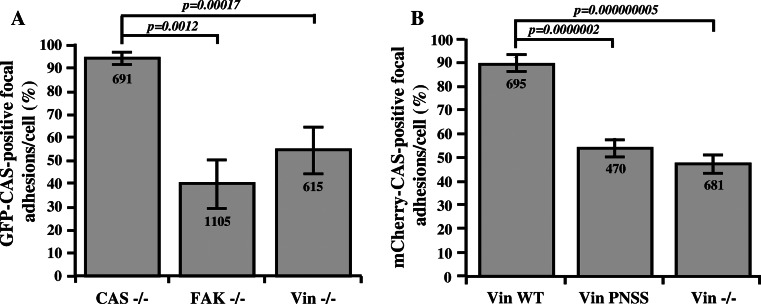
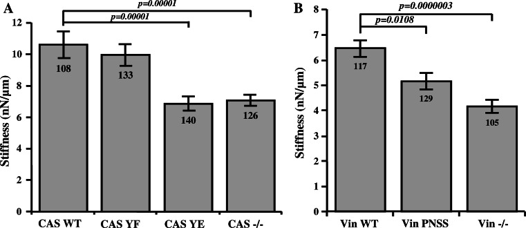
Focal adhesions are cellular structures through which both mechanical forces and regulatory signals are transmitted. Two focal adhesion-associated proteins, Crk-associated substrate (CAS) and vinculin, were both independently shown to be crucial for the ability of cells to transmit mechanical forces and to regulate cytoskeletal tension. Here, we identify a novel, direct binding interaction between CAS and vinculin. This interaction is mediated by the CAS SRC homology 3 domain and a proline-rich sequence in the hinge region of vinculin. We show that CAS localization in focal adhesions is partially dependent on vinculin, and that CAS-vinculin coupling is required for stretch-induced activation of CAS at the Y410 phosphorylation site. Moreover, CAS-vinculin binding significantly affects the dynamics of CAS and vinculin within focal adhesions as well as the size of focal adhesions. Finally, disruption of CAS binding to vinculin reduces cell stiffness and traction force generation. Taken together, these findings strongly implicate a crucial role of CAS-vinculin interaction in mechanosensing and focal adhesion dynamics.


Figures

Fig. 1
Fig. 1
The CAS SH3 domain interacts with the polyproline region of vinculin. a Binding of vinculin to SH3 domains of CAS WT, CAS Y12E, and CAS Y12F fused with GST was analyzed with pull-down assays by immunoblotting. Vinculin was detected with an anti vinculin antibody. GST fused SH3 domains were detected by Ponceau S staining. Aliquots of total cell lysates (total) were used as a control. b GFP CAS was immunoprecipitated from CAS−/− MEFs expressing CAS Y12 variants, and binding of vinculin and FAK (as a control) was analyzed using vinculin and FAK antibodies. c GFP CAS WT was immunoprecipitated from FAK−/− MEFs and binding of vinculin was analyzed using vinculin antibody. d In a far-Western experiment, Vin WT GFP or GFP immunoprecipitated from Vin−/− MEFs expressing the GFP constructs were transferred to nitrocellulose membranes and incubated with recombinant CAS–GST, followed by detection with anti-GST antibody. Loading controls of GFP constructs were analyzed by anti-GFP antibody. As a positive control for anti-GST reactivity, purified CAS–GST was run alongside. e GFP vinculin was immunoprecipitated from Vin−/− MEFs re-expressing GFP-fused Vin WT or Vin PNSS (PKPP sequence in the proline-rich region changed to PNSS), and binding of CAS and paxillin was detected with CAS and paxillin antibodies. f GFP vinculin was immunoprecipitated from Vin−/− MEFs re-expressing GFP-fused Vin WT or Vin PNSS (PKPP sequence in proline-rich region changed to PNSS), and binding of Arp2 was detected with Arp2 antibody
Fig. 2
Fig. 2
CAS localization in focal adhesions is dependent on FAK and vinculin. The bar graphs show the percentage of focal adhesions stained positive for GFP–CAS (a) or for mCherry-CAS (b). Focal adhesions were considered CAS-positive if the GFP–CAS or mCherry-CAS signal is at least double the signal in cytoplasm, adjacent to the focal adhesions, indicated by paxillin staining. Numbers in columns indicate number of analyzed focal adhesions and error bars represent standard deviation
Fig. 3
Fig. 3
CAS–vinculin interaction affects focal adhesion size. a Vin−/− MEFs re-expressing either Vin WT or Vin PNSS C-terminally fused with GFP were grown on fibronectin-coated coverslips and stained for paxillin (focal adhesion marker) and F-actin. Focal adhesion size was determined using ImageJ software. Scale bar 10 μm (b). The histogram bars represent average size of adhesion structures in cells deficient in vinculin, or re-expressing either Vin WT or Vin PNSS mutant. Numbers in columns indicate number of analyzed focal adhesions
Fig. 4
Fig. 4
Dependence of CAS dynamics on FAK and vinculin within focal adhesions. a FRAP curves of CAS-Venus associated with focal adhesions in MEFs lacking FAK or vinculin, or re-expressing either Vin WT or mutated Vin PNSS. Numbers indicate average half-maximum recovery times (t1/2). b The bar plot shows average half-maximum recovery times of CAS-Venus in different MEFs. Error bars represent standard errors
Fig. 5
Fig. 5
Dependence of vinculin dynamics on CAS–vinculin interaction. a FRAP curves of GFP-vinculin WT (left side) or GFP-vinculin PNSS (right side) associated with focal adhesions in CAS−/− MEFs re-expressing indicated CAS variants. Numbers in plot indicate average half-maximum recovery times (t1/2). b The bar plot shows half-maximum recovery times of GFP-fused Vin WT (left) and GFP-vinculin PNSS (right) in CAS−/− MEFs re-expressing indicated CAS variants. Error bars represent standard errors
Fig. 6
Fig. 6
Stretch-mediated phosphorylation of CAS is dependent on CAS–vinculin interaction. a Vin−/− MEFs or Vin−/− MEFS re-expressing indicated vinculin variants, b CAS−/− MEFs re-expressing indicated CAS variants and c MEFs transformed by constitutively active Src (MEFs + Src527F) were seeded on fibronectin-coated flexible membrane, incubated for 24 h, and then subjected to 20 % static stretch for 10 min (a, b) or for indicated times (c). Subsequently, cells were lysed and analyzed by Western blotting against phosphorylated Y410 in the CAS substrate domain and Y12 in CAS SH3 domain. Numbers indicate fold-change (mean ± SD) in CAS Y410 and Y12 phosphorylation after stretching. The immunoblots are representative of three independent experiments
Fig. 7
Fig. 7
Effect of CAS–vinculin interaction on cell mechanical properties. Stiffness of MEFs analyzed at 6nN force using magnetic tweezers. The bar plots show stiffness of a CAS−/− MEFs re-expressing indicated CAS variants and b Vin−/− MEFs re-expressing indicated vinculin variants. Numbers in columns indicate number of analyzed cells, and error bars represent standard error
Fig. 8
Fig. 8
Traction force generation is modulated by phosphorylation of Y12 in the CAS SH3 domain. a Bright field (upper row), traction field (middle row), and fluorescent (bottom row) images of CAS−/− MEFs re-expressing indicated CAS variants. Scale bar 10 μm. b The bar plot shows the strain energy generated by single cells (mean ± SE). Numbers in columns indicate number of analyzed cells
Fig. 9
Fig. 9
Disruption of CAS–vinculin interaction results in impaired traction forces generation. a Upper row cell image represented by GFP fluorescence (Vin Wt, Vin PNNS) and bright field image (Vin−/−) traction field (middle row), and fluorescent (bottom row) images of Vin−/− MEFs re-expressing different vinculin variants. Scale bar 10 μm. b The bar plot shows the strain energy generated by single cells (mean ± SE). Numbers in columns indicate number of analyzed cells
Fig. 10
Fig. 10
Model for regulation of CAS-dependent mechanosensing. CAS is anchored in focal adhesions by N-terminal SH3 and C-terminal CCH domains [22, 24]. In quiescent cells, the substrate domain of CAS adopts a compact structure (left). Mechanical stretch leads to extension of the CAS substrate domain and subsequent phosphorylation by Src, which activates CAS-mediated mechanotransduction signals (middle). Src phosphorylation of CAS on tyrosine 12 blocks CAS–vinculin binding, and the substrate domain returns to a compact structure. CAS-mediated mechanotransduction is attenuated (right) either by a gradual loss of substrate domain phosphorylation or by making the phosphorylated tyrosines in the substrate domain inaccessible for downstream signaling proteins

References

    1. Defilippi P, Di Stefano P, Cabodi S. p130Cas: a versatile scaffold in signaling networks. Trends Cell Biol. 2006;16:257–263. doi: 10.1016/j.tcb.2006.03.003. - DOI - PubMed
    1. Brabek J, Constancio BS, Shin NY, Pozzi A, Weaver AM, Hanks SK. CAS promotes invasiveness of Src-transformed cells. Oncogene. 2004;23:7406–7415. doi: 10.1038/sj.onc.1207965. - DOI - PubMed
    1. Brabek J, Constancio SS, Siesser PF, Shin NY, Pozzi A, Hanks SK. Crk-associated substrate tyrosine phosphorylation sites are critical for invasion and metastasis of Src-transformed cells. Mol Cancer Res. 2005;3:307–315. doi: 10.1158/1541-7786.MCR-05-0015. - DOI - PubMed
    1. Brinkman A, van der Flier S, Kok EM, Dorssers LCJ. BCAR1, a human homologue of the adapter protein p130Cas, and antiestrogen resistance in breast cancer cells. J Natl Cancer Inst. 2000;92:112–120. doi: 10.1093/jnci/92.2.112. - DOI - PubMed
    1. Dorssers LCJ, Grebenchtchikov N, Brinkman A, Look MP, Klijn AGM, Geurts-Moespot A, Span PN, Foekens JA, Sweep CGJ. Application of a newly developed ELISA for BCAR1 protein for prediction of clinical benefit of tamoxifen therapy in patients with advanced breast cancer. Clin Chem. 2004;50:1445–1447. doi: 10.1373/clinchem.2004.035493. - DOI - PubMed

Publication types

MeSH terms

LinkOut - more resources