

Platelet size and density affect shear-induced thrombus formation in tortuous arterioles
- PMID: 23974300
- PMCID: PMC3814027
- DOI: 10.1088/1478-3975/10/5/056003
Platelet size and density affect shear-induced thrombus formation in tortuous arterioles
Abstract
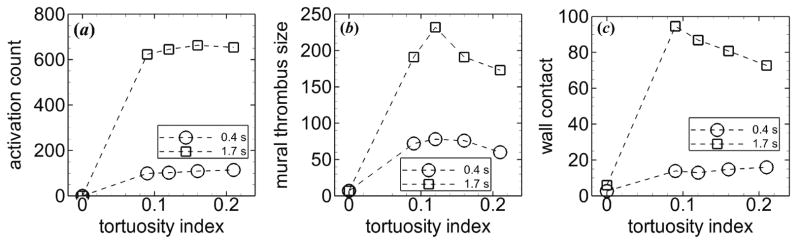
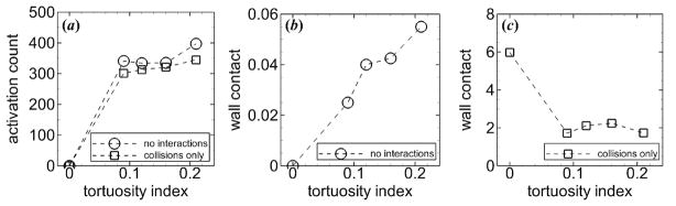
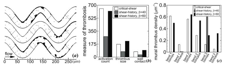
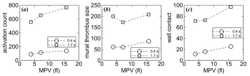
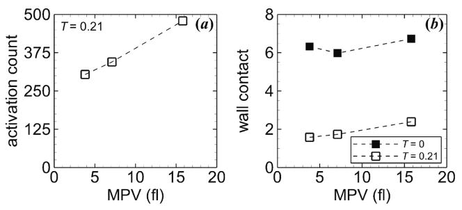
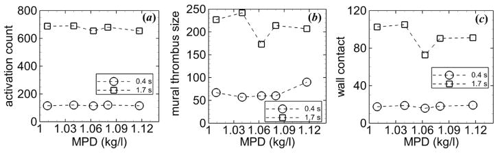
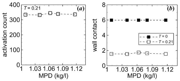
Thrombosis accounts for 80% of deaths in patients with diabetes mellitus. Diabetic patients demonstrate tortuous microvessels and larger than normal platelets. Large platelets are associated with increased platelet activation and thrombosis, but the physical effects of large platelets in the microscale processes of thrombus formation are not clear. Therefore, the objective of this study was to determine the physical effects of mean platelet volume (MPV), mean platelet density (MPD) and vessel tortuosity on platelet activation and thrombus formation in tortuous arterioles. A computational model of the transport, shear-induced activation, collision, adhesion and aggregation of individual platelets was used to simulate platelet interactions and thrombus formation in tortuous arterioles. Our results showed that an increase in MPV resulted in a larger number of activated platelets, though MPD and level of tortuosity made little difference on platelet activation. Platelets with normal MPD yielded the lowest amount of mural thrombus. With platelets of normal MPD, the amount of mural thrombus decreased with increasing level of tortuosity but did not have a simple monotonic relationship with MPV. The physical mechanisms associated with MPV, MPD and arteriole tortuosity play important roles in platelet activation and thrombus formation.
Figures
Similar articles
-
Effect of Red Blood Cells on Platelet Activation and Thrombus Formation in Tortuous Arterioles.Front Bioeng Biotechnol. 2013 Dec 3;1:18. doi: 10.3389/fbioe.2013.00018. eCollection 2013. Front Bioeng Biotechnol. 2013. PMID: 25022613 Free PMC article.
-
Numerical Simulation of Thrombotic Occlusion in Tortuous Arterioles.J Cardiol Cardiovasc Med. 2017;2(1):95-111. Epub 2017 Dec 6. J Cardiol Cardiovasc Med. 2017. PMID: 29327739 Free PMC article.
-
Tortuosity triggers platelet activation and thrombus formation in microvessels.J Biomech Eng. 2011 Dec;133(12):121004. doi: 10.1115/1.4005478. J Biomech Eng. 2011. PMID: 22206421 Free PMC article.
-
[Platelet--vessel wall interactions].Therapie. 2006 Sep-Oct;61(5):371-8. doi: 10.2515/therapie:2006068. Therapie. 2006. PMID: 17243265 Review. French.
-
Interactions of platelets, blood-borne tissue factor, and fibrin during arteriolar thrombus formation in vivo.Microcirculation. 2005 Apr-May;12(3):301-11. doi: 10.1080/10739680590925682. Microcirculation. 2005. PMID: 15814438 Review.
Cited by
-
Quantifying Platelet Margination in Diabetic Blood Flow.Biophys J. 2018 Oct 2;115(7):1371-1382. doi: 10.1016/j.bpj.2018.08.031. Epub 2018 Aug 30. Biophys J. 2018. PMID: 30224049 Free PMC article.
-
Effect of Red Blood Cells on Platelet Activation and Thrombus Formation in Tortuous Arterioles.Front Bioeng Biotechnol. 2013 Dec 3;1:18. doi: 10.3389/fbioe.2013.00018. eCollection 2013. Front Bioeng Biotechnol. 2013. PMID: 25022613 Free PMC article.
-
Lysyl oxidase is associated with increased thrombosis and platelet reactivity.Blood. 2016 Mar 17;127(11):1493-501. doi: 10.1182/blood-2015-02-629667. Epub 2016 Jan 11. Blood. 2016. PMID: 26755713 Free PMC article.
-
Metabolomic Analysis of Biochemical Changes in the Plasma of High-Fat Diet and Streptozotocin-Induced Diabetic Rats after Treatment with Isoflavones Extract of Radix Puerariae.Evid Based Complement Alternat Med. 2016;2016:4701890. doi: 10.1155/2016/4701890. Epub 2016 Mar 3. Evid Based Complement Alternat Med. 2016. PMID: 27042190 Free PMC article.
-
Numerical Simulation of Thrombotic Occlusion in Tortuous Arterioles.J Cardiol Cardiovasc Med. 2017;2(1):95-111. Epub 2017 Dec 6. J Cardiol Cardiovasc Med. 2017. PMID: 29327739 Free PMC article.
References
-
- Carr ME. Diabetes mellitus: A hypercoagulable state. J Diabetes Complications. 2001;15:44–54. - PubMed
-
- Hendra TJ, Oswald GA, Yudkin JS. Increased mean platelet volume after acute myocardial infarction relates to diabetes and to cardiac failure. Diabetes Res Clin Pract. 1988;5:63–9. - PubMed
-
- Tschoepe D, Roesen P, Esser J, Schwippert B, Nieuwenhuis HK, Kehrel B, Gries FA. Large platelets circulate in an activated state in diabetes mellitus. Semin Thromb Hemost. 1991;17:433–8. - PubMed
-
- Sharpe PC, Trinick T. Mean platelet volume in diabetes mellitus. Q J Med. 1993;86:739–42. - PubMed
Publication types
MeSH terms
Grants and funding
LinkOut - more resources
Full Text Sources
Other Literature Sources
Medical