. 2013 Oct;45(10):1244-1248.
doi: 10.1038/ng.2739. Epub 2013 Aug 25.

Desmoglein 1 deficiency results in severe dermatitis, multiple allergies and metabolic wasting

Affiliations

Desmoglein 1 deficiency results in severe dermatitis, multiple allergies and metabolic wasting

Liat Samuelov et al. Nat Genet. 2013 Oct.

Abstract

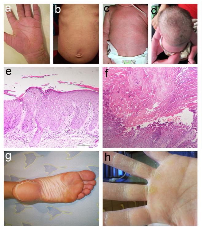
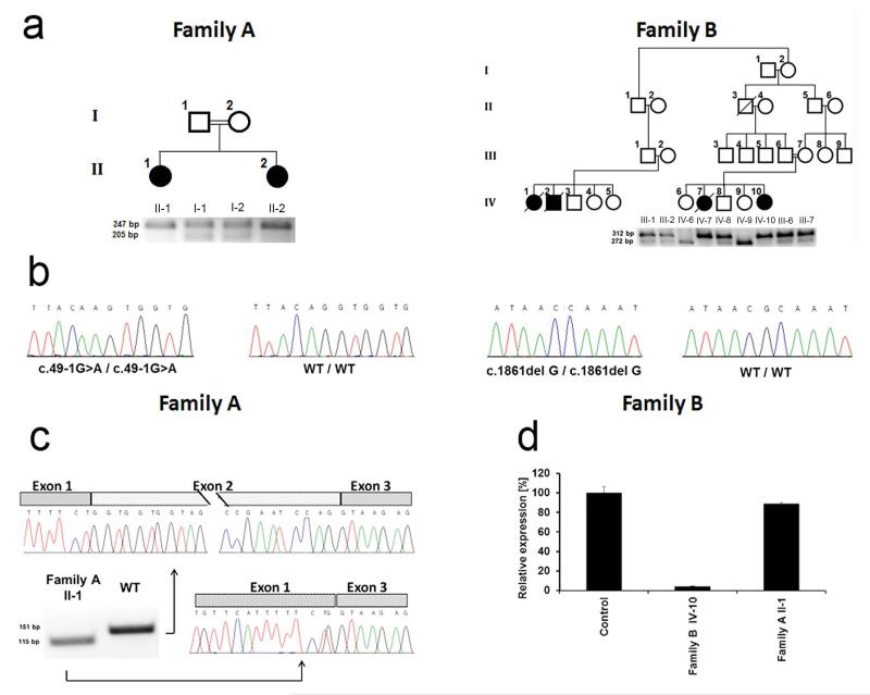
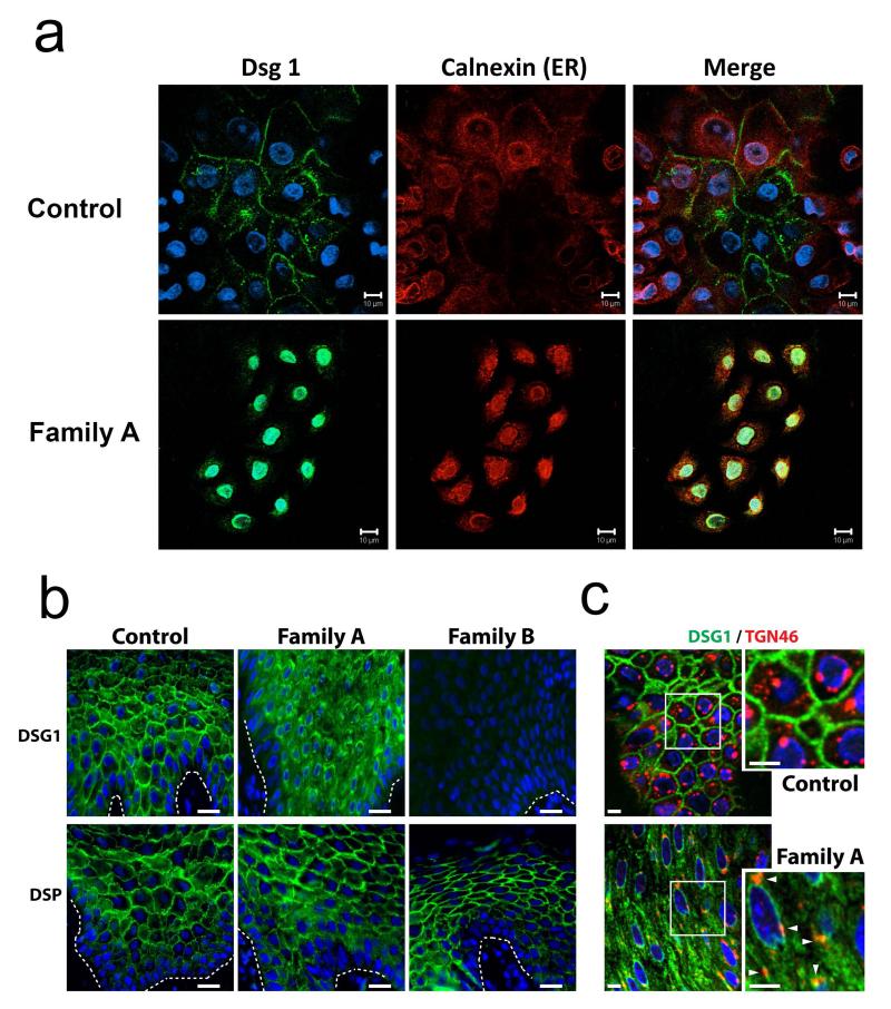
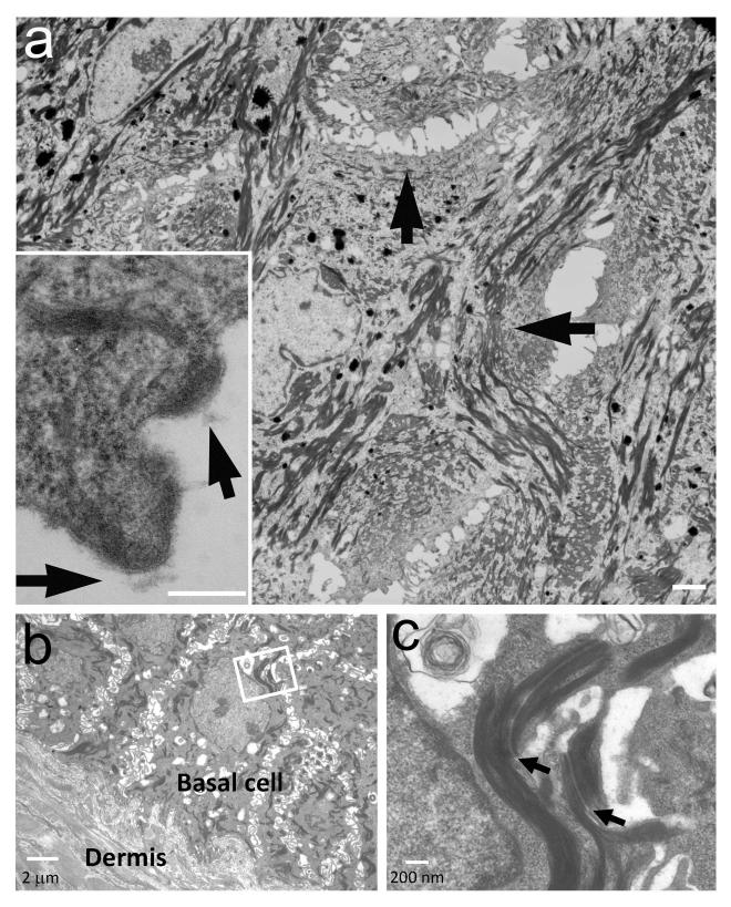
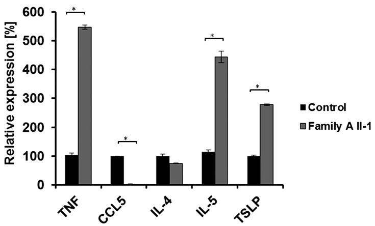
The relative contribution of immunological dysregulation and impaired epithelial barrier function to allergic diseases is still a matter of debate. Here we describe a new syndrome featuring severe dermatitis, multiple allergies and metabolic wasting (SAM syndrome) caused by homozygous mutations in DSG1. DSG1 encodes desmoglein 1, a major constituent of desmosomes, which connect the cell surface to the keratin cytoskeleton and have a crucial role in maintaining epidermal integrity and barrier function. Mutations causing SAM syndrome resulted in lack of membrane expression of DSG1, leading to loss of cell-cell adhesion. In addition, DSG1 deficiency was associated with increased expression of a number of genes encoding allergy-related cytokines. Our deciphering of the pathogenesis of SAM syndrome substantiates the notion that allergy may result from a primary structural epidermal defect.


Figures

Figure 1
Figure 1
Clinical and pathological features. (a) Individual II-2 of family A displays diffusely red and fissured palms covered with hyperkeratotic yellowish papules and plaques, which are arranged linearly over the fingers. (b,c) Body skin is reddish and covered with scales and erosions in individual II-1 of family A (b) and in individual IV-10 of family B (c). (d) Hypotrichosis is evident in individual IV-10 of family B. (e,f) Skin biopsies obtained from the popliteal area in individual IV-10 of family B (e) and from the palms of individual II-1 of family A (f) demonstrate subcorneal separation associated with acantholysis within the spinous and granular layers (hematoxylin and eosin; bars = 100 μm). (g,h) Heterozygous carriers of DSG1 mutations c.49-1G>A (individual I-2, family A) (g) and c.1861delG (individual III-3, family B) (h) display diffuse plantar and focal palmar keratoderma, respectively.
Figure 2
Figure 2
Molecular and immunohistochemical analysis. (a) Family pedigrees are presented in the upper panels. Black symbols denote affected individuals. PCR-RFLP assays (as described in the Online Methods) were used in each family to confirm co-segregation of the mutation with the disease phenotype (lower panels). Mutation c.49-1G>A is associated with the presence of a 247 bp fragment in family A. while mutation c.1861delG results in a 312bp fragment in family B. (b) Direct sequencing of DSG1 identified a homozygous G>A transition at position c.49-1 of the cDNA sequence in patients of family A and a homozygous single nucleotide G deletion at cDNA position c.1861 in family B. The wild type sequences (WT/WT) are given for comparison. (c) cDNA was reverse transcribed from an RNA sample extracted from patient II-1 (family A) skin and from a healthy individual. PCR amplification of a cDNA fragment spanning exons 1-3 demonstrates in the patient skin the presence of an aberrant PCR product shorter than the expected 151-bp fragment. Direct sequencing of both fragment indicated that c.49-1G>A results in skipping of exon 2. (d) Quantitative real-time PCR analysis for gene expression of DSG1 was performed using cDNA derived from skin biopsies of patients II-1 (family A) and IV-10 (family B) as well as a healthy individual. Results are provided as percentage of expression relative to gene expression in control + standard error normalized to ACTB RNA levels.
Figure 3
Figure 3
DSG1 and DSP expression in patient skin. (a) Keratinocytes were harvested from skin biopsies obtained from a healthy individual (Normal KC) and from individual I-1, family A (Family A). Healthy individual-derived keratinocytes demonstrate normal membrane localization of DSG1 (upper panels). There is no co-localization of DSG1 with the endoplasmic reticulum (ER) chaperone protein calnexin; in contrast, patient keratinocytes (Family A) display no membrane staining of DSG1; instead nuclear and cytoplasmic punctuate DSG1 staining is observed (lower panels). DSG1 occasionally co-localizes with calnexin (scale bar = 10 μm); (b) Immunofluorescence analysis of skin sections from control and affected individuals shows cytoplasmic accumulation of DSG1 and loss of DSG1 staining in patients carrying mutations c.49-1G>A (Family A) and c.1861delG (Family B), respectively, compared with the plasma membrane localization in control samples (upper panels) (scale bars = 20 μm). DSP staining is retained in both c.49-1G>A and c.1861delG-carrying individuals (lower panels). (c) Double label immunofluorescence analysis of skin sections obtained from a healthy individual (Control) and from an individual carrying c.49-1G>A (Family A) showed partial co-localization of cytoplasmic DSG1 with TGN46, a Golgi and endosome marker, in the patient skin (scale bars = 5 μm).
Figure 4
Figure 4
Electron microscopy. (a) Electron microscopy demonstrates large areas lacking mature desmosomes in the upper spinous and granular layers with retracted keratin filaments (arrows) (bar = 4 μm). In these areas, half split desmosomes can be seen (insert, arrows, bar = 200 nm). (b) Acantholysis is absent in the basal cell layers (bar = 2μm); (c) Desmosomes (arrows) are normal in the basal and lower spinous cells (bar = 200 nm).
Figure 5
Figure 5
Cytokine gene expression. Cytokine gene expression in keratinocytes isolated from patient II-1 (Family A) or from a healthy control was measured using qRT-PCR. Results are expressed as percentage of expression relative to gene expression in control cells + standard error (two sided t-test; *P < 0.01). Results are normalized to ACTB RNA levels.

References

    1. Menon GK, Cleary GW, Lane ME. The structure and function of the stratum corneum. Int. J. Pharm. 2012;435:3–9. - PubMed
    1. Kubo A, Nagao K, Amagai M. Epidermal barrier dysfunction and cutaneous sensitization in atopic diseases. J. Clin. Invest. 2012;122:440–447. - PMC - PubMed
    1. Simpson CL, Patel DM, Green KJ. Deconstructing the skin: cytoarchitectural determinants of epidermal morphogenesis. Nat. Rev. Mol. Cell. Biol. 2011;12:565–580. - PMC - PubMed
    1. Desai BV, Harmon RM, Green KJ. Desmosomes at a glance. J. Cell Sci. 2009;122:4401–4407. - PMC - PubMed
    1. Amagai M, Stanley JR. Desmoglein as a target in skin disease and beyond. J. Invest. Dermatol. 2012;132:776–784. - PMC - PubMed

Publication types

Associated data