. 2013 Aug 23;2(4):e000365.
doi: 10.1161/JAHA.113.000365.

Hypotension due to Kir6.1 gain-of-function in vascular smooth muscle

Affiliations

Hypotension due to Kir6.1 gain-of-function in vascular smooth muscle

Anlong Li et al. J Am Heart Assoc. .

Abstract

Background: KATP channels, assembled from pore-forming (Kir6.1 or Kir6.2) and regulatory (SUR1 or SUR2) subunits, link metabolism to excitability. Loss of Kir6.2 results in hypoglycemia and hyperinsulinemia, whereas loss of Kir6.1 causes Prinzmetal angina-like symptoms in mice. Conversely, overactivity of Kir6.2 induces neonatal diabetes in mice and humans, but consequences of Kir6.1 overactivity are unknown.

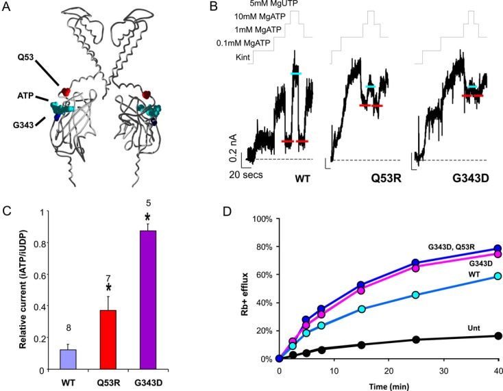
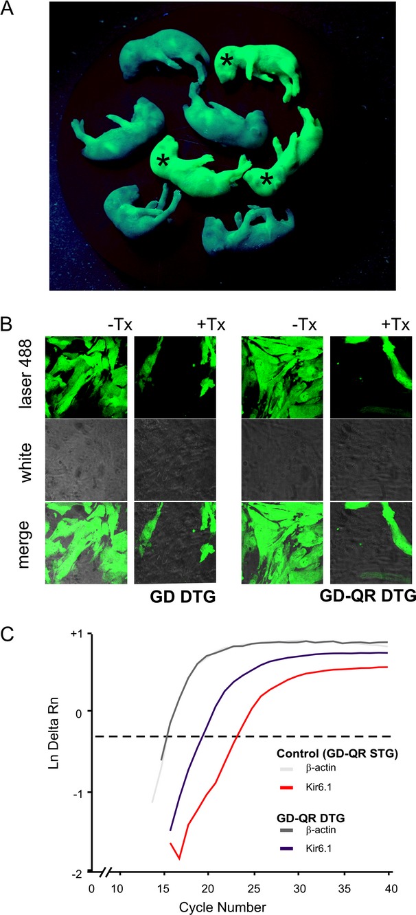
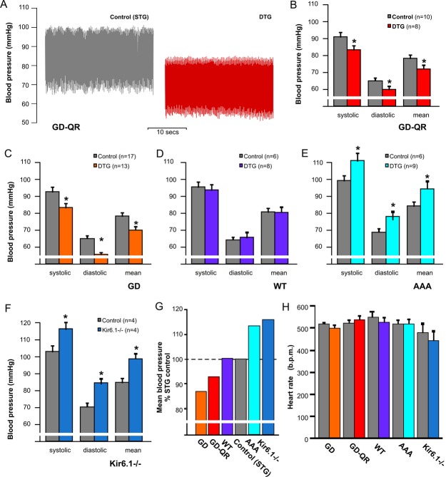
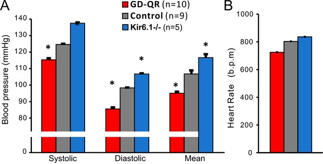
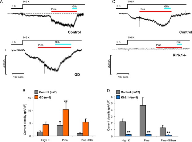
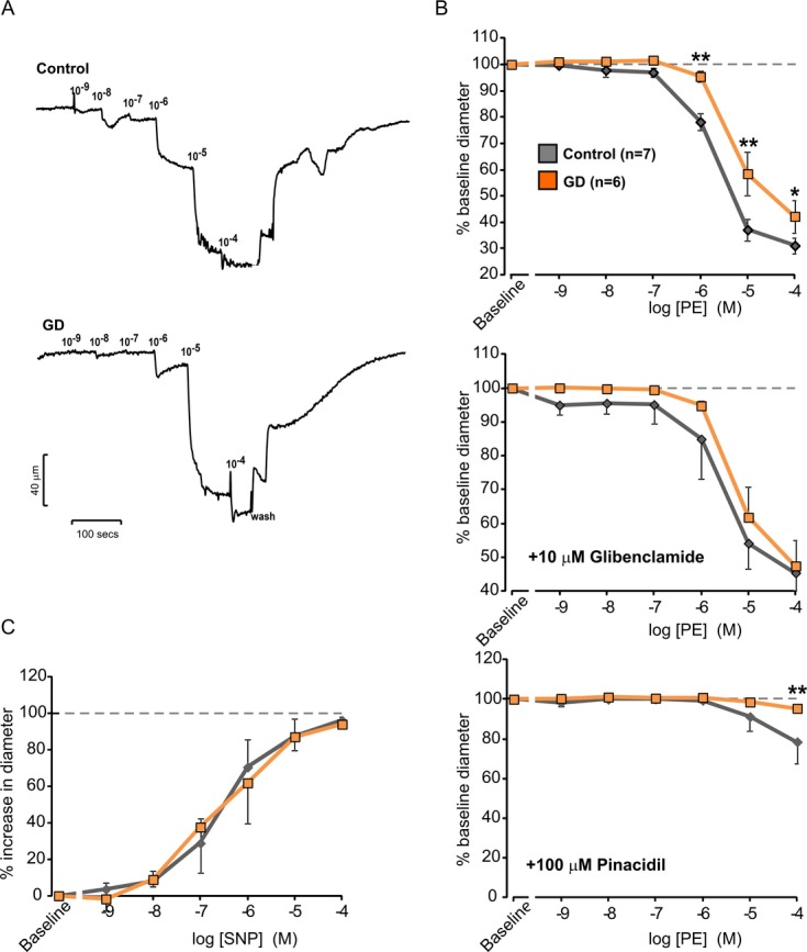
Methods and results: We generated transgenic mice expressing wild-type (WT), ATP-insensitive Kir6.1 [Gly343Asp] (GD), and ATP-insensitive Kir6.1 [Gly343Asp,Gln53Arg] (GD-QR) subunits, under Cre-recombinase control. Expression was induced in smooth muscle cells by crossing with smooth muscle myosin heavy chain promoter-driven tamoxifen-inducible Cre-recombinase (SMMHC-Cre-ER) mice. Three weeks after tamoxifen induction, we assessed blood pressure in anesthetized and conscious animals, as well as contractility of mesenteric artery smooth muscle and KATP currents in isolated mesenteric artery myocytes. Both systolic and diastolic blood pressures were significantly reduced in GD and GD-QR mice but normal in mice expressing the WT transgene and elevated in Kir6.1 knockout mice as well as in mice expressing dominant-negative Kir6.1 [AAA] in smooth muscle. Contractile response of isolated GD-QR mesenteric arteries was blunted relative to WT controls, but nitroprusside relaxation was unaffected. Basal KATP conductance and pinacidil-activated conductance were elevated in GD but not in WT myocytes.

Conclusions: KATP overactivity in vascular muscle can lead directly to reduced vascular contractility and lower blood pressure. We predict that gain of vascular KATP function in humans would lead to a chronic vasodilatory phenotype, as indeed has recently been demonstrated in Cantu syndrome.

Keywords: ABCC9; KATP; KCNJ8; hypotension; mice; transgenic.


Figures

Figure 1.
Figure 1.
Reiteration of GOF phenotype from mutations in Kir6.1. A, Homology model of kir6.1 indicating location of mutated residues. Residue Gly343 is found in the predicted ATP binding site, whereas Gln53 is located in the amino‐terminus and is involved in transduction. B, Excised patch currents from SUR1/Kir6.1 subunits coexpressed in COSm6 cells, in nucleotide‐free Kint solution or in Mg‐nucleotides as indicated. WT channels are activated at low [ATP] and high [UTP]. WT channels become strongly inhibited at high (10 mmol/L) ATP, but Gln53Arg (Q53R) and Gly343Asp (G343D) are insensitive to ATP inhibition. C, Averaged data from experiments as in (B) (currents in 10 mmol/L MgATP/current in 5 mmol/L MgUTP, mean±SEM, n as indicated, 1‐way ANOVA followed by post‐hoc Tukey's test). D, Representative data from an 86Rb efflux experiment on untransfected (Unt) COS‐m6 cells and cells transfected with Kir6.1WT, (G343D) or G343D, Q53R mutant channels, plus SUR1. Basal activity of GD or GD‐QR channels was significantly higher than WT in the intact cell. GOF indicates gain‐of‐function; WT, wild‐type.
Figure 2.
Figure 2.
Generation of Cx1‐Kir6.1[WT, GD, and GD‐QR] transgenic mice, and targeted transgene expression in specific tissue. A, CX1 mice crossed with C57 WT mice, under UV light, newborn mice carrying the CX1 transgene were green (*), whereas nontransgenic mice were dark. B, Primary cultured mesenteric artery smooth muscle cells from the GD and GD‐QR DTG offspring of CX1 transgenic mice and SMMHC‐Cre‐ER mice. Confocal images show that VSM cells from DTG with tamoxifen‐induction lose green fluorescence but other cells remain green. C, In transgene targeted vascular tissue, quantitative real time PCR shows the copy number of transgene in DTG mice is significantly higher than that of their STG littermates. DTG indicates double‐transgenic; PCR, polymerase chain reaction; SMMHC‐Cre‐ER, smooth muscle myosin heavy chain promoter‐driven tamoxifen inducible Cre recombinase; STG, single‐transgenic; VSM, vascular smooth muscle.
Figure 3.
Figure 3.
Kir6.1 GOF results in hypotension in mice, whereas Kir6.1 LOF results in hypertensive phenotype. A, Typical blood pressure measurement traces show that both systolic and diastolic blood pressure are markedly lower in anesthetized mice expressing GD‐QR in smooth muscle cells than that of control littermates. B through F, Averaged systolic, diastolic, and mean blood pressures from each genotype studied in GD‐QR (B), GD (C), WT (D) and AAA (E) DTG mice, and control littermates, as well as Kir6.1−/− and Kir6.1+/+ littermates (F). *P < .05 versus control by unpaired Student's t tests in (B through F). G, Normalized averaged mean blood pressures of each genotype to the average value of their STG control littermates illustrates the marked progression from hypotensive GD and GD‐QR DTG to hypertensive Kir6.1−/− and Kir6.1[AAA] animals. H, Heart rates from each condition demonstrate no significant difference by 1‐way ANOVA. DTG indicates double‐transgenic; GOF, gain‐of‐function; LOF, loss of function; WT, wild‐type.
Figure 4.
Figure 4.
Kir6.1 GOF results in hypotension in conscious mice, whereas Kir6.1 LOF results in hypertensive phenotype (A) Averaged systolic, diastolic, and mean blood pressures from GD‐QR DTG (n=10), Kir6.1−/− mice (n=5), and control mice (n=9). B, Averaged heart rates demonstrate no significant difference between the 3 groups. *P < .05 versus control by one‐way ANOVA followed by post‐hoc Tukey's test. DTG indicates double‐transgenic; GOF, gain‐of‐function; LOF, loss of function.
Figure 5.
Figure 5.
Kir6.1 GOF results in enhanced KATP channel currents in isolated arterial smooth muscle cells. A and C, Representative whole‐cell recordings of KATP currents in control (top) and GD DTG (A) or Kir6.1−/− (C) (bottom) VSMCs. VSMCs were sequentially exposed to low (6 mmol/L) K+, high (140 mmol/L) K+, pinacidil, and glibenclamide. B and D, Summarized data show KATP current density in high K+, pinacidil and glibenclamide for GD DTG (B) or Kir6.1−/− (D) and control littermate VSMCs. **P < .01 versus control at the same condition, by 2‐way ANOVA followed by Bonferroni's posttests. DTG indicates double‐transgenic; GOF, gain‐of‐function; VSMCs, vascular smooth muscle cells.
Figure 6.
Figure 6.
Kir6.1 GOF results in decreased vascular reactivity of Kir6.1 GOF mouse mesenteric arteries. A, Representative recordings of vessel diameter of (top) WT control and GD‐QR DTG (bottom) mesenteric arteries in response to increasing phenylephrine (PE) concentrations. B, Concentration‐response of PE‐induced vasoconstriction from experiments as in (A) (n=6 in each case) expressed as percent decrease in arterial diameter after application of each concentration of PE (10−9 to 10−4 mmol/L) as indicated (top) and in the additional presence of glibenclamide 10 μmol/L (center) or pinacidil 100 μmol/L (bottom). C, Concentration‐dependent vasodilatation of control and GD‐QR DTG mesenteric arteries in response to sodium nitroprusside (SNP) following constriction with 5 μmol/L PE. As indicated, percent relaxation shows no difference between control (n=4) and GD‐QR DTG (n=5) arterial diameter after application of PE and increasing concentration of SNP. Data are expressed as mean±SE in all cases. *P < .05, **P < .01 versus control at the same condition (2‐way repeated‐measures ANOVA followed by Bonferroni's posttests). DTG indicates double‐transgenic; GOF, gain‐of‐function; WT, wild‐type.

Similar articles

Cited by

References

    1. Noma A. ATP‐regulated K+ channels in cardiac muscle. Nature. 1983; 305:147-148 - PubMed
    1. Standen NB, Quayle JM, Davies NW, Brayden JE, Huang Y, Nelson MT. Hyperpolarizing vasodilators activate ATP‐sensitive K+ channels in arterial smooth muscle. Science. 1989; 245:177-180 - PubMed
    1. Katnik C, Adams DJ. Characterization of ATP‐sensitive potassium channels in freshly dissociated rabbit aortic endothelial cells. Am J Physiol. 1997; 272:H2507-H2511 - PubMed
    1. Kurata HT, Marton LJ, Nichols CG. The polyamine binding site in inward rectifier K+ channels. J Gen Physiol. 2006; 127:467-480 - PMC - PubMed
    1. Yamada M, Isomoto S, Matsumoto S, Kondo C, Shindo T, Horio Y, Kurachi Y. Sulphonylurea receptor 2B and Kir6.1 form a sulphonylurea‐sensitive but ATP‐insensitive K+ channel. J Physiol. 1997; 499:715-720 - PMC - PubMed

Publication types

MeSH terms