. 2014 Jan;63(1):132-41.
doi: 10.2337/db13-0940. Epub 2013 Aug 23.

Mitochondrial respiratory capacity and content are normal in young insulin-resistant obese humans

Affiliations

Mitochondrial respiratory capacity and content are normal in young insulin-resistant obese humans

Kelsey H Fisher-Wellman et al. Diabetes. 2014 Jan.

Abstract

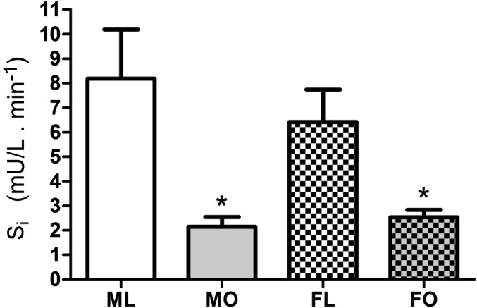
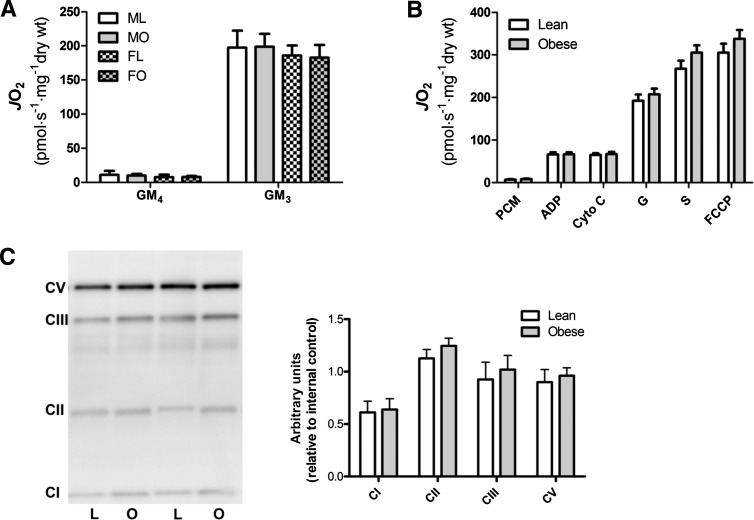
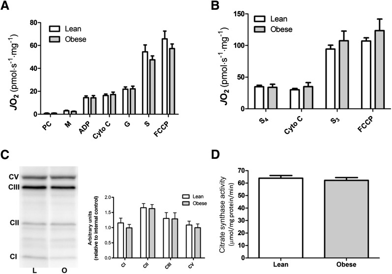
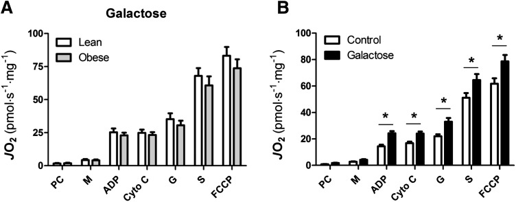
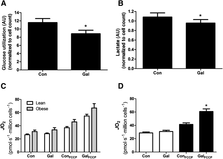
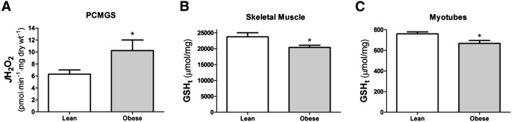
Considerable debate exists about whether alterations in mitochondrial respiratory capacity and/or content play a causal role in the development of insulin resistance during obesity. The current study was undertaken to determine whether such alterations are present during the initial stages of insulin resistance in humans. Young (∼23 years) insulin-sensitive lean and insulin-resistant obese men and women were studied. Insulin resistance was confirmed through an intravenous glucose tolerance test. Measures of mitochondrial respiratory capacity and content as well as H(2)O(2) emitting potential and the cellular redox environment were performed in permeabilized myofibers and primary myotubes prepared from vastus lateralis muscle biopsy specimens. No differences in mitochondrial respiratory function or content were observed between lean and obese subjects, despite elevations in H(2)O(2) emission rates and reductions in cellular glutathione. These findings were apparent in permeabilized myofibers as well as in primary myotubes. The results suggest that reductions in mitochondrial respiratory capacity and content are not required for the initial manifestation of peripheral insulin resistance.


Figures

Figure 1
Figure 1
Impact of sex and obesity on insulin sensitivity. Insulin sensitivity index was calculated in response to an IVGTT. Data are mean ± SEM. *Different from ML or FL (P < 0.05). FL, female lean (n = 11); FO, female obese (n = 11); ML, male lean (n = 13); MO, male obese (n =10); Si, insulin sensitivity index.
Figure 2
Figure 2
Mitochondrial respiratory capacity is not different in permeabilized myofibers from young lean and obese humans. A and B: Mitochondrial oxygen consumption rate (JO2) was assessed in permeabilized myofibers prepared from vastus lateralis muscle of lean and obese human subjects. A: JO2 in the presence of glutamate (10 mmol/L) and malate (2 mmol/L) (GM) under basal (state 4 [GM4]) and maximal ADP (4 mmol/L)–stimulated (state 3 [GM3]) conditions. B: JO2 in response to palmitoyl-l-carnitine (25 μmol/L) and malate (2 mmol/L) (PCM), ADP (4 mmol/L), cytochrome C (Cyto C) (10 μmol/L), glutamate (G) (10 mmol/L), succinate (S) (10 mmol/L), and FCCP (2 μmol/L). With the exception of A, male and female data were pooled to compare lean vs. obese. C: Western blot analysis of mitochondrial OXPHOS proteins (MitoSciences) prepared from vastus lateralis frozen tissue homogenate. Data are mean ± SEM; n = 7–10 (A); n = 16–17 (B); n = 10 (C). CI, complex I; CII, complex II; CIII, complex III; CV, complex V; FL, female lean; FO, female obese; L, lean; ML, male lean; MO, male obese; O, obese; wt, weight.
Figure 3
Figure 3
Respiratory capacity and mitochondrial content in primary myotubes are not different in primary myotubes from young lean and obese humans. Mitochondrial oxygen consumption rate (JO2) was assessed in permeabilized primary human myotubes prepared from vastus lateralis muscle of lean and obese subjects (A and B). A: JO2 in response to palmitoyl-l-carnitine (PC) (25 μmol/L), malate (M) (2 mmol/L), ADP (4 mmol/L), cytochrome C (Cyto C) (10 μmol/L), glutamate (G) (10 mmol/L), succinate (S) (10 mmol/L), and FCCP (2 μmol/L). B: JO2 in the presence of S (10 mmol/L) plus rotenone (1 μmol/L), under basal (S4), ADP-stimulated (S3), and uncoupled (FCCP) conditions. C: Western blot analysis of mitochondrial OXPHOS proteins (MitoSciences) prepared from cell lysate. Note the representative image was spliced to remove sample lanes not relevant to the current data set. The image is from a single gel. D: Citrate synthase activity. Data are mean ± SEM; n = 9–10 (A); n = 5 (B); n = 10 (C and D). CI, complex I; CII, complex II; CIII, complex III; CV, complex V; L, lean; O, obese.
Figure 4
Figure 4
Myotubes from young lean and obese humans show similar adaptive increases in respiratory capacity in response to metabolic challenge. A and B: Fully differentiated myotubes were incubated for 24 h in the presence of galactose, which was added directly to the differentiation media. After this 24-h incubation, myotubes were harvested and permeabilized, and oxygen consumption was assessed. B: Data from lean and obese subjects were pooled to illustrate the effects of galactose. Data are mean ± SEM; n = 9–10 (A); n = 19 (B). *Different from vehicle control (P < 0.05). Cyto C, cytochrome C; G, glutamate; JO2, rate of mitochondrial oxygen consumption; M, malate; PC, palmitoyl-l-carnitine; S, succinate.
Figure 5
Figure 5
Basal and FCCP-stimulated respiration within intact primary human myotubes. Primary human myotubes were incubated for 24 h in differentiation media alone (control) or in differentiation media supplemented with galactose. A and B: Glucose utilization and lactate production during the 24-h incubations. C and D: Basal and FCCP (5 μmol/L)–stimulated respiration was assessed in intact primary human myotubes after the 24-h incubation. D: Pooled data from lean and obese subjects. Data are mean ± SEM; n = 14–16 (A, B, and D); n = 7–8 (C). *Different from corresponding vehicle control condition (P < 0.05). AU, arbitrary units; Con, control; Gal, galactose; JO2, mitochondrial oxygen consumption rate.
Figure 6
Figure 6
Elevations in H2O2 emitting potential and depressed total glutathione (GSH) within young obese human subjects. A: Mitochondrial rates of H2O2 emission (JH2O2) were assessed in permeabilized fibers in the presence of palmitoyl-l-carnitine (25 μmol/L), malate (2 mmol/L), glutamate (5 mmol/L), and succinate (10 mmol/L) (PCMGS). B and C: Total GSH (GSHt) was assessed in tissue homogenate from vastus lateralis muscle of human subjects and cell lysate from primary human myotubes. Data are mean ± SEM; n = 11/15 (A), n = 10 (B and C). *Different from lean (P < 0.05).

Comment in

Similar articles

Cited by

References

    1. Kelley DE, He J, Menshikova EV, Ritov VB. Dysfunction of mitochondria in human skeletal muscle in type 2 diabetes. Diabetes 2002;51:2944–2950 - PubMed
    1. Ritov VB, Menshikova EV, He J, Ferrell RE, Goodpaster BH, Kelley DE. Deficiency of subsarcolemmal mitochondria in obesity and type 2 diabetes. Diabetes 2005;54:8–14 - PubMed
    1. Ritov VB, Menshikova EV, Azuma K, et al. Deficiency of electron transport chain in human skeletal muscle mitochondria in type 2 diabetes mellitus and obesity. Am J Physiol Endocrinol Metab 2010;298:E49–E58 - PMC - PubMed
    1. Petersen KF, Dufour S, Befroy D, Garcia R, Shulman GI. Impaired mitochondrial activity in the insulin-resistant offspring of patients with type 2 diabetes. N Engl J Med 2004;350:664–671 - PMC - PubMed
    1. Petersen KF, Befroy D, Dufour S, et al. Mitochondrial dysfunction in the elderly: possible role in insulin resistance. Science 2003;300:1140–1142 - PMC - PubMed

Publication types