. 2013 Aug 22;14(9):17256-78.
doi: 10.3390/ijms140917256.

Assessing the effect of loop mutations in the folding space of β2-microglobulin with molecular dynamics simulations

Affiliations

Assessing the effect of loop mutations in the folding space of β2-microglobulin with molecular dynamics simulations

Sílvia G Estácio et al. Int J Mol Sci. .

Abstract

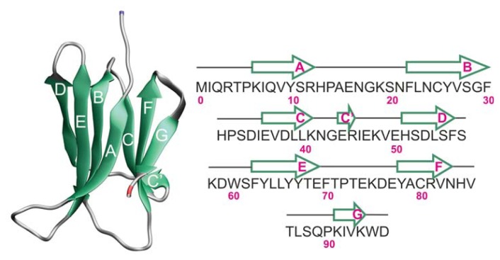
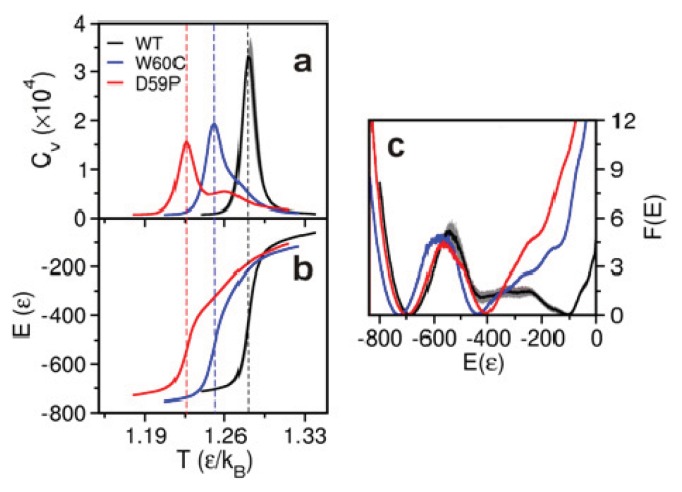
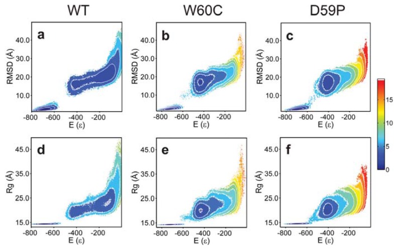
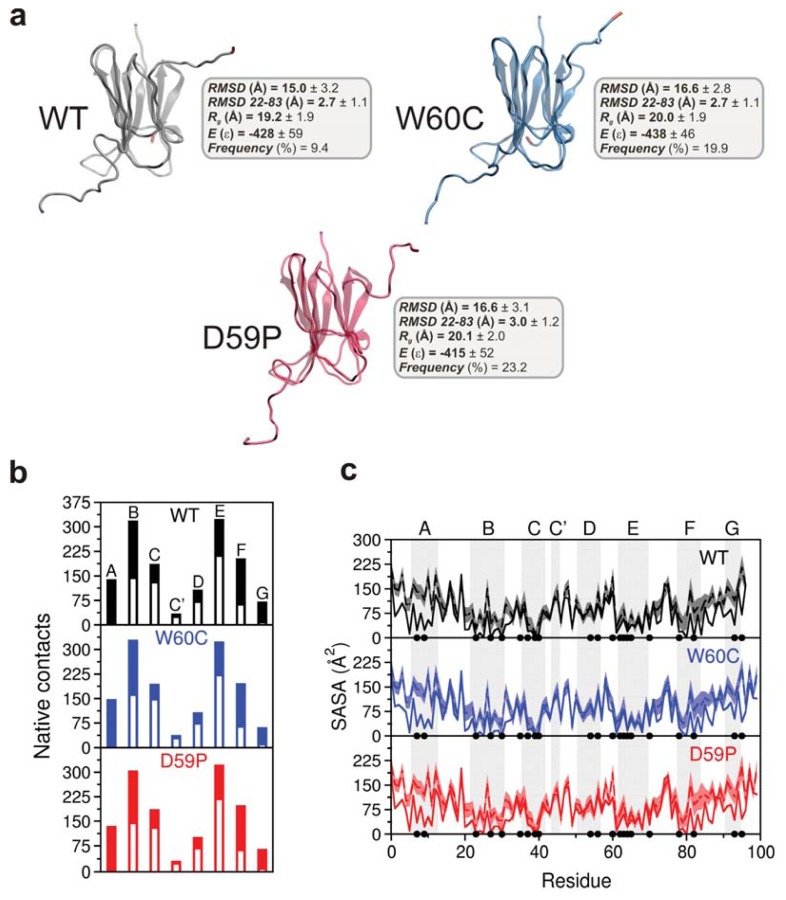
We use molecular dynamics simulations of a full atomistic Gō model to explore the impact of selected DE-loop mutations (D59P and W60C) on the folding space of protein human β2-microglobulin (Hβ2m), the causing agent of dialysis-related amyloidosis, a conformational disorder characterized by the deposition of insoluble amyloid fibrils in the osteoarticular system. Our simulations replicate the effect of mutations on the thermal stability that is observed in experiments in vitro. Furthermore, they predict the population of a partially folded state, with 60% of native internal free energy, which is akin to a molten globule. In the intermediate state, the solvent accessible surface area increases up to 40 times relative to the native state in 38% of the hydrophobic core residues, indicating that the identified species has aggregation potential. The intermediate state preserves the disulfide bond established between residue Cys25 and residue Cys80, which helps maintain the integrity of the core region, and is characterized by having two unstructured termini. The movements of the termini dominate the essential modes of the intermediate state, and exhibit the largest displacements in the D59P mutant, which is the most aggregation prone variant. PROPKA predictions of pKa suggest that the population of the intermediate state may be enhanced at acidic pH explaining the larger amyloidogenic potential observed in vitro at low pH for the WT protein and mutant forms.


Figures

Figure 1
Figure 1
The native structure of wild-type (WT) human beta-2 microglobulin (Hβ2m) (PDB ID: 1LDS) and its primary sequence. The location of each β-strand along the protein sequence is also shown. In the single-point mutants, the secondary structure assignment is identical. Hβ2m exhibits a sandwich-like structure formed by two sheets of anti-parallel β-strands. One of the sheets comprises strands A-B-E-D with the second sheet being formed by strands C-F-G. The native structure is stabilized by a disulfide bond established between Cys25 (on strand B) and Cys80 (on strand F). Another key structural feature is the existence of a peptidyl-prolyl bond between His31 and Pro32 (on the BC-loop) which adopts a thermodynamically unfavorable cis isomerization in the native structure.
Figure 2
Figure 2
Folding thermodynamics. Temperature dependence of the heat capacity (a) and energy E (b) of the three Hβ2m variants and free energy profile along the reaction coordinate E at Tm (c). The melting (or folding) temperature, Tm, is that corresponding to the peak of the heat capacity curve.
Figure 3
Figure 3
Free energy surfaces. Projection of the free energy on the energy (E) and root-mean-square deviation (RMSD) (ac) and on the energy (E) and radius of gyration (Rg) (df) at Tm.
Figure 4
Figure 4
Characterization of the intermediate state identified for each of the three Hβ2m variants. (a) Three-dimensional structures of the intermediate state fitted to the original X-ray native structure. The (starting) N-terminus is colored in blue while the C-terminus is shown in red. The intermediate state populates 9.4%, 19.9%, and 23.2% of the equilibrium ensembles of Hβ2m (WT), W60C, and D59P, respectively, at their respective Tm; (b) Comparison between the average number of native contacts established by each β-strand in the intermediate (white bars) and the number of native contacts by β-strand in the respective PDB native structure; (c) Mean SASA per residue in the intermediate state compared with that of the native structure (black line). The shaded area stands for the standard error bars for the mean SASA ensemble average. The black dots represent the 21 hydrophobic core amino acids: Leu7, Val9, Leu23, Val27, Phe30, Ile35, Val37, Leu39, Leu40, Leu54, Phe56, Trp60, Phe62, Tyr63, Leu64, Leu65, Phe70, Tyr78, Val82, Val93, and Trp95.
Figure 5
Figure 5
(Cα) covariance matrices (non-mass-weighted) for the native (ac) and intermediate states (df). A red point corresponding to a positive covariance value indicates that the two Cα atoms move in a correlated manner (i.e., together) while the blue color indicates that the atoms move into opposite directions. White spots indicate null covariance values, which mean that the Cα movements are not correlated.
Figure 6
Figure 6
Contribution of each Cα atom to the total RMSF along the first five eigenvectors for the sampled native (a), intermediate (b), and native and intermediate (c) ensembles of the three Hβ2m forms.

Similar articles

Cited by

References

    1. Trinh C.H., Smith D.P., Kalverda A.P., Phillips S.E.V., Radford S.E. Crystal structure of monomeric human β-2-microglobulin reveals clues to its amyloidogenic properties. Proc. Natl. Acad. Sci. USA. 2002;99:9771–9776. - PMC - PubMed
    1. Hasegawa K., Ohhashi Y., Yamaguchi I., Takahashi N., Tsutsumi S., Goto Y., Gejyo F., Naiki H. Amyloidogenic synthetic peptides of β2-microglobulin—A role of the disulfide bond. Biochem. Biophys. Res. Commun. 2003;304:101–106. - PubMed
    1. Yamamoto K., Yagi H., Ozawa D., Sasahara K., Naiki H., Goto Y. Thiol compounds inhibit the formation of amyloid fibrils by β2-microglobulin at neutral pH. J. Mol. Biol. 2008;376:258–268. - PubMed
    1. Bellotti V., Gallieni M., Giorgetti S., Brancaccio D. Dynamic of β2-microglobulin fibril formation and reabsorption: The role of proteolysis. Semin. Dial. 2001;14:117–122. - PubMed
    1. Eakin C.M., Miranker A.D. From chance to frequent encounters: Origins of β2-microglobulin fibrillogenesis. BBA-Proteins Proteom. 2005;1753:92–99. - PubMed

Publication types

Substances

LinkOut - more resources