Clinical Trial
. 2014 Feb;99(2):267-75.
doi: 10.3324/haematol.2013.090076. Epub 2013 Aug 23.

Resveratrol accelerates erythroid maturation by activation of FoxO3 and ameliorates anemia in beta-thalassemic mice

Affiliations
Clinical Trial

Resveratrol accelerates erythroid maturation by activation of FoxO3 and ameliorates anemia in beta-thalassemic mice

Sara Santos Franco et al. Haematologica. 2014 Feb.

Abstract

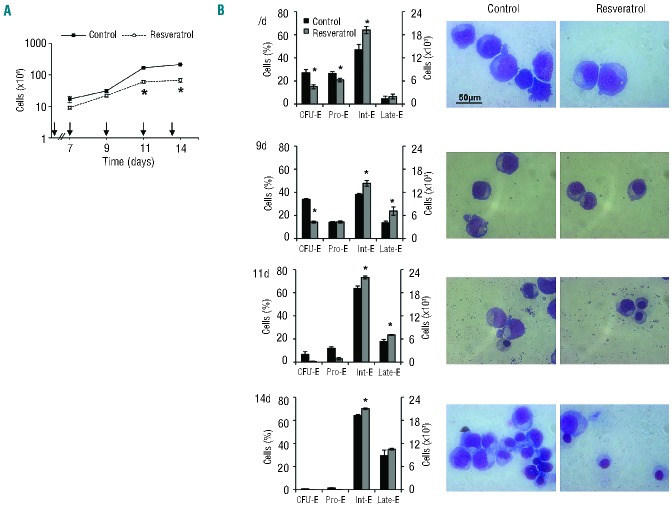
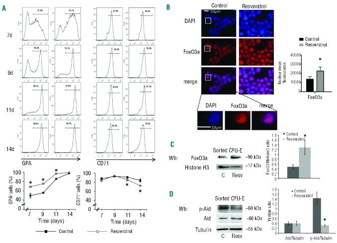
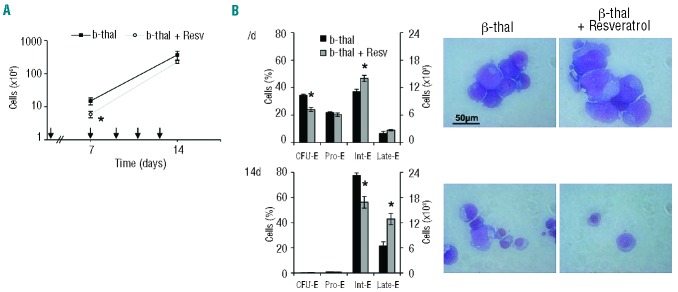
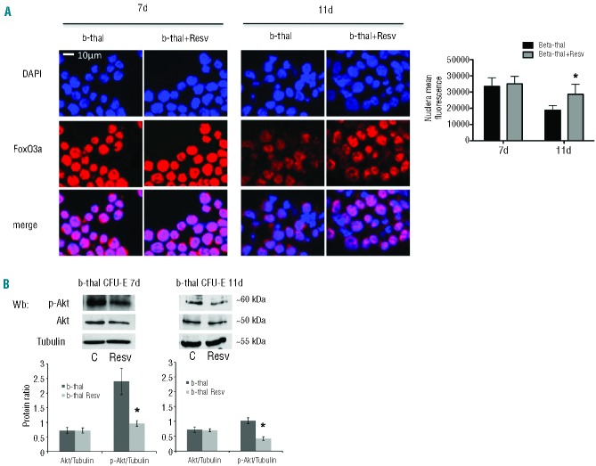
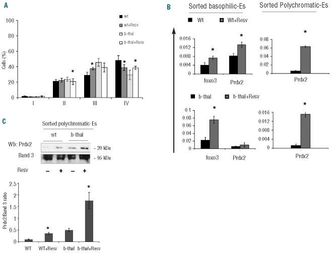
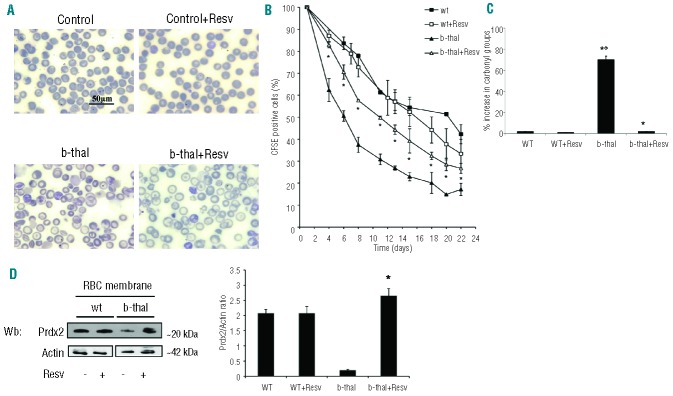
Resveratrol, a polyphenolic-stilbene, has received increased attention in the last decade due to its wide range of biological activities. Beta(β)-thalassemias are inherited red cell disorders, found worldwide, characterized by ineffective erythropoiesis and red cell oxidative damage with reduced survival. We evaluated the effects of low-dose-resveratrol (5 μM) on in vitro human erythroid differentiation of CD34(+) from normal and β-thalassemic subjects. We found that resveratrol induces accelerated erythroid-maturation, resulting in the reduction of colony-forming units of erythroid cells and increased intermediate and late erythroblasts. In sorted colony-forming units of erythroid cells resveratrol activates Forkhead-box-class-O3, decreases Akt activity and up-regulates anti-oxidant enzymes as catalase. In an in vivo murine model for β-thalassemia, resveratrol (2.4 mg/kg) reduces ineffective erythropoiesis, increases hemoglobin levels, reduces reticulocyte count and ameliorates red cell survival. In both wild-type and β-thalassemic mice, resveratrol up-regulates scavenging enzymes such as catalase and peroxiredoxin-2 through Forkhead-box-class-O3 activation. These data indicate that resveratrol inhibits Akt resulting in FoxO3 activation with upregulation of cytoprotective systems enabling the pathological erythroid precursors to resist the oxidative damage and continue to differentiate. Our data suggest that the dual effect of resveratrol on erythropoiesis through activation of FoxO3 transcriptional factor combined with the amelioration of oxidative stress in circulating red cells may be considered as a potential novel therapeutic strategy in treating β-thalassemia.


Figures

Figure 1.
Figure 1.
Low-dose resveratrol hamper s cell growth and affects the pattern of erythroid maturation in normal erythropoiesis. (A) Cell proliferation of erythroid precursors derived by in vitro liquid culture of CD34+ cells isolated from peripheral blood of normal (control cells) subjects with or without resveratrol (n=10). Arrows indicate when resveratrol 5 μM was added to the culture medium. Data are presented as means± SD; *P<0.05 compared to untreated cells. (B) (Left). Cytofluorimetric analysis of maturation pattern of erythroid precursors at different times of cell culture, 7, 9, 11, and 14 days (d) using the following surface marker s: CD36, glycophorin-A and CD71 (see also Online Supplementary Methods). This cytofluorimetric strategy allows the identification of the following homogenous cell populations: pro-erythroblasts (Pro-E), basophilic erythroblasts corresponding to intermediate erythroblasts (Int-E) and polychromatic and orthochromatic erythroblasts as late erythroblasts (Late). Data are expressed as percentages or as absolute cell counts and shown as means ± SD (n=10); *P< 0.05 compared to untreated cells (Right) morphology of erythroid precursors with or without resveratrol. Cytospins were stained with May-Grunwald-Giemsa. Cells were imaged under oil at 100× magnification using a Panfluor objective with 1.30 numeric aperture on a Nikon Eclipse DS-5M camera and processed with Digital Slide (DS-L1) Nikon. One representative image from a total of 10 for each condition at the different time points is shown.
Figure 2.
Figure 2.
Low-dose resveratrol induces early erythroid maturation, activates FOxO3a and inhibits Akt pathway (A) (Upper panel). Flow cytometric analysis of expression of transmembrane glycophorin-A (GPA) CD71 during erythropoiesis at days (d) 7, 9, 11, and 14 of culture with or without resveratrol. Note the early appearance of GPA and the early reduction of CD71 in resveratrol-treated cells compared to untreated cells. One representative image from a total of 10 for each condition different time points is shown. Lower panel. Kinetic of GPA appearance and reduction of CD71 in resveratrol treated cells compared to untreated ones. Data are presented as means ± SD (n=10); *P< 0.05 compared to untreated cells. (B) FoxO3 immunostaining of CFU-E cells at Day 7 (7d) of culture with or without resveratrol. Cells were FACS-sorted, cytospun onto glass slides and immunostained with anti-FoxO3a antibody and counterstained with DAPI. The mean fluorescence was measured in the nucleus of 30 cells using Image J software. *P<0.05 compared to untreated cells (n=6). (C) Immunoblot analysis of FoxO3a on nucleus of sorted CFU-E at Day 7 of culture. Histone-H3 was used as loading control. representative gel from the other 6 with similar results is presented. (Right). Relative quantification of immunoreactivity of FoxO3 and Histone-sorted CFU-E cells. Data are presented as FoxO3/Histone-H3 ratio and shown as means ±SD (n=6). *P<0.05 compared to untreated cells. (D) Western-blot (Wb) analysis of phospho-Akt (p-Akt) and Akt in sorted CFU-E cells at Day 7 of culture with resveratrol (Resv) or without (control, C). Tubulin was used as protein loading control. One representative gel from the other 6 with similar results is presented./Right). Relative quantification of immunoreactivity of phospho-Akt (p-Akt), Akt and tubulin in sorted CFU-E cells. Data are presented as p-Akt/tubulin or Akt/tubulin ratio and shown as means ±SD (n=6); *P<0.05 compared to untreated cells.
Figure 3.
Figure 3.
β-thalassemic erythropoiesis is affected by low-dose resveratrol, which induces early erythroid maturation. (A) Cell proliferation of β-thalassemic (b-thal) erythroid precursors derived by in vitro liquid culture of CD34+ cells isolated from peripheral blood of β-thalassemic (b-thal) subjects with or without resveratrol (Resv) (n=10). Arrows indicated when resveratrol 5 μM was added to the culture medium. Data are presented as means± SD; *P<0.05 compared to untreated β-thalassemic cells. (B) Cytofluorimetric analysis of maturation pattern of β-thalassemic erythroid precursors at different times of cell culture, 7, and 14 days (d), using the following surface markers: CD36, glycophorin-A and CD71(23) (Online Supplementary Appendix). This cytofluorimetric strategy allows the identification of the following homogenous cell populations: pro-erythroblasts (Pro-E), basophilic erythroblasts corresponding to intermediate erythroblasts (Int-E) and polychromatic and orthochromatic erythroblasts as late erythroblasts (Late E). Data are expressed as percentages or as absolute cell counts and shown as means ± SD (n=10); *P< 0.05 compared to untreated β-thalassemic cells. (Right) morphology of β-thalassemic (b-thal) erythroid precursors with or without resveratrol (Resv). Cytospins were stained with May-Grunwald-Giemsa. Cells were imaged under oil at 100× magnification using a Panfluor objective with 1.30 numeric aperture on a Nikon Eclipse DS-5M camera and processed with Digital Slide (DS-L1) Nikon. One representative image of the other 10 for each condition at the different time points is shown.
Figure 4.
Figure 4.
β-thalassemic sorted CFU-E from cells cultured with low dose resveratrol results in the upregulation and activation of FoxO3a and inhibition of Akt. (A) FoxO3 immunostaining of β-thalassemic CFU-E cells at Days 7 (7d) and 11 (11d) of culture with and without resveratrol. Cells are FACS-sorted, cytospun onto glass slides, and immunostained with anti-FoxO3a antibody and counterstained with DAPI. (Right panel). The mean fluorescence was measured in the nucleus of 30 cells using Image J software. Data are presented as means ±SD; *P<0.05 compared to untreated cells (n=5). (B) Western-blot (Wb) analysis of phospho-Akt (p-Akt) and Akt in sorted β-thalassemic CFU-E cells at Days 7 and of culture with resveratrol (Resv) or without (control, C). Tubulin was used as protein loading control. One representative gel from the other 6 with similar results is presented. (Right panel). Relative quantification of immunoreactivity of phospho-Akt (p-Akt), Akt and tubulin in sorted CFU-E cells. Data are presented as p-Akt/tubulin or Akt/tubulin ratio and shown as means ±SD (n=6). *P<0.05 compared to untreated cells.
Figure 5.
Figure 5.
In vivo supplementation with resveratrol ameliorates β-thalassemic ineffective erythropoiesis, up-regulates Foxo3 and peroxiredoxin-2 (Prdx2). (A) Cytofluorimetric analysis of maturation pattern of wild-type (wt) and β-thalassemic (b-thal) erythroid precursors from the bone marrow of mice with or without resveratrol supplementation using the following surface markers: CD44 and TER119 (Online Supplementary Appendix and Figure S5B). This cytofluorimetric strategy allows the identification of the following homogenous cell populations: population I corresponding to pro-erythroblasts, population II corresponding to basophilic erythroblasts, population III corresponding to polychromatic erythroblasts and population IV corresponding to orthochromatic erythroblsts. Data presented as means ± SD (absolute cell counts are shown in Online Supplementary Figure S5C) (n=10); *P<0.05 compared to untreated mice. (B) (Left): RT-PCR expression of Foxo3 and peroxiredoxin-2 (Prdx2) on sorted basophilic erythroblasts from bone marrow of mice with or without resveratrol (Resv) supplementation. Sorted basophilic erythroblasts and polychromatic erythroblasts from 10 different mice from each mouse group were analyzed. Experiments were performed in triplicate. Error bars represent standard deviations (mean ± SD; *P<0.05 compared to untreated mice, n=10). (Right): RT-PCR expression of peroxiredoxin-2 (Prdx2) on sorted polychromatic erythroblasts from bone marrow of mice with or without resveratrol (Resv) supplementation. Sorted polychromatic erythroblasts from 10 different mice from each group were analyzed. Experiments were performed in triplicate. Error bars represent the standard deviations (mean ± SD; *P<0.05 compared to untreated mice; n=10). (D) Immunoblot analysis of PRDX2 in sorted basophilic and polychromatic erythroblasts of wild-type (wt) and β-thalassemic mice with or without resveratrol (Resv) supplementation. Band 3 was used as loading control. One representative experiment of 4 others with similar results. (Right): relative quantification of immunoreactivity of peroxiredoxin-2 (Prdx2) and band 3 in sorted polychromatic erythroblasts of wild-type (wt) and β-thalassemic (b-thal) mice with or without resveratrol (Resv) supplementation. Data expressed as Prdx2/band 3 ratio and presented as means ±SD (n=5); *P<0.05 compared to untreated mice.
Figure 6.
Figure 6.
In vivo supplementation with resveratrol ameliorates red cell morphology, increases red cell lifespan and reduces red cell membrane oxidative damage in β-thalassemic mice. (A) Morphology of red cells from wild-type (wt) and β-thalassemic (b-thal) mice with or without resveratrol supplementation. Cells were stained with May-Grunwald-Giemsa. Cells were imaged under oil at 100× magnification using a Panfluor objective with 1.30 numeric aperture on a Nikon Eclipse DS-5M camera and processed with Digital Slide (DS-L1) Nikon. We show one representative image from a total of 12 for each condition. (B) Red cell survival (see also Methods) of CFSE labeled red cells from wild-type (wt) and β-thalassemic (b-thal) mice with or without resveratrol (Resv) supplementation. Data presented as means ± SD (n=4) from each mouse group; *P<0.05 compared to untreated mice. (C) Percentage of carbonyl groups from red cell membranes from wild-type (wt) and β-thalassemic (b-thal) mice with or without resveratrol (Resv) supplementation. Data are presented as means ± SD (n= 6) from each group; *P<0.05 compared to untreated mice; °P<0.05 compared to wild-type mice. (D) Peroxiredoxin-2 (Prdx2) membrane association in wild-type (wt) and β-thalassemic (b-thal) mice with or without resveratrol (Resv) supplementation. Actin was used as loading control protein. (Right): relative quantification of immunoreactivity of peroxiredoxin-2 (Prdx2) and actin in red cell membrane from wild-type (wt) and β-thalassemic (b-thal) mice with or without resveratrol (Resv) supplementation. Data are presented as means ±SD (n=6); *P<0.05 compared to untreated mice.

References

    1. Bauer JH, Helfand SL. New tricks of an old molecule: lifespan regulation by p53. Aging cell. 2006;5(5):437–40 - PMC - PubMed
    1. Armour SM, Baur JA, Hsieh SN, Land-Bracha A, Thomas SM, Sinclair DA. Inhibition of mammalian S6 kinase by resveratrol suppresses autophagy. Aging. 2009;1(6):515–28 - PMC - PubMed
    1. Cucciolla V, Borriello A, Oliva A, Galletti P, Zappia V, Della Ragione F. Resveratrol: from basic science to the clinic. Cell Cycle. 2007; 6(20):2495–510 - PubMed
    1. Hubbard BP, Gomes AP, Dai H, Li J, Case AW, Considine T, et al. Evidence for a common mechanism of SIRT1 regulation by allosteric activators. Science. 2013;339(6124):1216–9 - PMC - PubMed
    1. Ferry-Dumazet H, Garnier O, Mamani-Matsuda M, Vercauteren J, Belloc F, Billiard C, et al. Resveratrol inhibits the growth and induces the apoptosis of both normal and leukemic hematopoietic cells. Carcinogenesis. 2002;23(8):1327–33 - PubMed

Publication types