. 2013 Sep;8(9):990-7.
doi: 10.4161/epi.25798. Epub 2013 Jul 24.

Methylation loss at H19 imprinted gene correlates with methylenetetrahydrofolate reductase gene promoter hypermethylation in semen samples from infertile males

Affiliations

Methylation loss at H19 imprinted gene correlates with methylenetetrahydrofolate reductase gene promoter hypermethylation in semen samples from infertile males

John C Rotondo et al. Epigenetics. 2013 Sep.

Abstract

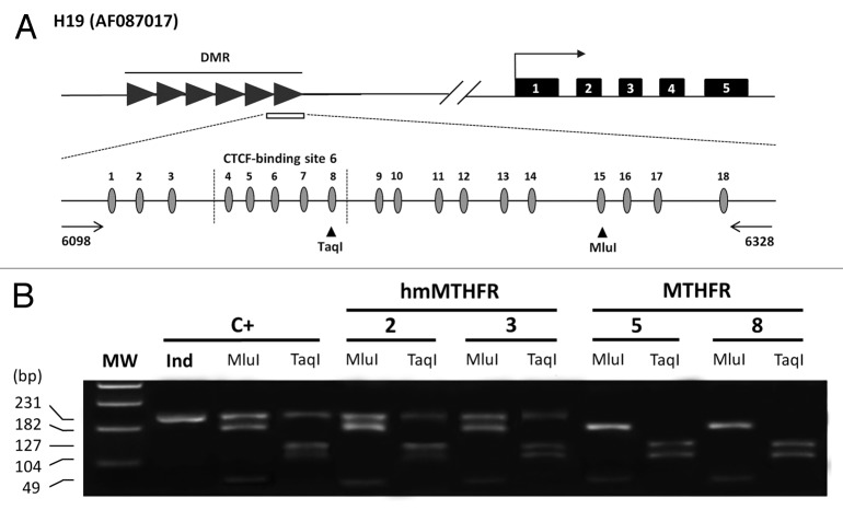
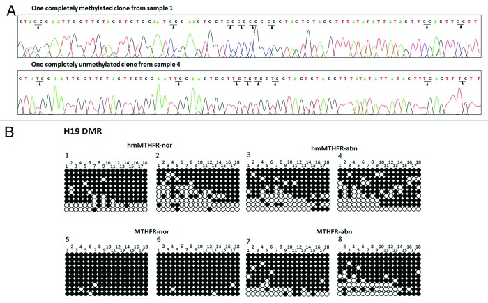
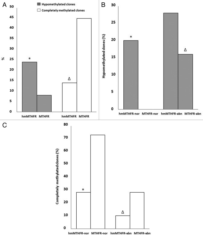
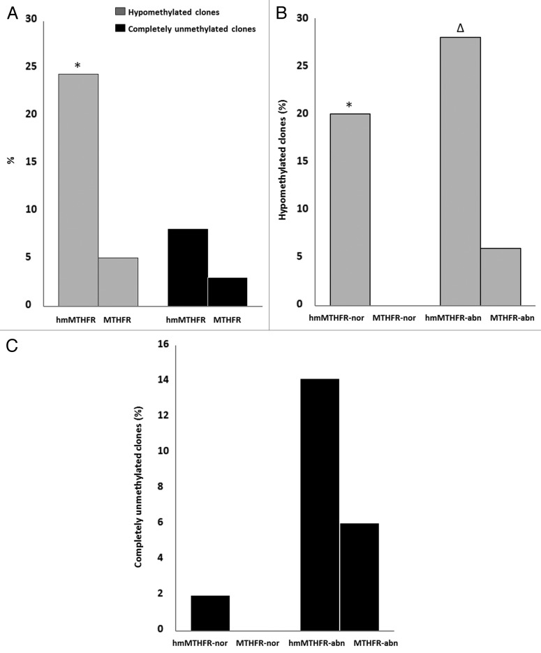
Aberrant methylation at the H19 paternal imprinted gene has been identified in different cohorts of infertile males. The causes of H19 methylation errors are poorly understood. In this study, we investigated the methylation status of the H19 gene in semen DNA samples from infertile males affected by MTHFR gene promoter hypermethylation. DNA from normal and abnormal semen samples harbouring MTHFR gene promoter hypermethylated, hmMTHFR-nor and hmMTHFR-abn, and without MTHFR methylation, MTHFR-nor and MTHFR-abn, were investigated for methylation status in the H19 locus using bisulfite-treated DNA PCR, followed by cloning and sequencing. The prevalence of H19 hypomethylated clones was 20% in hmMTHFR-nor and 0% in MTHFR-nor semen samples (p<0.05), and 28% in hmMTHFR-abn compared with 16% in MTHFR-abn semen samples (p>0.05). These results underscore the association between H19 methylation defects and hypermethylation of the MTHFR gene promoter in normal semen samples and suggest that aberrant methylation at H19 may occur in the normal sperm of infertile males affected by MTHFR gene dysfunction. These findings provide new insights into the mechanisms causing abnormal methylation in imprinted genes and, in turn, male infertility.

Keywords: DNA methylation; genetic imprinting; male infertility; spermatozoa.


Figures

None
Figure 1. Methylation status of the H19 imprinted gene in hmMTHFR and MTHFR semen DNA samples from infertile males. (A)Genomic structure of H19 locus and the GenBank accession number. Upper line: filled-in boxes and horizontal arrow indicate gene exons and orientation, respectively; filled-in horizontal arrows represent H19 DMR region and the white box represents the DMR sequence which was analyzed in this study. Lower line: DMR sequence includes 18 CpG islands (the CTCF-binding site 6 region from 4 to 8 CpG island is also shown). The horizontal arrows represent the primers. Vertical arrowheads indicate the unique bisulfite-PCR restriction enzyme sites, which were analyzed with Mlu I and Taq I. (B) Overall methylation status of H19 detected by COBRA assay in sperm from hmMTHFR and MTHFR semen DNA samples and in the control leukocyte DNA. The same bisulfite-treated DNA amplified by PCR and used for cloning and sequencing, was digested with Mlu I and Taq I restriction enzymes, which were cut only if the restriction site was methylated. Cases 2 and 3 from hmMTHFR semen samples show the unmethylated band with both Mlu I and Taq I enzyme digestion. MW, Molecular weight; C+, human leukocyte DNA.
None
Figure 2. Sequencing analyses of clones from hmMTHFR and MTHFR semen samples. (A) DNA sequencing of H19 in two different clones hmMTHFR-nor (sample 1) and hmMTHFR-abn (sample 4). Arrowheads indicate the methylated CpG islands in sample 1 and unmethylated CpG islands in sample 4. (B) Bisulfite-PCR sequencing for H19 in two representative semen samples endowed with the lowest and the highest prevalence of H19 hypomethylated clones, respectively, from hmMTHFR-nor and hmMTHFR-abn, as well as MTHFR-nor and hmMTHFR-abn semen samples. Filled-in and clear circles represent methylated and unmethylated CpG islands, respectively. The 18 CpG islands within the H19 locus are numbered on the upper side of the circles.
None
Figure 3. Frequency of H19 hypomethylated and completely methylated clones. (A) Frequency of hypomethylated (gray bars) and completely methylated (white bars) clones of H19 in hmMTHFR and MTHFR groups. * H19 frequency significantly different from MTHFR group; H19 frequency significantly different from MTHFR group. (B) Frequency of H19 hypomethylated clones in hmMTHFR-nor and MTHFR-nor groups and in hmMTHFR-abn and MTHFR-abn groups. * H19 frequency significantly different from MTHFR-nor group; H19 frequency significantly different from MTHFR-nor group; (C) Frequency of H19 completely methylated clones in hmMTHFR-nor and MTHFR-nor groups and in hmMTHFR-abn and MTHFR-abn groups. * H19 frequency significantly different from MTHFR-nor and hmMTHFR-abn groups; H19 frequency significantly different from MTHFR-abn group.
None
Figure 4. Frequency of clones with hypomethylation and complete unmethylation of CTCF-binding site 6. (A) Frequency of clones with hypomethylation (gray bars) and complete unmethylation (black bars) of CTCF binding site 6 in hmMTHFR and MTHFR groups. *The frequency of clones with hypomethylation of CTCF binding site 6 is significantly different from MTHFR group; (B) Frequency of clones with hypomethylation of CTCF binding site 6 in hmMTHFR-nor and MTHFR-nor groups and in hmMTHFR-abn and MTHFR-abn groups. * The frequency of clones with hypomethylation of CTCF binding site 6 is significantly different from MTHFR-nor group; The frequency of clones with hypomethylation of CTCF binding site 6 is significantly different from MTHFR-abn group; (C) Frequency of clones with complete unmethylation of CTCF binding site 6 in hmMTHFR-nor and MTHFR-nor groups and in hmMTHFR-abn and MTHFR-abn groups.

Similar articles

Cited by

References

    1. Koerner MV, Barlow DP. Genomic imprinting-an epigenetic gene-regulatory model. Curr Opin Genet Dev. 2010;20:164–70. doi: 10.1016/j.gde.2010.01.009. - DOI - PMC - PubMed
    1. Georgiades P, Watkins M, Burton GJ, Ferguson-Smith AC. Roles for genomic imprinting and the zygotic genome in placental development. Proc Natl Acad Sci U S A. 2001;98:4522–7. doi: 10.1073/pnas.081540898. - DOI - PMC - PubMed
    1. Piedrahita JA. The role of imprinted genes in fetal growth abnormalities. Birth Defects Res A Clin Mol Teratol. 2011;91:682–92. doi: 10.1002/bdra.20795. - DOI - PMC - PubMed
    1. Serman L, Vlahović M, Sijan M, Bulić-Jakus F, Serman A, Sincić N, et al. The impact of 5-azacytidine on placental weight, glycoprotein pattern and proliferating cell nuclear antigen expression in rat placenta. Placenta. 2007;28:803–11. doi: 10.1016/j.placenta.2007.04.001. - DOI - PubMed
    1. Diplas AI, Lambertini L, Lee MJ, Sperling R, Lee YL, Wetmur J, et al. Differential expression of imprinted genes in normal and IUGR human placentas. Epigenetics. 2009;4:235–40. - PubMed

Publication types

MeSH terms

LinkOut - more resources