. 2013 Oct;41(19):e184.
doi: 10.1093/nar/gkt755. Epub 2013 Aug 23.

Heterogeneous staining: a tool for studies of how fluorescent dyes affect the physical properties of DNA

Affiliations

Heterogeneous staining: a tool for studies of how fluorescent dyes affect the physical properties of DNA

Lena Nyberg et al. Nucleic Acids Res. 2013 Oct.

Abstract

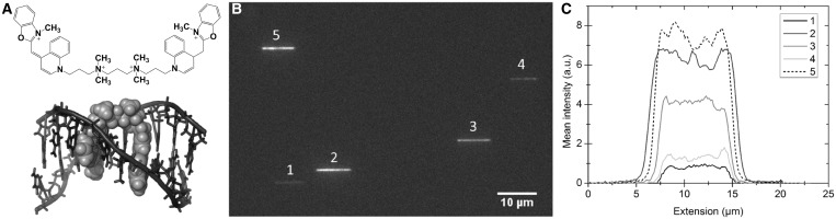
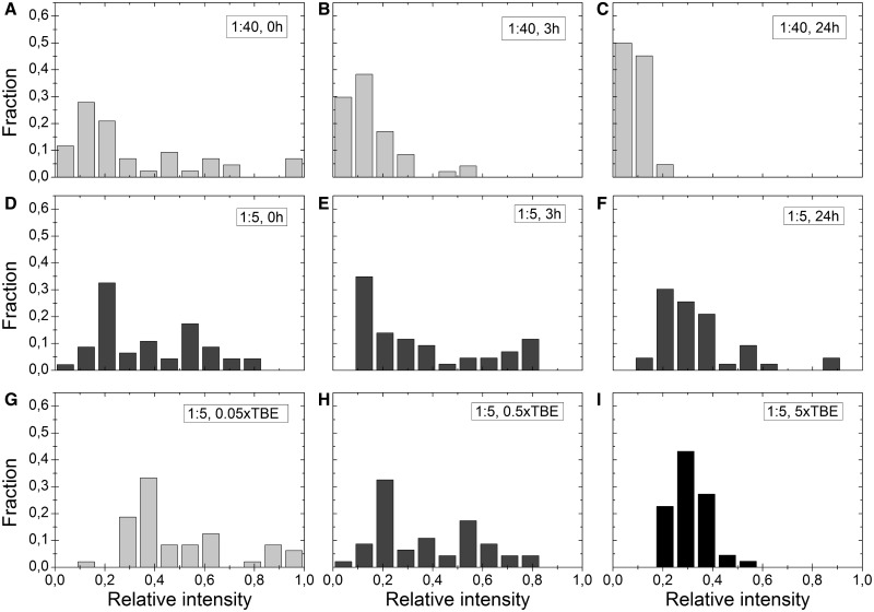
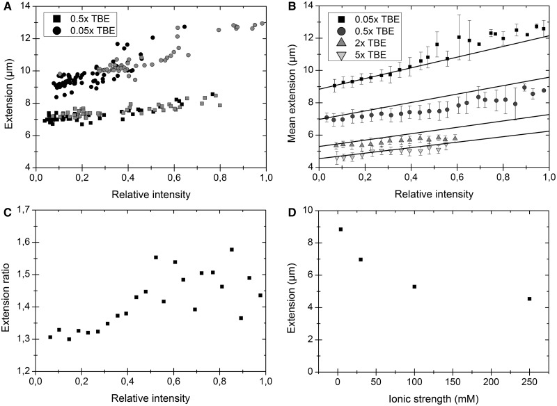
The commonly used fluorescent dye YOYO-1 (YOYO) has, using bulk techniques, been demonstrated to stain DNA heterogeneously at substoichiometric concentrations. We here, using nanofluidic channels and fluorescence microscopy, investigate the heterogeneous staining on the single DNA molecule level and demonstrate that the dye distribution is continuous. The equilibration of YOYO on DNA is extremely slow but can be accelerated by increasing the ionic strength and/or the temperature. Furthermore, we demonstrate how to use the heterogeneous staining as a tool for detailed and time-efficient studies of how fluorescent dyes affect the physical properties of DNA. We show that the relative increase in extension of DNA with increasing amount of YOYO bound is higher at low ionic strengths and also extrapolate the extension of native DNA. Our study reveals important information on how YOYO affects the physical properties of DNA, but it also has broader applications. First, it reveals how cationic intercalators, such as potential DNA drugs, affect DNA under strong confinement. Second, the strategy of using heterogeneous staining is of general use for single molecule studies of DNA interacting with proteins or ligands.


Figures

Figure 1.
Figure 1.
(A) Chemical structure of YOYO-1. (B) NMR structure of the bis-intercalating YOYO-analogue TOTO-1 bound to DNA, visualized with PyMOL (28). (C) Fluorescence microscopy image of five λ-DNA molecules in the same field of view trapped in nanochannels with the dimensions 100 × 150 nm2. (D) Corresponding intensity traces for the five λ-DNA molecules in (C) averaged over 200 consecutive images for each molecule at a dye:bp ratio of 1:10 in 0.5× TBE buffer.
Figure 2.
Figure 2.
(A–C) Intensity fractions for λ-DNA molecules with a dye:bp ratio of 1:40 in 0.5× TBE. (D–F) Corresponding data for a dye:bp ratio of 1:5 in 0.5× TBE. (A) and (D) are samples that have not been heated (0 h), samples in (B) and (E) have been heated for 3 h at 50°C, and samples in (C) and (F) have been heated for 24 h at 50°C. (G–I) Intensity distributions at three different ionic strengths – 0.05×, 0.5× and 5× TBE – and a dye:bp ratio of 1:5. None of the samples were heated. Each set (A–I) comprises 40–50 molecules and corresponds to a single experiment. All experiments were repeated at least twice, and the results are in qualitative agreement. The graphs in D and H are identical; they are shown as duplicates to increase the readability of the figure.
Figure 3.
Figure 3.
(A) Extension versus intensity for λ-DNA molecules in 0.05× TBE (circles) and 0.5× TBE (squares) at two different dye:bp ratios; 1:20 (black) and 1:5 (gray). (B) Extension and intensity distributions for ∼2650 λ-DNA molecules (heated and non-heated) at four different ionic strengths. The samples are sorted and binned according to relative intensity in steps of 0.04. The solid lines correspond to the expected increase in extension if the contour length of a fully intercalated DNA (1 dye per 4 bp) is extended with 0.51 nm per YOYO molecule and no other effects are considered (21,22). (C) Extension ratio, i.e. the extension at 0.05× TBE divided by the extension at 0.5× TBE, at different degrees of YOYO binding. (D) The extracted extension (from B) for native DNA at four different ionic strengths, converted from TBE according to ref (14).

Similar articles

Cited by

References

    1. Bustamante C. In singulo biochemistry: when less is more. Annu. Rev. Biochem. 2008;77:45–50. - PubMed
    1. van Oijen AM. Cutting the forest to see a single tree. Nat. Chem. Biol. 2008;4:440–443. - PubMed
    1. Chaurasiya KR, Paramanathan T, McCauley MJ, Williams MC. Biophysical characterization of DNA binding from single molecule force measurements. Phys. Life Rev. 2010;7:299–341. - PMC - PubMed
    1. De Vlaminck I, Dekker C. Recent advances in magnetic tweezers. Annu. Rev. Biophys. 2012;41:453–472. - PubMed
    1. Levy SL, Craighead HG. DNA manipulation, sorting, and mapping in nanofluidic systems. Chem. Soc. Rev. 2010;39:1133. - PubMed

Publication types

MeSH terms

Substances