Prolonged hemodynamic response during incidental facial emotion processing in inter-episode bipolar I disorder
- PMID: 23975275
- PMCID: PMC3944373
- DOI: 10.1007/s11682-013-9246-z
Prolonged hemodynamic response during incidental facial emotion processing in inter-episode bipolar I disorder
Abstract
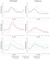
This fMRI study examined whether hemodynamic responses to affectively-salient stimuli were abnormally prolonged in remitted bipolar disorder, possibly representing a novel illness biomarker. A group of 18 DSM-IV bipolar I-diagnosed adults in remission and a demographically-matched control group performed an event-related fMRI gender-discrimination task in which face stimuli had task-irrelevant neutral, happy or angry expressions designed to elicit incidental emotional processing. Participants' brain activation was modeled using a "fully informed" SPM5 basis set. Mixed-model ANOVA tested for diagnostic group differences in BOLD response amplitude and shape within brain regions-of-interest selected from ALE meta-analysis of previous comparable fMRI studies. Bipolar-diagnosed patients had a generally longer duration and/or later-peaking hemodynamic response in amygdala and numerous prefrontal cortex brain regions. Data are consistent with existing models of bipolar limbic hyperactivity, but the prolonged frontolimbic response more precisely details abnormalities recognized in previous studies. Prolonged hemodynamic responses were unrelated to stimulus type, task performance, or degree of residual mood symptoms, suggesting an important novel trait vulnerability brain dysfunction in bipolar disorder. Bipolar patients also failed to engage pregenual cingulate and left orbitofrontal cortex-regions important to models of automatic emotion regulation-while engaging a delayed dorsolateral prefrontal cortex response not seen in controls. These results raise questions about whether there are meaningful relationships between bipolar dysfunction of specific ventromedial prefrontal cortex regions believed to automatically regulate emotional reactions and the prolonged responses in more lateral aspects of prefrontal cortex.
Figures


Similar articles
-
Amygdala activity and prefrontal cortex-amygdala effective connectivity to emerging emotional faces distinguish remitted and depressed mood states in bipolar disorder.Bipolar Disord. 2012 Mar;14(2):162-74. doi: 10.1111/j.1399-5618.2012.00999.x. Bipolar Disord. 2012. PMID: 22420592 Free PMC article.
-
Dissociable patterns of medial prefrontal and amygdala activity to face identity versus emotion in bipolar disorder.Psychol Med. 2012 Sep;42(9):1913-24. doi: 10.1017/S0033291711002935. Epub 2012 Jan 25. Psychol Med. 2012. PMID: 22273442 Free PMC article.
-
Emotion processing influences working memory circuits in pediatric bipolar disorder and attention-deficit/hyperactivity disorder.J Am Acad Child Adolesc Psychiatry. 2010 Oct;49(10):1064-80. doi: 10.1016/j.jaac.2010.07.009. J Am Acad Child Adolesc Psychiatry. 2010. PMID: 20855051 Free PMC article.
-
Emotional processing in bipolar disorder: behavioural and neuroimaging findings.Int Rev Psychiatry. 2009;21(4):357-67. doi: 10.1080/09540260902962156. Int Rev Psychiatry. 2009. PMID: 20374149 Review.
-
Emotion processing and regulation in bipolar disorder: a review.Bipolar Disord. 2012 Jun;14(4):326-39. doi: 10.1111/j.1399-5618.2012.01021.x. Bipolar Disord. 2012. PMID: 22631618 Review.
Cited by
-
Neural correlates of implicit emotion regulation in mood and anxiety disorders: an fMRI meta-analytic review.Sci Rep. 2025 Jun 4;15(1):19564. doi: 10.1038/s41598-025-03828-5. Sci Rep. 2025. PMID: 40467619 Free PMC article. Review.
-
Reliability of neural activation and connectivity during implicit face emotion processing in youth.Dev Cogn Neurosci. 2018 Jun;31:67-73. doi: 10.1016/j.dcn.2018.03.010. Epub 2018 Mar 27. Dev Cogn Neurosci. 2018. PMID: 29753993 Free PMC article.
-
Accounting for Dynamic Fluctuations across Time when Examining fMRI Test-Retest Reliability: Analysis of a Reward Paradigm in the EMBARC Study.PLoS One. 2015 May 11;10(5):e0126326. doi: 10.1371/journal.pone.0126326. eCollection 2015. PLoS One. 2015. PMID: 25961712 Free PMC article.
References
-
- American Psychiatric Association, A. P. Diagnostic and statistical manual of mental disorders: DSM-IV-TR. Washington, DC: American Psychiatric Association; 2000.
-
- Blumberg HP, Charney DS, Krystal JH. Frontotemporal neural systems in bipolar disorder. Semin Clin Neuropsychiatry. 2002;7(4):243–254. [pii] - PubMed
-
- Blumberg HP, Donegan NH, Sanislow CA, Collins S, Lacadie C, Skudlarski P, et al. Preliminary evidence for medication effects on functional abnormalities in the amygdala and anterior cingulate in bipolar disorder. Psychopharmacology (Berl) 2005;183(3):308–313. doi: 10.1007/s00213-005-0156-7. - DOI - PubMed
-
- Blumberg HP, Leung HC, Skudlarski P, Lacadie CM, Fredericks CA, Harris BC, et al. A functional magnetic resonance imaging study of bipolar disorder: state- and trait-related dysfunction in ventral prefrontal cortices. Arch Gen Psychiatry. 2003;60(6):601–609. doi: 10.1001/archpsyc.60.6.601. 60/6/601 [pii] - DOI - PubMed
Publication types
MeSH terms
Substances
Grants and funding
LinkOut - more resources
Full Text Sources
Other Literature Sources
Medical

