. 2014 Nov;219(6):2083-101.
doi: 10.1007/s00429-013-0625-4. Epub 2013 Aug 23.

The human cuneate nucleus contains discrete subregions whose neurochemical features match those of the relay nuclei for nociceptive information

Affiliations

The human cuneate nucleus contains discrete subregions whose neurochemical features match those of the relay nuclei for nociceptive information

Marina Del Fiacco et al. Brain Struct Funct. 2014 Nov.

Abstract

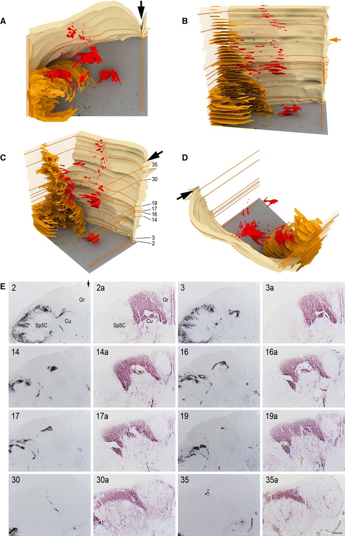
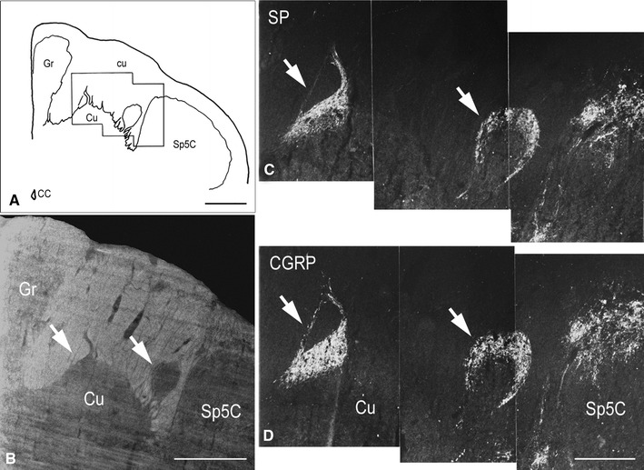
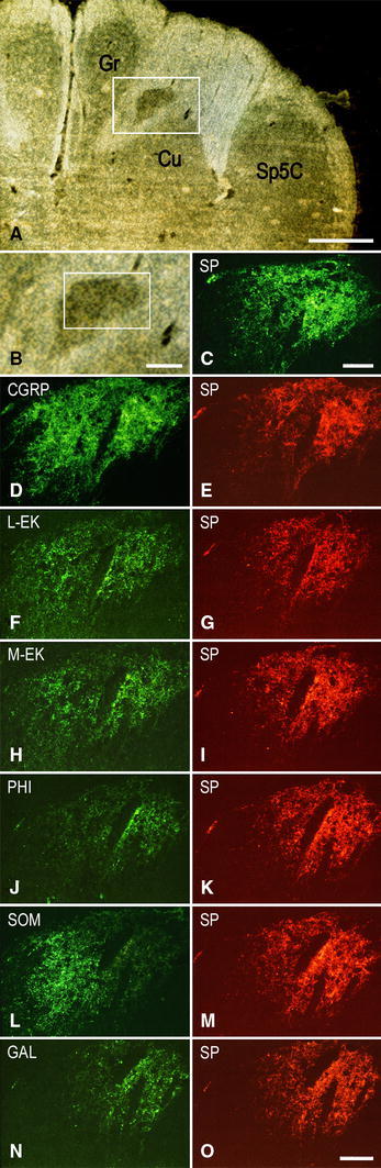
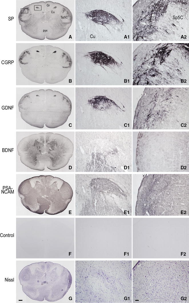
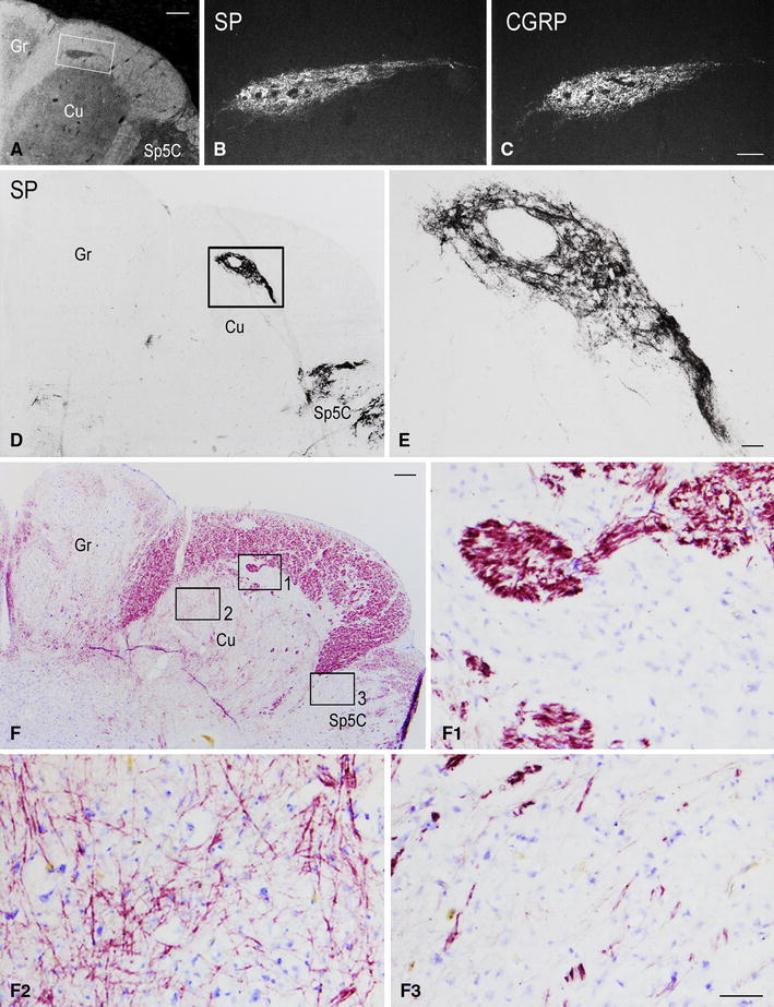
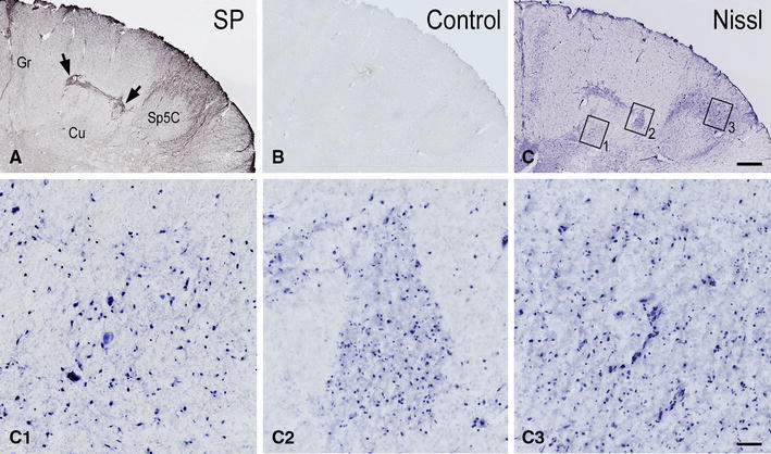
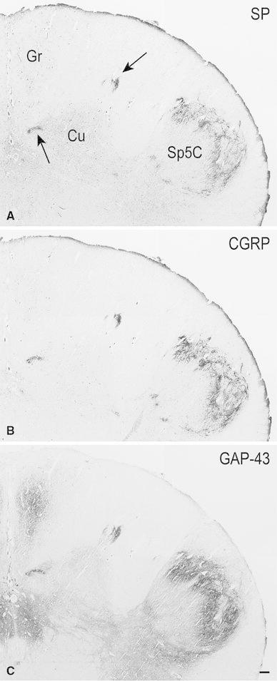
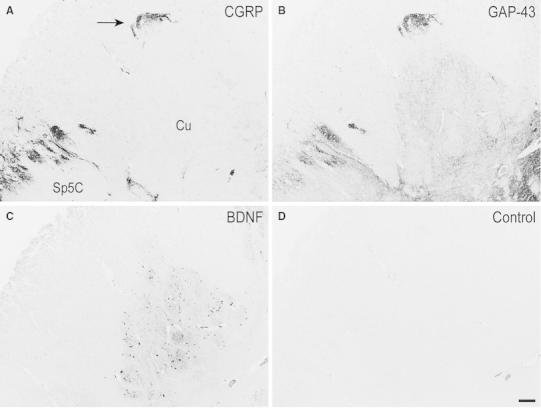
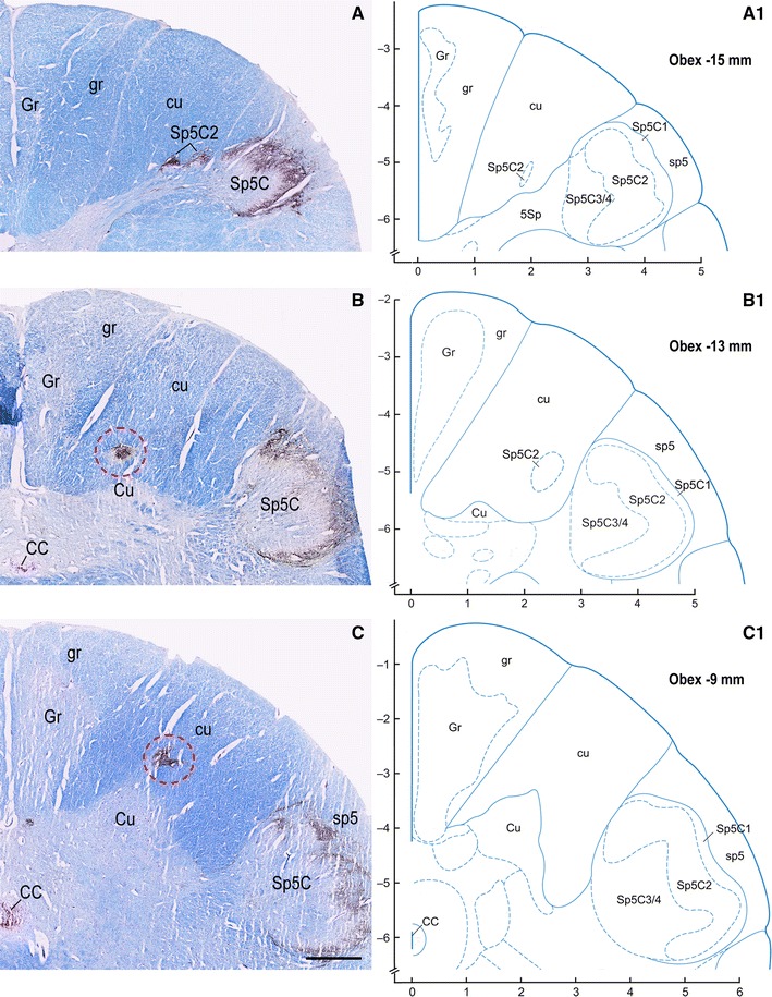
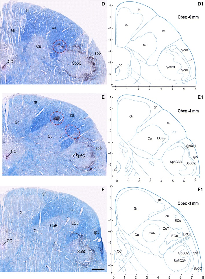
The present paper is aimed at defining distinctive subdivisions of the human cuneate nucleus (Cu), evident from prenatal to old life, whose occurrence has never been clearly formalized in the human brain, or described in other species so far. It extends our early observations on the presence of gray matter areas that host strong substance P (SP) immunoreactivity in the territory of the human Cu and adjacent cuneate fascicle. Here we provide a three-dimensional reconstruction of the Cu fields rich in SP and further identify those areas by means of their immunoreactivity to the neuropeptides SP, calcitonin gene-related peptide, methionine- and leucine-enkephalin, peptide histidine-isoleucine, somatostatin and galanin, to the trophins glial cell line-derived neurotrophic factor and brain-derived neurotrophic factor, and to the neuroplasticity proteins polysialylated neural cell adhesion molecule and growth-associated protein-43. The presence, density and distribution of immunoreactivity for each of these molecules closely resemble those occurring in the superficial layers of the caudal spinal trigeminal nucleus (Sp5C). Myelin and Nissl stainings suggest that those Cu subregions and the Sp5C superficial layers share a similar histological aspect. This work establishes the existence of definite subregions, localized within the Cu territory, that bear the neurochemical and histological features of sensory nuclei committed to the neurotransmission of protopathic stimuli, including pain. These findings appear of particular interest when considering that functional, preclinical and clinical studies show that the dorsal column nuclei, classical relay station of fine somatic tactile and proprioceptive sensory stimuli, are also involved in pain neurotransmission.


Figures

Fig. 1
Fig. 1
Full-term newborn, case 9. AD 3D rendering of SP-immunoreactive areas in the territory of the cuneate nucleus and fascicle (red) and in the substantia gelatinosa of the spinal trigeminal nucleus, caudal part (yellow) as seen in thirty-seven 12 μm thick serial sections of the medulla oblongata left dorsal quadrant. Sections are distant 120 μm from one another, for a total thickness of about 4.5 mm; the top level is about 740 μm caudal to the obex. Arrows in A, C, D and E 2 point to the posterior median sulcus and septum; the midsagittal plane was used to align the section images. Views from the top (A), anterior (B), anteromedial-superior (C), dorsolateral-superior (D). Levels of sections 2, 3, 14, 16, 17, 19, 30, and 35 in caudo-rostral sequence are indicated by orange lines and are numbered in C. Corresponding sections and paired, 36 μm distant, sections stained for myelin are shown in E. Orange arrow in B indicates the level of the caudal pole of the external cuneate nucleus. E Cu cuneate nucleus, Gr gracile nucleus, Sp5C spinal trigeminal nucleus, caudal part. Scale bar in E 35a = 500 μm applies to all micrographs
Fig. 2
Fig. 2
Full-term newborn, case 11. A Schematic drawing of the dorsal right quadrant of the caudal medulla oblongata transverse section shown in B and C; the black frame indicates the field shown in C and D. B Bright field orientation image of the territory of the cuneate nucleus (Cu) in a section immunostained for SP (detail in C). C, D Two adjacent sections immunostained for SP and CGRP, respectively, showing the dorsal part of the caudal Cu and the dorsomedial part of the spinal trigeminal nucleus, caudal part (Sp5C). Arrows in BD indicate the immunoreactive Cu regions. CC central canal, cu cuneate fascicle, Gr gracile nucleus. Scale bars A, B = 1 mm, C = D = 500 μm
Fig. 3
Fig. 3
Full-term newborn, case 8. Series of seven consecutive sections of caudal medulla oblongata immunostained for SP (A, B, C) and double immunostained for CGRP (D), L-EK (F), M-EK (H), PHI (J), SOM (L), GAL (H) (DTAF fluorochrome), and SP (E, G, I, K, M, O, respectively) (TRITC fluorochrome). Images A, B and C belong to the same section immunostained for SP. A Bright field orientation image of the right dorsal quadrant: white box delineates the area shown in B at higher magnification. B Region of gray matter dorsomedial to the main cuneate nucleus (Cu); white box delineates the area shown in C. C Dark field image of the SP immunoreactivity. D, E to N, O Dark field paired micrographs of the area shown in C in six consecutive double immunostained sections. Gr gracile nucleus, Sp5C spinal trigeminal nucleus, caudal part. Scale bars A = 1 mm, B = 250 μm, C = 100 μm, DN = O = 100 μm
Fig. 4
Fig. 4
Pre-term newborn, case 5. AG Series of seven consecutive sections of caudal medulla oblongata immunostained for SP (A), CGRP (B), GDNF (C), BDNF (D), PSA-NCAM (E) and as control (preabsorbed anti-PSA-NCAM primary antibody; preabsorbed antibodies against the other molecules yielded a similar outcome), and Nissl stained with cresyl violet (G). Boxes in A delineate the areas shown in A 1G 1 and A 2G 2. A 1G 1 Higher magnification of the dorsal gray matter area in the left side cuneate nucleus (Cu) detectable in AG sections, respectively. A 2G 2 Detail at higher magnification of the left side substantia gelatinosa of the spinal trigeminal nucleus, caudal part (Sp5C), detectable in AG sections, respectively. Gr gracile nucleus, pyx pyramidal decussation. Scale bars AF = G = 500 μm, A 1G 1 and A 2F 2 = G 2 = 50 μm
Fig. 5
Fig. 5
Full-term newborn, case 11 (AC) and 9 (DF 3). A Bright field orientation image of the territory of the right cuneate nucleus (Cu) in a section of caudal medulla oblongata immunostained for SP (detail in darkfield in B): white box indicates area shown at higher magnification in B. B, C Two adjacent sections immunostained for SP and CGRP, respectively, showing the immunoreactive region of gray matter dorsal to the main Cu. D Right dorsal quadrant of a section immunostained for SP; the strongly immunoreactive area present along the dorsal border of the Cu (box) is shown at higher magnification in E. F Right dorsal quadrant of a section consecutive to D stained for myelin and Nissl; boxes 1, 2 and 3 outline fields of the SP-immunoreactive Cu subregion, main Cu and caudal spinal trigeminal nucleus (Sp5C) substantia gelatinosa, respectively, shown at higher magnification in F 1– F 3. Gr gracile nucleus. Scale bars A = 500 μm, B = C = 100 μm, D = F = 250 μm, E = 50 μm, F 1, F 2 = F 3 = 250 μm
Fig. 6
Fig. 6
Adult, case 18. AC Right dorsal quadrant of three consecutive sections of caudal medulla oblongata immunostained for SP (A) and as control (preabsorbed anti-SP primary antibody) (B), and Nissl stained (C). Details at higher magnifications of the Nissl stained main cuneate nucleus (Cu) (box 1 in C), a region of the Cu territory immunoreactive to SP (box 2 in C, compare to A), and the caudal spinal trigeminal nucleus (Sp5C) substantia gelatinosa (box 3 in C) are shown in C 1– C 3, respectively. Arrows in A point to two SP-immunostained regions connected by a bridge of immunoreactive gray matter. Gr gracile nucleus. Scale bars A, B = C = 500 μm; C 1, C 2 = C 3 = 50 μm
Fig. 7
Fig. 7
Adult, case 19. AC Right dorsal quadrant of three consecutive sections of caudal medulla oblongata immunostained for SP (A), CGRP (B) and GAP-43 (C). Immunoreactvity for the three molecules occurs in two regions of the cuneate nucleus (Cu) (pointed out by arrows in A), as in the superficial layers of the spinal trigeminal nucleus, caudal part (Sp5C). Gr gracile nucleus. Scale bar A, B = C = 250 μm
Fig. 8
Fig. 8
Adult, case 14. A–D Left side cuneate nucleus (Cu) and dorsomedial part of the caudal spinal trigeminal nucleus (Sp5C) substantia gelatinosa in four consecutive sections of caudal medulla oblongata immunostained for CGRP (A), GAP-43 (B), BDNF (C), and as Control (preabsorbed anti-BDNF primary antibody; preabsorbed antibodies against the other molecules yielded a similar outcome). Note how the immunoreactivity of the dorsal region of the Cu (arrow in A) goes in parallel to that of the substantia gelatinosa of the Sp5C for the three molecules. Scale bar AC = D = 250 μm
Fig. 9
Fig. 9
Adult, case 15. AF Right dorsal quadrant of caudal medulla oblongata sections immunostained for CGRP and counterstained with Kluver-Barrera. A 1F 1 diagrams modified from Paxinos et al. (2012) at levels indicated as distance from obex, matching those shown in AF. Gray matter regions endowed with CGRP immunoreactivity and located medially (B), dorsally (C, E) and laterally (D, E) in the cuneate fascicle (cu) near the cuneate nucleus (Cu) border are encircled in red. Note that similar regions do not appear in the matching level diagrams. CC central canal; CuR cuneate nucleus, pars rotunda; CuT cuneate nucleus, pars triangularis; ECu external cuneate nucleus; Gr gracile nucleus; gr gracile fasciculus; sp5 spinal trigeminal tract; Sp5C spinal trigeminal nucleus, caudal part; Sp5C1 spinal trigeminal nucleus, caudal part, lamina 1; Sp5C2 spinal trigeminal nucleus, caudal part, lamina 2; Sp5C3/4 spinal trigeminal nucleus, caudal part, lamina 3/4. Scale bar A, B = C = 1 mm; D, E = F = 1 mm
Fig. 9
Fig. 9
Adult, case 15. AF Right dorsal quadrant of caudal medulla oblongata sections immunostained for CGRP and counterstained with Kluver-Barrera. A 1F 1 diagrams modified from Paxinos et al. (2012) at levels indicated as distance from obex, matching those shown in AF. Gray matter regions endowed with CGRP immunoreactivity and located medially (B), dorsally (C, E) and laterally (D, E) in the cuneate fascicle (cu) near the cuneate nucleus (Cu) border are encircled in red. Note that similar regions do not appear in the matching level diagrams. CC central canal; CuR cuneate nucleus, pars rotunda; CuT cuneate nucleus, pars triangularis; ECu external cuneate nucleus; Gr gracile nucleus; gr gracile fasciculus; sp5 spinal trigeminal tract; Sp5C spinal trigeminal nucleus, caudal part; Sp5C1 spinal trigeminal nucleus, caudal part, lamina 1; Sp5C2 spinal trigeminal nucleus, caudal part, lamina 2; Sp5C3/4 spinal trigeminal nucleus, caudal part, lamina 3/4. Scale bar A, B = C = 1 mm; D, E = F = 1 mm

Similar articles

Cited by

References

    1. Al-Chaer ED, Feng Y, Willis WD. Comparative study of viscerosomatic input onto postsynaptic dorsal column and spinothalamic tract neurons in the primate. J Neurophysiol. 1999;82:1876–1882. - PubMed
    1. Anand P, Gibson SJ, Yiangou Y, Christofides ND, Polak JM, Bloom SR. PHI-like immunoreactivity co-locates with the VIP-containing system in human lumbosacral spinal cord. Neurosci Lett. 1984;46:191–196. - PubMed
    1. Benowitz LI, Apostolides PJ, Perrone-Bizzozero N, Finklestein SP, Zwiers H. Anatomical distribution of the growth-associated protein GAP-43/B-50 in the adult rat brain. J Neurosci. 1988;8:339–352. - PMC - PubMed
    1. Berkley KJ, Hubscher CH. Are there separate central nervous system pathways for touch and pain? Nat Med. 1995;1:766–773. - PubMed
    1. Bisby MA. Dependence of GAP43 (B50, Fl) transport on axonal regeneration in rat dorsal root ganglion neurons. Brain Res. 1988;458:157–161. - PubMed

MeSH terms

LinkOut - more resources