. 2013 Dec 12;32(50):5602-13.
doi: 10.1038/onc.2013.349. Epub 2013 Aug 26.

Par-4/THAP1 complex and Notch3 competitively regulated pre-mRNA splicing of CCAR1 and affected inversely the survival of T-cell acute lymphoblastic leukemia cells

Affiliations
Free PMC article

Par-4/THAP1 complex and Notch3 competitively regulated pre-mRNA splicing of CCAR1 and affected inversely the survival of T-cell acute lymphoblastic leukemia cells

C Lu et al. Oncogene. .
Free PMC article

Abstract

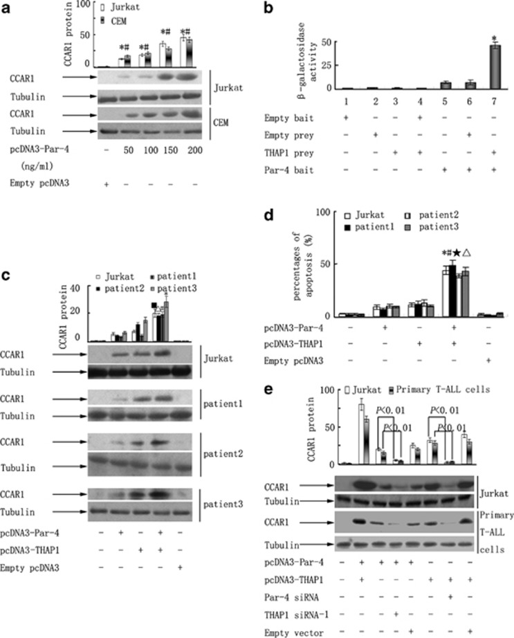
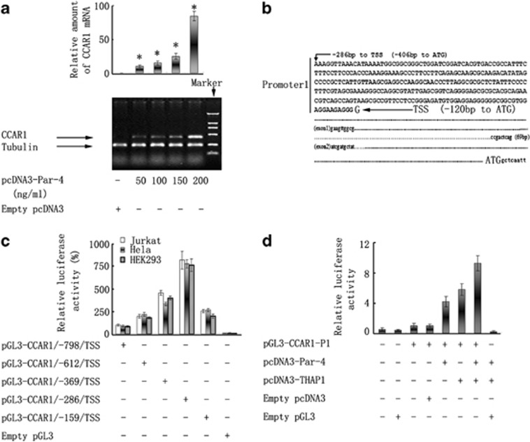
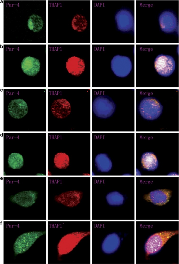
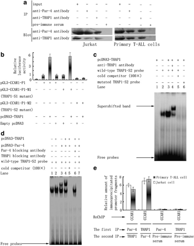
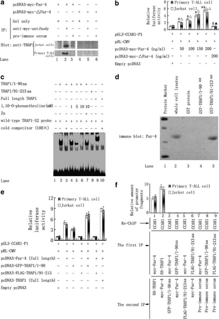
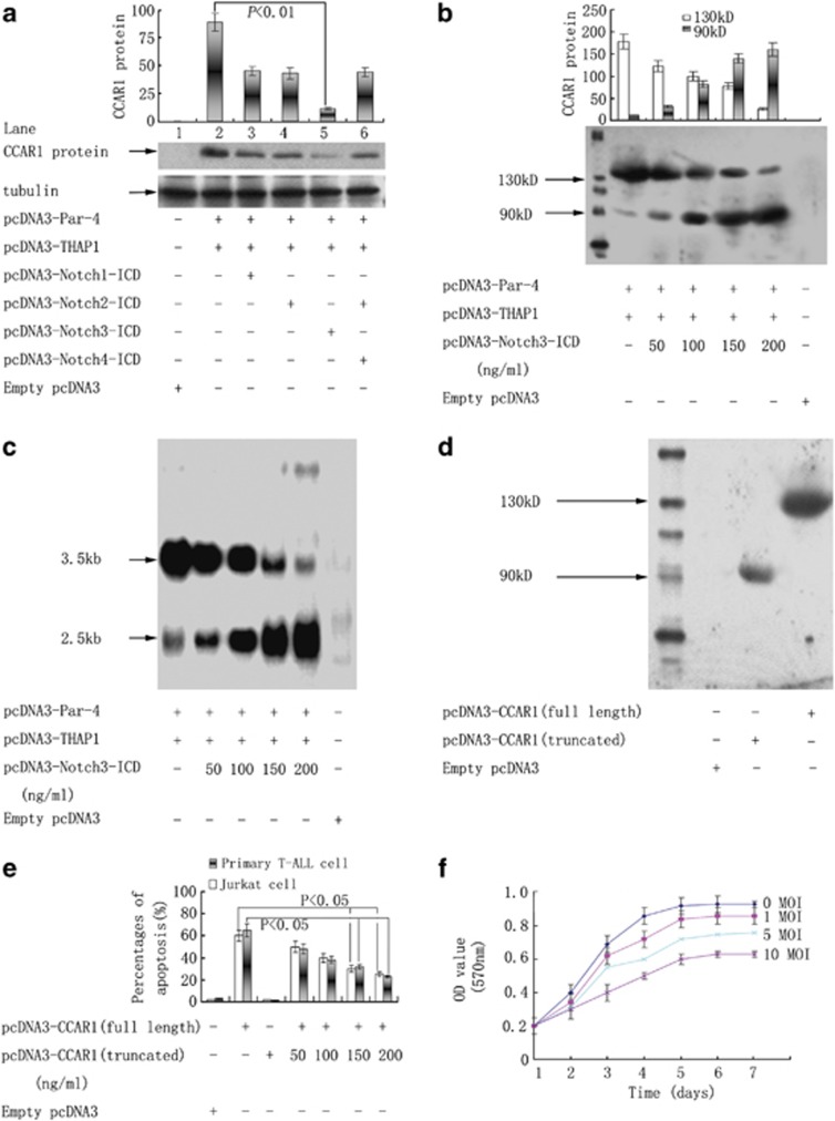
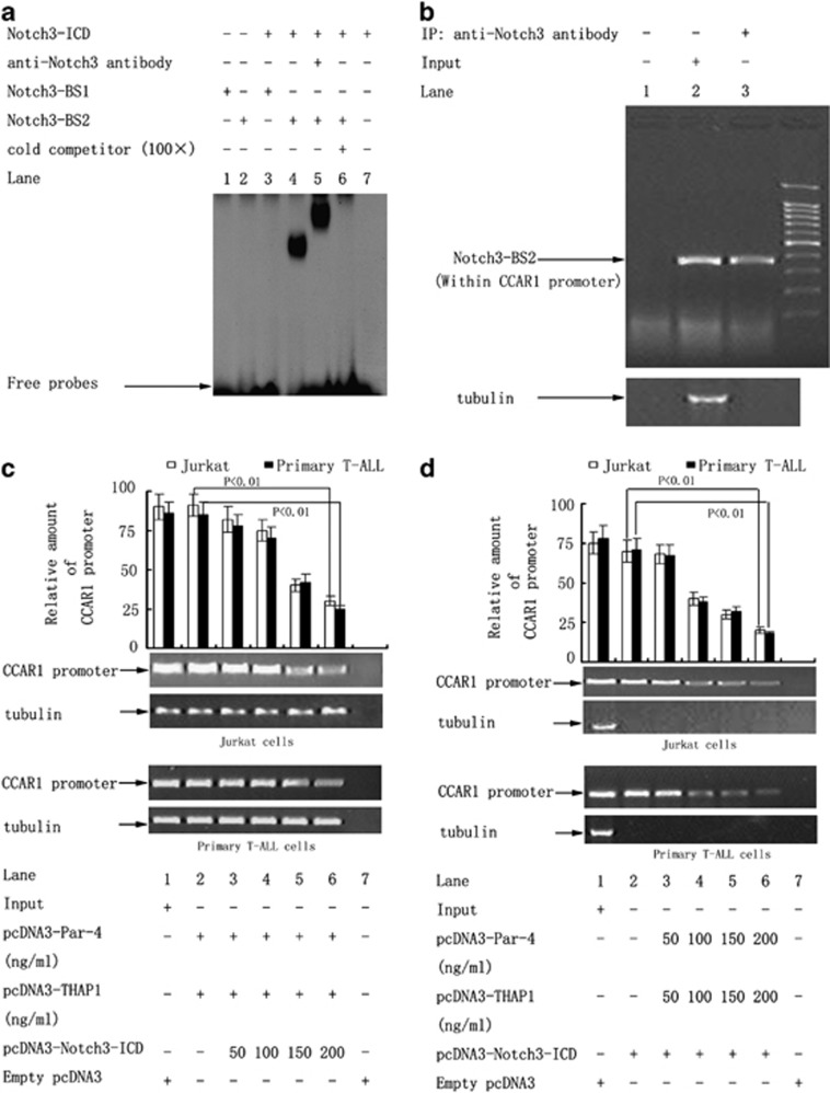
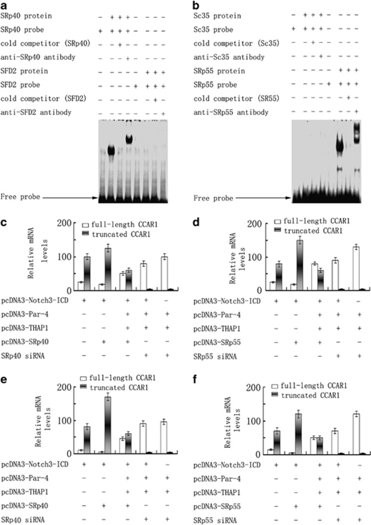
Although the intensification of therapy for children with T-cell acute lymphoblastic leukemia (T-ALL) has substantially improved clinical outcomes, T-ALL remains an important challenge in pediatric oncology. Here, we report that the cooperative synergy between prostate apoptosis response factor-4 (Par-4) and THAP1 induces cell cycle and apoptosis regulator 1 (CCAR1) gene expression and cellular apoptosis in human T-ALL cell line Jurkat cells, CEM cells and primary cultured neoplastic T lymphocytes from children with T-ALL. Par-4 and THAP1 collaborated to activate the promoter of CCAR1 gene. Mechanistic investigations revealed that Par-4 and THAP1 formed a protein complex by the interaction of their carboxyl termini, and THAP1 bound to CCAR1 promoter though its zinc-dependent DNA-binding domain at amino terminus. Par-4/THAP1 complex and Notch3 competitively bound to CCAR1 promoter and competitively modulated alternative pre-mRNA splicing of CCAR1, which resulted in two different transcripts and played an opposite role in T-ALL cell survival. Despite Notch3 induced a shift splicing from the full-length isoform toward a shorter form of CCAR1 mRNA by splicing factor SRp40 and SRp55, Par-4/THAP1 complex strongly antagonized this inductive effect. Our finding revealed a mechanistic rationale for Par-4/THAP1-induced apoptosis in T-ALL cells that would be of benefit to develop a new therapy strategy for T-ALL.


Figures

Figure 1
Figure 1
Par-4 and THAP1 cooperatively induced CCAR1 gene expression and cellular apoptosis in T-ALL cells. (a) pcDNA3-Par-4 was transfected into Jurkat cells and CEM cells with increasing dosages and CCAR1 protein was detected with western blotting. * and #: P<0.05, compared with transfection with empty pcDNA3. (b) Interaction of Par-4 with THAP1 was determined by two-hybrid testing. The β-galactosidase activity of reporter gene was measured. *P<0.01, compared with Bar 1∼6. (c) Jurkat cells and primary neoplastic T lymphocytes from three patients were transfected with either pcDNA3-Par-4 or pcDNA3-THAP1 or a combination of both. CCAR1 protein was detected with western blotting. Compared with transfection with empty pcDNA3, ▪, Δ, *, #: P<0.01. This figure shows the results of three representatives of patients with T-ALL. (d) Jurkat cells and primary neoplastic T lymphocyes were transfected with either pcDNA3-Par-4 or pcDNA3-THAP1 or a combination of both. The percentage of apoptotic cells was measured by FACS (fluorescence-activated cell sorter) analysis. *, #, ★, Δ: P<0.01, compared with transfection with empty pcDNA3. This figure shows the results of three representatives of patients with T-ALL. (e) Both pcDNA3-Par-4 and THAP1 siRNA were co-transfected into Jurkat cells and the primary neoplastic T lymphocyes. Similarly, Jurkat cells and the primary neoplastic T lymphocyes were transfected with pcDNA3-THAP1 together with Par-4 siRNA. CCAR1 protein was detected with western blotting.
Figure 2
Figure 2
Par-4 and THAP1 cooperated to activate an alternative promoter of CCAR1. (a) pcDNA3-Par-4 was transfected into Jurkat cells with increasing dosages, and relative amount of CCAR1 mRNA was detected with RT–PCR. *P<0.05, compared with transfection with empty pcDNA3. (b) 5′ RACE experiments were performed to map the transcription start sites (TSS) of CCAR1 in Jurkat cells. Nucleotide sequences of a promoter P1 of CCAR1 are shown. (c) Various deletion constructs of the putative CCAR1 promoter P1 were transfected into Jurkat cells, Hela cells and HEK293 cell lines. Luciferase activity assays were performed for characterization of CCAR1 promoter P1. The reporter activities were expressed as the percentage of transfection of the pGL3-CCAR1/-798/TSS, in which the DNA fragment spanning from −798 bp to TSS was contained. (d) Jurkat cells were co-transfected with pGL3-CCAR1-P1 reporter. Luciferase activity was determined and normalized for expression of Renilla luciferase and expressed as X-fold induction relative to the activity of pGL3-CCAR1-P1 reporter co-transfected with empty pcDNA3.
Figure 3
Figure 3
Co-localization of Par-4 and THAP1 in cell nuclei. Jurkat cells (b), primary T-ALL cells (d) and 293T cells (f) were co-transfected with both pcDNA3-Par-4 and pcDNA3-THAP1. Par-4 and THAP1 were stained with anti-Par-4 (Green) and anti-THAP1 (Red) antibodies, respectively. DAPI (4′,6-diamidino-2-phenylindole; Blue) was used to show cell nucleus. Co-localization is shown in Merge. Blue, green and red overlap results in white. Jurkat cells (a), primary T-ALL cells (c) and 293T cells (e) were co-transfected with empty plasmids as controls.
Figure 4
Figure 4
Par-4 and THAP1 formed a protein complex and bound to CCAR1 promoter P1 by a THAP1-binding motif. (a) Jurkat cells and the primary T-ALL cells were transfected with pcDNA3-Par-4 and pcDNA3-THAP1. Immunoprecipitations and western blotting analysis were performed with anti-Par-4 antibody and anti-THAP1 antibody. (b) Mutation analysis of two candidate DNA-binding sites for THAP1, THAP1-S1 (−387 bp head of ATG) and THAP1-S2 (−270 bp ahead of ATG) within the region of CCAR1 promoter P1. The reporter vectors having mutations (THAP1-S1 mutant or THAP1-S2 mutant) were transiently transfected into Jurkat cells together with pcDNA3-THAP1. The reporter activities were expressed as the fold increases over the co-transfection of the wild-type pGL3-CCAR1-P1 and empty pcDNA3 vectors. (c) DNA-binding activities of THAP1-S2 within CCAR1 promoter P1 assessed by EMSA. After the transfection of pcDNA3-THAP1, nuclear extracts were incubated with either wild-type or mutated THAP1-S2 probes in the presence or absence of anti-THAP1 antibody. The arrows indicate supershifts of the bands by antibodies against THAP1. (d) Jurkat cells were transfected with pcDNA3-Par-4 and pcDNA3-THAP1 alone or together. Nuclear extracts were incubated with THAP1-S2 probe along with blocking antibodies directed against Par-4 or THAP1. DNA-binding activity was detected by EMSA. (e) Jurkat cells and the primary T-ALL cells were transfected with pcDNA3-Par-4 and pcDNA3-THAP1. With anti-Par-4, anti-THAP1 or control pre-immune antibodies, ChIP-reChIP assay was carried out as described under MATERIALS AND METHODS. The immunoprecipitated DNA fragments were analyzed by real-time PCR with primers for CCAR1 promoter P1. The relative amounts of immunoprecipitated promoter fragments after normalizing to their respective levels in the input are shown. Data are presented as mean±s.e. of three separate experiments.
Figure 5
Figure 5
The death domain at the C-terminus of Par-4 and the zinc-dependent DNA-binding domain of THAP1 were both required for Par-4/THAP1 protein complex to activate CCAR1 promoter P1. (a) The pcDNA3-myc-ΔPar-4 was constructed, which encoded a myc fusion protein lacking the death domain at the COOH terminus of Par-4. Jurkat cells and the primary T-ALL cells were transfected with either pcDNA3-myc-ΔPar-4 or pcDNA3-myc-Par-4, of which the latter encoding the full-length Par-4 protein. Co-immunoprecipitation assays and western blot were performed with anti-myc antibody and anti-THAP1 antibody. (b) Both pGL3-CCAR1-P1 reporter and pcDNA3-THAP1 were co-transfected into Jurkat cells and the primary T-ALL cells with pcDNA3-myc-ΔPar-4 or pcDNA3-myc-Par-4. Luciferase activities were measured and pGL3-CCAR1-P1 reporter activities were normalized to pRL-CMV internal standard. All values are expressed as X-fold induction relative to the activity of pGL3-CCAR1-P1 under conditions where empty pcDNA3 was co-transfected. *P<0.05, **P<0.01, compared with the transfection with empty pcDNA3. #, ▴: P>0.05, compared with the transfection of empty pcDNA3. (c) EMSA was performed to evaluate the DNA–protein interaction in vitro between 32P-end-labeled THAP1-S2 probes and the recombinant THAP1/1-90aa and THAP1/91-213aa. The DNA-binding activity was detected after the incubation of the THAP1/1-90aa with increasing amounts of metal-chelating agent 1, 10-o-phenanthroline in the absence and presence of zinc pre-incubation. (d) GST pull-down experiment was performed with recombinant GST, GST-THAP1/1-90aa and GST-THAP1/91-213aa. Co-precipitation of Par-4 was detected by western blotting using an anti-Par-4 antibody. (e) The pGL3-CCAR1-P1 reporter and pcDNA3-Par-4 were co-transfected into Jurkat cells and the primary T-ALL cells with pcDNA3-THAP1, pcDNA3-GFP-THAP1/1-90 (amino acids 1–90) or pcDNA3-FLAG-THAP1/91-213 (amino acids 91–213). Luciferase activity assays were performed to detect the activity of CCAR1 promoter P1. All values were normalized for the expression of Renilla luciferase and expressed as X-fold induction relative to the activity of pGL3-CCAR1-P1. (f) Jurkat cells and the primary T-ALL cells were transfected with pcDNA3-myc-Par-4, pcDNA3-HA-THAP1, pcDNA3-GFP-THAP/1-90 and pcDNA3-FLAG-THAP1/91-213 alone or in combination. Pre-immune serum was used as a control. ChIP-reChIP assay were carried out as described under Materials and methods. The immunoprecipitated DNA fragments were analyzed by real-time PCR with primers for CCAR1 promoter P1. The relative amounts of immunoprecipitated promoter fragments after normalizing to their respective levels in the input are shown.
Figure 6
Figure 6
Par-4/THAP1 complex and Notch3 competitively regulated alternative pre-mRNA splicing of CCAR1 and affected inversely T-ALL cell survival. (a) Jurkat cells were co-transfected with both pcDNA3-Par-4 and pcDNA3-THAP1, together with plasmid expressing Notch1-ICD, Notch2-ICD, Notch3-ICD or Notch4-ICD. CCAR1 protein was detected by western blotting. (b) Jurkat cells were co-transfected with both pcDNA3-Par-4 and pcDNA3-THAP1, together with an increasing amount of pcDNA3-Notch3-ICD. CCAR1 protein was detected by western blotting. (c) Jurkat cells were co-transfected with both pcDNA3-Par-4 and pcDNA3-THAP1, together with an increasing amount of pcDNA3-Notch3-ICD. Northern blotting analysis was used to determine CCAR1 mRNA. (d) The full-length CCAR1 mRNA (3.5 kb) and the truncated transcript (2.5 kb) were cloned into expression plasmid pcDNA3 and were transfected into Jurkat cells. Western blotting was used to detect CCAR1 protein. (e) Jurkat cells and the primary T-ALL cells were transfected with both pcDNA3-CCAR1 (full length) and an increasing amount of pcDNA3-CCAR1 (truncated). The percentages of apoptosis were detected with FACS (fluorescence-activated cell sorter) analysis. (f) Jurkat cells were transducted with both recombinant lentivirus containing CCAR1 (truncated) cDNA and an increasing amount of recombinant lentivirus containing CCAR1 (full length) cDNA with a multiplicity of infection of 0, 1, 5 or 10. The cell proliferation was assessed with the MTT assay, and the growth curves are shown.
Figure 7
Figure 7
Notch3-ICD and Par-4/THAP1 complex competitively and exclusively bind to CCAR1 promoter. (a) DNA-binding activity of Notch3-BS within CCAR1 promoter P1 was assessed by EMSA. After the transfection of pcDNA3-Notch3-ICD, nuclear extracts were obtained from the Jurkat cells. Nuclear extracts were incubated with either Notch3-BS1 or Notch3-BS2 probes in the presence or absence of anti-Notch3 antibody. In competitive studies, a 100-fold molar excess of unlabeled probes was added to the binding reaction mixture before the addition of the labeled probes. Results shown are representative of three independent experiments. (b) Jurkat cells were transfected with pcDNA3-Notch3-ICD. ChIP experiments were performed with nuclear extracts. The precipitated DNA fragment containing Notch3-BS2 within the region of CCAR1 promoter was amplified by PCR. (c) Jurkat cells and primary T-ALL cells were co-transfected with both pcDNA3-Par-4 and pcDNA3-THAP1, together with an increasing amount of pcDNA3-Notch3-ICD. The precipitated THAP1-associated DNA fragment of CCAR1 promoter was amplified by PCR. (d) Jurkat cells and primary T-ALL cells were co-transfected with both pcDNA3-Notch3-ICD and the increasing amounts of pcDNA3-Par-4 and pcDNA3-THAP1. The precipitated Notch3-ICD-binded DNA fragment of CCAR1 promoter was amplified by PCR.
Figure 8
Figure 8
Splicing factor SRp40 and SRp55 contributed to Notch3-induced shift in alternative splicing of CCAR1 pre-mRNA, which was antagonized by Par-4/THAP1. RNA EMSAs were used to determine the binding of splicing factor SRp40 (a) and SRp55 (b) to CCAR1 mRNA. Further verification of RNA–protein complexes was accomplished by supershift assays using anti-SRp40 and anti-SRp55 antibodies. SRp40 and SRp55 were either overexpressed or knocked down with either co-transfection of pcDNA3-SRp40 and pcDNA3-SRp55 or co-transfection of SRp40 siRNA and SRp55 siRNA. The real-time RT–PCR was used to detect the relative mRNA levels of full-length and truncated CCAR1 in the Jurkat cells (c, d) and the primary T-ALL cells (e, f) exposed to transfection with pcDNA3-Notch3-ICD alone or together with pcDNA3-Par-4 and pcDNA3-THAP1.
Figure 9
Figure 9
Schematic drawing showing the spliced structure of the CCAR1 pre-mRNA.

Similar articles

Cited by

References

    1. Schrappe M, Hunger SP, Pui CH, Saha V, Gaynon PS, Baruchel A. Outcomes after induction failure in childhood acute lymphoblastic leukemia. N Engl J Med. 2012;366:1371–1381. - PMC - PubMed
    1. Lu C, Chen JQ, Zhou GP, Wu SH, Guan YF, Yuan CS. Multimolecular complex of Par-4 and E2F1 binding to Smac promoter contributes to glutamate-induced apoptosis in human bone mesenchymal stem cells. Nucleic Acids Res. 2008;36:5021–5032. - PMC - PubMed
    1. Hebbar N, Wang C, Rangnekar VM. Mechanisms of apoptosis by the tumor suppressor Par-4. J Cell Physiol. 2012;227:3715–3721. - PMC - PubMed
    1. Glienke W, Chow KU, Bauer N, Bergmann L. Down-regulation of wt1 expression in leukemia cell lines as part of apoptotic effect in arsenic treatment using two compounds. Leuk Lymphoma. 2006;47:1629–1638. - PubMed
    1. Ou CY, Kim JH, Yang CK, Stallcup MR. Requirement of cell cycle and apoptosis regulator 1 for target gene activation by Wnt and beta-catenin and for anchorage-independent growth of human colon carcinoma cells. J Biol Chem. 2009;284:20629–20637. - PMC - PubMed

Publication types

MeSH terms