. 2013 Oct 1;191(7):3884-95.
doi: 10.4049/jimmunol.1301344. Epub 2013 Aug 23.

Modulation of inflammasome-mediated pulmonary immune activation by type I IFNs protects bone marrow homeostasis during systemic responses to Pneumocystis lung infection

Affiliations

Modulation of inflammasome-mediated pulmonary immune activation by type I IFNs protects bone marrow homeostasis during systemic responses to Pneumocystis lung infection

Steve Searles et al. J Immunol. .

Abstract

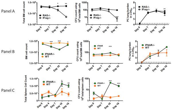
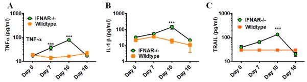
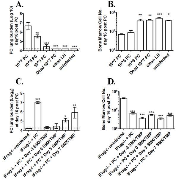
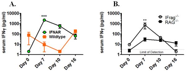
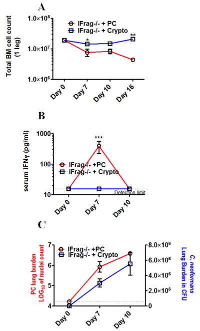
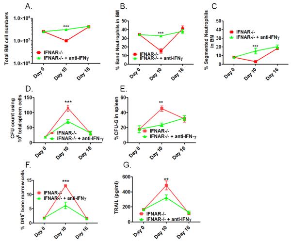
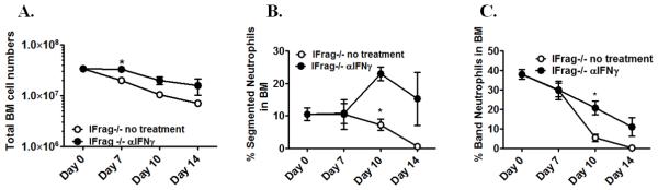
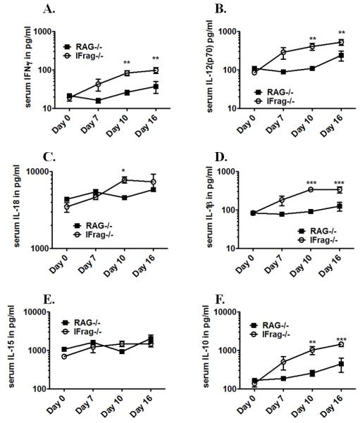
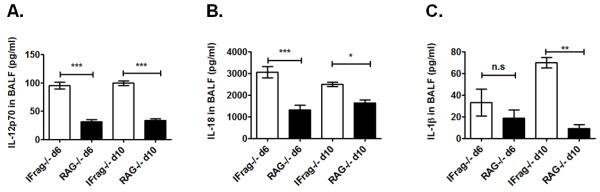
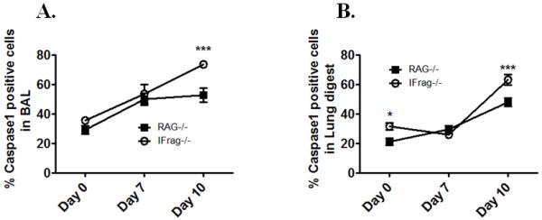
Although acquired bone marrow failure (BMF) is considered a T cell-mediated autoimmune disease, possible innate immune defects as a cause for systemic immune deviations in response to otherwise innocuous infections have not been extensively explored. In this regard, we recently demonstrated an important role of type I IFNs in protecting hematopoiesis during systemic stress responses to the opportunistic fungal pathogen Pneumocystis in lymphocyte-deficient mice. Mice deficient in both lymphocytes and type I IFN receptor (IFrag(-/-) mice) develop rapidly progressing BMF due to accelerated bone marrow (BM) cell apoptosis associated with innate immune deviations in the BM in response to Pneumocystis lung infection. However, the communication pathway between lung and BM eliciting the induction of BMF in response to this strictly pulmonary infection has been unclear. In this study, we report that absence of an intact type I IFN system during Pneumocystis lung infection not only causes BMF in lymphocyte-deficient mice but also transient BM stress in lymphocyte-competent mice. This is associated with an exuberant systemic IFN-γ response. IFN-γ neutralization prevented Pneumocystis lung infection-induced BM depression in type I IFN receptor-deficient mice and prolonged neutrophil survival time in BM from IFrag(-/-) mice. IL-1β and upstream regulators of IFN-γ, IL-12, and IL-18 were also upregulated in lung and serum of IFrag(-/-) mice. In conjunction, there was exuberant inflammasome-mediated caspase-1 activation in pulmonary innate immune cells required for processing of IL-18 and IL-1β. Thus, absence of type I IFN signaling during Pneumocystis lung infection may result in deregulation of inflammasome-mediated pulmonary immune activation, causing systemic immune deviations triggering BMF in this model.


Figures

Figure 1
Figure 1. Type-I-IFN-signaling is critical in regulating hematopoiesis during systemic responses to Pneumocystis lung infection in both lymphocyte-deficient and lymphocyte competent mice
Panel A. Shown are total bone marrow cell numbers, hematopoietic colony forming activity (CFU-counts) of bone marrow cells and PC lung burden from IFrag−/− (IFNAR−/−/RAG−/−) and RAG−/− mice (IFNAR+/+/RAG−/−) over the course of Pneumocystis lung infection at day 0, 7, 10 and 16 post infection. Panel B. Comparative analysis of total bone marrow cell numbers, hematopoietic colony forming activity (CFU-counts) of bone marrow cells and PC lung burden from lymphocyte-competent IFNAR−/− (IFNAR−/−/RAG+/+) mice and wildtype mice (IFNAR+/+/RAG+/+) over the course of Pneumocystis lung infection at day 0, 7, 10 and 16 post infection. Panel C. Demonstrated are total spleen counts and hematopoietic colony forming activity (CFU-counts) of spleen cells from IFNAR−/− and wildtype mice as a measure for extramedullary hematopoiesis at day 0, 7 10 and 16 post infection. Statistical analysis was performed using a 2-way ANOVA. Comparisons were made between groups at the same time point. P values are marked as * p< 0.05, ** p<0.01, *** p<0.001.
Figure 2
Figure 2. Transient bone marrow depression during Pneumocystis lung infection in IFNAR−/− mice is associated with deviated cytokine responses as previously seen in IFrag−/− mice during progression of bone marrow failure
Comparative cytokine analysis was performed in bone marrow lysates isolated from IFNAR−/− and wildtype mice at day 0, 7, 10 and 16 post Pneumocystis lung infection using an ELISA-based multiplex assay system. Shown are data for TNF-α (A), IL-1β (B) and TRAIL (C). Statistical analysis was performed using a 2-way ANOVA. Comparisons were made between groups at the same time point. P values are marked as * p< 0.05, ** p<0.01, *** p<0.001.
Figure 3
Figure 3. Induction of bone marrow failure in lymphocyte-deficient IFrag−/− mice depends on the exposure to a critical dose of 105 live pathogen nuclei and cannot be prevented by antibiotic treatment
Groups of IFrag−/− mice were intratracheally inoculated with either 107, 105, 103 live Pneumocystis (PC) nuclei, or 107 dead PC nuclei and bone marrow responses compared to animals either inoculated with clean lung homogenate at day 16 post infection and uninfected animals. Figure 3A shows Pneumocystis (PC) lung burden at day 16 post infection following microscopic enumeration. Figure 3 B shows total bone marrow counts of all IFrag−/− comparison groups at day 16 post infection. Antibiotic treatment was initiated in groups of IFrag−/− mice three days prior to (day -3) and at day 0, 3 and 7 post inoculations with 107 PC nuclei. Bone marrow cell numbers and PC lung burden were evaluated and compared to uninfected mice at day 16 post infection. Figure 3C shows PC lung burden and Figure 3D shows total bone marrow counts at day 16 post infection. Statistical analysis was performed using a 1-way ANOVA. All groups were compared to those mice receiving 107 PC nuclei. P values are marked as * p< 0.05, ** p<0.01, *** p<0.001.
Figure 4
Figure 4. IFNAR−/− and IFrag−/− show elevated serum IFNγ levels during early responses to Pneumocystis lung infection which correlate with induction of bone marrow stress
IFNγ serum evaluated over the course of Pneumocystis lung infection in IFNAR−/−, IFrag−/−, wildtype and RAG−/− mice at day 0, 7 10 and 16 post infection using a plate based ELISA assay. Figure 4A compares serum IFNγ responses between lymphocyte-competent IFNAR−/− and wildtype mice, Figure 4B compares serum IFNγ responses between lymphocyte-competent IFrag−/− and RAG−/− mice. Statistical analysis was performed using a 2-way ANOVA. Comparisons were made between groups at the same time point. P values are marked as * p< 0.05, ** p<0.01, *** p<0.001.
Figure 5
Figure 5
Crypotococcus neoformans lung infection does not induce bone marrow failure in IFrag−/− mice and also does not induce a systemic IFNγ serum response. Figure 5A compares total bone marrow cell numbers of IFrag−/− mice either infected with Cryptococcus neoformans (strain H99) or Pneumocystis at day 0, 7, 10 and 16 post infection. Figure 5B compares serum IFNγ levels between Cryptococcus and Pneumocystis-infected IFrag−/− mice at day 0, 7 and 10 post infection. Statistical analysis was performed using a 2-way ANOVA. Comparisons were made between groups at the same time point. P values are marked as * p< 0.05, ** p<0.01, *** p<0.001. Figure 5C shows the respective pulmonary fungal burden for Pneumocystis (left axis) and Cryptococcus (right axis) over the respective time course. Statistical analysis was performed using a 1-way ANOVA comparing fungal burdens within the respective groups. P values are marked as * p< 0.05, ** p<0.01, *** p<0.001.
Figure 6
Figure 6. Anti-IFNγ treatment prevents the transient bone marrow crisis in lymphocyte-competent IFNAR−/− mice
IFNAR−/− mice were infected with Pneumocystis and received either neutralizing anti-IFNγ (3× weekly 250μg, clone R4-6A2) or remained untreated. Bone marrow and spleen responses were analyzed at day 0, 10 and 16 post infections. Shown in Figure 6A-C are comparative analyses of total bone marrow cell numbers (A), and bone marrow cell differentiation with percentage of band neutrophils (B) and segmented neutrophils (C) between treated and untreated IFNAR−/− mice. Figure 6D shows comparative total CFU-counts of spleen cells as a measure of extramedullary hematopoiesis. Figure 6E shows the percentage of G-CFU within total CFUs counted as a measure for extramedullary granulopoiesis. Figure 6 F shows the percentage of TRAIL-receptor (DR5+) bone marrow cells plotted as % positive cells over the course of infection. Figure 6G shows TRAIL-protein levels in bone marrow cell lysates of comparison groups over the course of infection (Figure 6G). Statistical analysis was performed using a 2-way ANOVA. Comparisons were made between the groups at the same time point. P values are marked as * p< 0.05, ** p<0.01, *** p<0.001.
Figure 7
Figure 7. Anti-IFNγ treatment ameliorates loss of mature neutrophils but cannot prevent the progression of bone marrow failure in lymphocyte-deficient IFrag−/− mice
IFrag−/− mice were infected with Pneumocystis and received either neutralizing anti-IFNγ (3× weekly 250μg, clone R4-6A2) or no treatment. Total bone marrow cell numbers and cell differentiation was performed and compared between the treated (open circle) and untreated group (closed circle) at day 0, 7, 10 and 16 post infection. Figure 7A shows total bone marrow cell numbers, Figure 7B and C show the percentage of band and segmented neutrophils in the bone marrow of the comparison groups. Statistical analysis was performed using a 2-way ANOVA. Comparisons between the groups were made at each time point. P values are marked as * p< 0.05, ** p<0.01, *** p<0.001.
Figure 8
Figure 8. Cytokines considered inducers and regulators of IFNγ are concomitantly upregulated in the serum of IFrag−/− but not RAG−/− mice in response to Pneumocystis lung infection
Comparative serum cytokine analysis was performed on IFrag−/− and RAG−/− mice at day 0, 7 10 and 16 post Pneumocystis lung infection using a multiplex ELISA-based assay to evaluate if other pro-inflammatory and IFNγ-regulatory cytokines are uniquely induced in IFrag−/− mice. Shown are comparative data for IFNγ (A), IL-12p70 (B), IL-18 (C), IL-1β (D), IL-15 (E) and IL-10 (F). Statistical analysis was performed using a 2-way ANOVA. Comparisons reflect differences between the groups at each individual time point. P values are marked as * p< 0.05, ** p<0.01, *** p<0.001.
Figure 9
Figure 9. Cytokine profile of broncho alveolar lavage (BAL) fluid in part mirrors serum cytokine profile
Comparative cytokine analysis of BAL fluid from IFrag−/− and RAG−/− mice with focus on pro-inflammatory cytokines and upstream regulators of IFNγ using a multiplex assay system. Demonstrated are comparative results for IL12p70 (A), IL-18, (B) and IL-1β (C). Statistical analysis was performed using a 1-way ANOVA. Comparisons were made between the groups at each time point. P values are marked as * p< 0.05, ** p<0.01, *** p<0.001.
Figure 10
Figure 10. Increased BAL and serum IL-18 and IL-1β levels in IFrag−/− mice are associated with increased induction of inflammasome-mediated Caspase-1 activity in cellular sources of BAL and lung digest
Caspase-1 activity was evaluated on a single cell level in cellular sources of BAL fluid and lung digest of IFrag−/− and RAG−/− mice at day 0, 7, 10 and 16 of Pneumocystis lung infection using a live cell assay in which fluorescently-labeled Caspase-1 inhibitor (FAM-Flicka) enters the cell and binds irreversibly to activated caspase-1, allowing subsequent FACS analysis in combination with cell surface marker staining. Figure 10 A shows the percentage of active Caspase-1+ cells in BAL fluid and Figure 10B shows the percentage of caspase-1+ cells in lung digest single cell suspensions. Statistical analysis was performed using a 2-way ANOVA. Comparisons were made between groups at each time point. P values are marked as * p< 0.05, ** p<0.01, *** p<0.001.

Similar articles

Cited by

References

    1. Leguit RJ, van den Tweel JG. The pathology of bone marrow failure. Histopathology. 2010;57:655–670. - PubMed
    1. Young NS, Calado RT, Scheinberg P. Current concepts in the pathophysiology and treatment of aplastic anemia. Blood. 2006;108:2509–2519. - PMC - PubMed
    1. Bagby GC, Lipton JM, Sloand EM, Schiffer CA. Marrow failure. Hematology Am Soc Hematol Educ Program. 2004:318–336. - PubMed
    1. Bijangi-Vishehsaraei K, Saadatzadeh MR, Werne A, McKenzie KA, Kapur R, Ichijo H, Haneline LS. Enhanced TNF-alpha-induced apoptosis in Fanconi anemia type C-deficient cells is dependent on apoptosis signal-regulating kinase 1. Blood. 2005;106:4124–4130. - PMC - PubMed
    1. Du W, Adam Z, Rani R, Zhang X, Pang Q. Oxidative stress in Fanconi anemia hematopoiesis and disease progression. Antioxid Redox Signal. 2008;10:1909–1921. - PMC - PubMed

Publication types

MeSH terms