. 2013 Sep 10;110(37):15007-12.
doi: 10.1073/pnas.1309648110. Epub 2013 Aug 23.

Anti-inflammatory and immunomodulatory properties of α1-antitrypsin without inhibition of elastase

Affiliations

Anti-inflammatory and immunomodulatory properties of α1-antitrypsin without inhibition of elastase

Danny Jonigk et al. Proc Natl Acad Sci U S A. .

Abstract

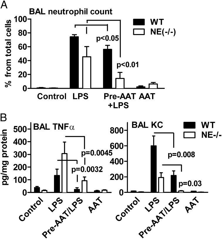
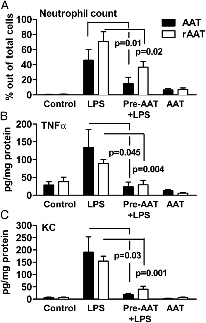
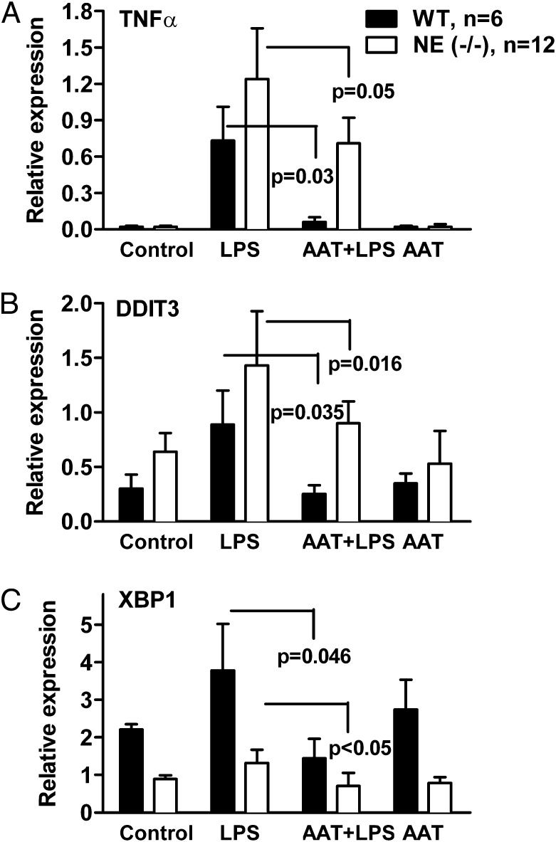
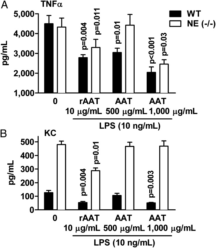
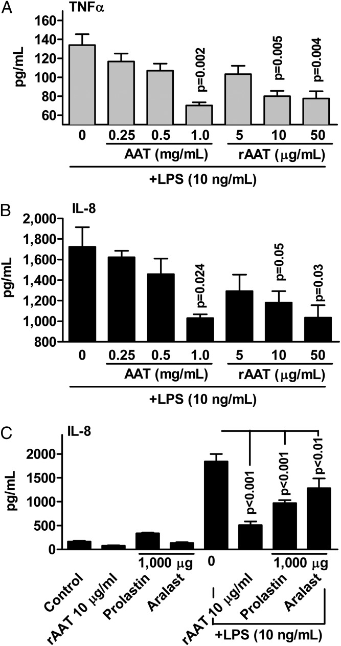
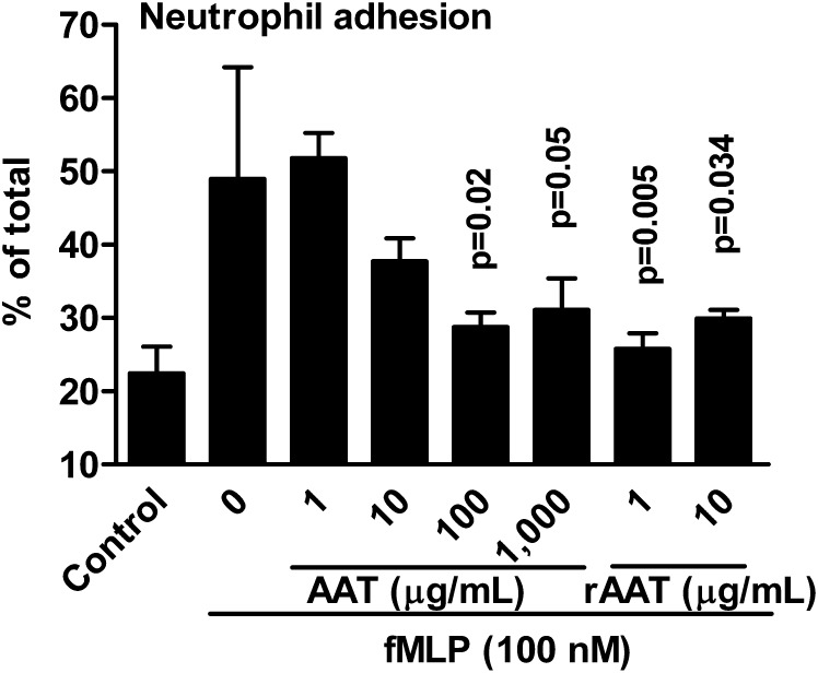
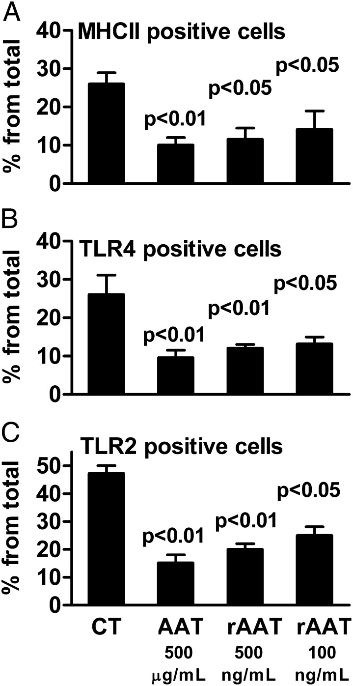
The rationale of α1-antitrypsin (AAT) augmentation therapy to treat progressive emphysema in AAT-deficient patients is based on inhibition of neutrophil elastase; however, the benefit of this treatment remains unclear. Here we show that clinical grade AAT (with elastase inhibitory activity) and a recombinant form of AAT (rAAT) without anti-elastase activity reduces lung inflammatory responses to LPS in elastase-deficient mice. WT and elastase-deficient mice treated with either native AAT or rAAT exhibited significant reductions in infiltrating neutrophils (23% and 68%), lavage fluid levels of TNF-α (70% and 80%), and the neutrophil chemokine KC (CXCL1) (64% and 90%), respectively. Lung parenchyma TNF-α, DNA damage-inducible transcript 3 and X-box binding protein-1 mRNA levels were reduced in both mouse strains treated with AAT; significantly lower levels of these genes, as well as IL-1β gene expression, were observed in lungs of AAT-deficient patients treated with AAT therapy compared with untreated patients. In vitro, LPS-induced cytokines from WT and elastase-deficient mouse neutrophils, as well as neutrophils of healthy humans, were similarly reduced by AAT or rAAT; human neutrophils adhering to endothelial cells were decreased by 60-80% (P < 0.001) with either AAT or rAAT. In mouse pancreatic islet macrophages, LPS-induced surface expression of MHC II, Toll-like receptor-2 and -4 were markedly lower (80%, P < 0.001) when exposed to either AAT or rAAT. Consistently, in vivo and in vitro, rAAT reduced inflammatory responses at concentrations 40- to 100-fold lower than native plasma-derived AAT. These data provide evidence that the anti-inflammatory and immunomodulatory properties of AAT can be independent of elastase inhibition.

Keywords: alpha 1-antitrypsin; immunomodulation; inflammation.


Conflict of interest statement

Conflict of interest statement: S.-H.K., E.C.L., and C.A.D. own stocks in Omni Bio Pharmaceutical.

Figures

Fig. 1.
Fig. 1.
Neutrophil infiltration and cytokine levels in BAL fluid in WT and NE-deficient mice. Twenty-four hours before LPS challenge, WT mice were treated with 2 mg of AAT (Prolastin). (A) Mean ± SD percent of neutrophils of total cells in WT (n = 8) and NE-deficient mice (n = 7). (B) Mean ± SD levels of TNF-α and KC in WT and NE-deficient mice (n = 8 per group). The statistical significance values are between LPS in the presence and absence of AAT treatment. The data are from the same samples shown in Table 1.
Fig. 2.
Fig. 2.
Comparison of AAT to rAAT in LPS-induced acute lung injury in NE-deficient mice. NE-deficient mice were intranasally pretreated with rAAT (50 µg per mouse, 13 per group, open bars) or AAT (2 mg Prolastin, 7 per group, filled bars) 24 h before LPS challenge and BAL fluid obtained after an additional 24 h. (A) Mean ± SD percent of neutrophils. (B) Mean ± SD TNF-α levels per milligram of BAL protein. (C) Mean ± SD KC levels. Data for AAT (Prolastin)-treated NE-deficient mice are taken from Fig. 1 and shown for comparison.
Fig. 3.
Fig. 3.
(AC) AAT reduces proinflammatory gene expression in lung tissue following LPS challenge in WT and NE-deficient mice. Before LPS challenge, WT mice (n = 6) and NE-deficient mice (n = 12) were pretreated for 24 h with 2 mg of intranasal AAT (Prolastin) per mouse. Twenty-four hours after LPS challenge, mRNA was prepared from whole lung tissue. The relative gene expression in each group is shown as the mean ± SD.
Fig. 4.
Fig. 4.
AAT and rAAT reduce LPS-induced release of cytokines from mouse neutrophils in vitro. Bone marrow-derived mouse neutrophils (2 × 10⁶ per mL) were preincubated for 1 h with and without AAT (Prolastin) or rAAT and then stimulated with LPS (10 ng/mL) for 8 h at 37 °C. Mean ± SE levels of (A) TNF-α and (B) KC released from neutrophils of WT (n = 6) and NE-deficient (n = 8) mice. Concentrations of Prolastin and rAAT are indicated. Zero indicates LPS without either AAT or rAAT.
Fig. 5.
Fig. 5.
AAT and rAAT reduce LPS-induced release of cytokines from human neutrophils in vitro. Human blood neutrophils (2 × 10⁶ per mL) isolated from six donors were preincubated with AAT (Prolastin in A and B) or rAAT for 1 h and then stimulated with LPS (10 ng/mL) for 8 h. (A) Mean ± SE TNF-α release at 8 h. (B) Mean ± SE IL-8 release. (C) Mean ± SE IL-8 release in an additional three donors comparing Prolastin, Aralast, and rAAT. Concentrations are micrograms per milliliter (µg/mL).
Fig. 6.
Fig. 6.
AAT and rAAT inhibit human neutrophil adhesion to primary human lung microvascular endothelial cells. Mean ± SE percent-change of fMLP activated human blood neutrophils adhering to the human lung HMVEC. The neutrophils were pretreated with or without AAT (Prolastin) or rAAT. Concentrations of Prolastin and rAAT are depicted under the horizontal axis. The data are derived from the neutrophils of three donors, each performed in quadruplicate. The P values are in comparison with fMLP-stimulated neutrophils without exposure to either AAT or rAAT.
Fig. 7.
Fig. 7.
Expression of macrophage surface receptors on islet cell macrophages exposed to AAT or rAAT. Mean ± SE change in the percent of cells from CD45+CD11b+ cells expressing surface markers for MHC II (A), TLR4 (B), and TLR2 (C) 72 h after exposure to LPS (10 ng/mL) in the presence of AAT (Aralast) or rAAT at the indicated concentrations. The data are one of three similar experiments, each performed in triplicate.

References

    1. Carrell RW, Lomas DA. Alpha1-antitrypsin deficiency—A model for conformational diseases. N Engl J Med. 2002;346(1):45–53. - PubMed
    1. Luisetti M, Seersholm N. Alpha1-antitrypsin deficiency. 1: Epidemiology of alpha1-antitrypsin deficiency. Thorax. 2004;59(2):164–169. - PMC - PubMed
    1. Gadek JE, Klein HG, Holland PV, Crystal RG. Replacement therapy of alpha 1-antitrypsin deficiency. Reversal of protease-antiprotease imbalance within the alveolar structures of PiZ subjects. J Clin Invest. 1981;68(5):1158–1165. - PMC - PubMed
    1. Miravitlles M. Alpha-1-antitrypsin and other proteinase inhibitors. Curr Opin Pharmacol. 2012;12(3):309–314. - PubMed
    1. Wewers MD, Crystal RG. Alpha-1 antitrypsin augmentation therapy. COPD. 2013;10(Suppl 1):64–67. - PubMed

Publication types

MeSH terms