. 2013;11(8):e1001635.
doi: 10.1371/journal.pbio.1001635. Epub 2013 Aug 20.

The HILDA complex coordinates a conditional switch in the 3'-untranslated region of the VEGFA mRNA

Affiliations

The HILDA complex coordinates a conditional switch in the 3'-untranslated region of the VEGFA mRNA

Peng Yao et al. PLoS Biol. 2013.

Abstract

Cell regulatory circuits integrate diverse, and sometimes conflicting, environmental cues to generate appropriate, condition-dependent responses. Here, we elucidate the components and mechanisms driving a protein-directed RNA switch in the 3'UTR of vascular endothelial growth factor (VEGF)-A. We describe a novel HILDA (hypoxia-inducible hnRNP L-DRBP76-hnRNP A2/B1) complex that coordinates a three-element RNA switch, enabling VEGFA mRNA translation during combined hypoxia and inflammation. In addition to binding the CA-rich element (CARE), heterogeneous nuclear ribonucleoprotein (hnRNP) L regulates switch assembly and function. hnRNP L undergoes two previously unrecognized, condition-dependent posttranslational modifications: IFN-γ induces prolyl hydroxylation and von Hippel-Lindau (VHL)-mediated proteasomal degradation, whereas hypoxia stimulates hnRNP L phosphorylation at Tyr(359), inducing binding to hnRNP A2/B1, which stabilizes the protein. Also, phospho-hnRNP L recruits DRBP76 (double-stranded RNA binding protein 76) to the 3'UTR, where it binds an adjacent AU-rich stem-loop (AUSL) element, "flipping" the RNA switch by disrupting the GAIT (interferon-gamma-activated inhibitor of translation) element, preventing GAIT complex binding, and driving robust VEGFA mRNA translation. The signal-dependent, HILDA complex coordinates the function of a trio of neighboring RNA elements, thereby regulating translation of VEGFA and potentially other mRNA targets. The VEGFA RNA switch might function to ensure appropriate angiogenesis and tissue oxygenation during conflicting signals from combined inflammation and hypoxia. We propose the VEGFA RNA switch as an archetype for signal-activated, protein-directed, multi-element RNA switches that regulate posttranscriptional gene expression in complex environments.


Conflict of interest statement

The authors have declared that no competing interests exist.

Figures
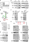
Figure 1
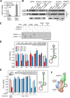
Figure 1. Heterotrimeric HILDA complex binds the VEGFA HSR in hypoxia.
(A) Recombinant hnRNP L by itself does not drive the VEGFA RNA switch and restore in vitro translation of the GAIT-element-bearing reporter. In vitro translation of capped and poly(A)-tailed firefly luciferase (FLuc)-VEGFA HSR-A30 reporter transcript was determined in a wheat germ extract in the presence of [35S]Met, cytosolic extracts from IFN-γ-treated U937 cells, and recombinant hnRNP L. FLuc expression was determined by activity assay, normalized by RLuc expression, and reported as mean ± standard deviation (SD, n = 3). (B) Schematic of HSR in VEGFA 3′UTR. CARE (red), GAIT element (green), extended CARE (CARE-E, dotted line), AUSL-A (dashed line), and AUSL-D (dashed and dotted line) are indicated. (C) Mass spectrometric analysis of CARE-binding proteins. U937 cells were treated with normoxia (Nmx.) or hypoxia (Hpx.) for 24 h and the S100 extracts, precleared, and incubated with biotinylated CARE-E (extended CARE, sequences in Materials and Methods) and magnetic streptavidin microbeads. Specifically bound proteins were subjected to SDS-PAGE and Coomassie staining. Bands specifically enriched in affinity-purified lysates from hypoxia-treated cells were trypsinized, and peptide sequences of hnRNP L, DRBP76, and hnRNP A2/B1 detected by mass spectrometry. (D) Hypoxia-inducible binding of hnRNP L, DRBP76, and hnRNP A2/B1 to CARE. Cells were treated with Nmx. or Hpx. for 24 h, and the precleared S100 extracts incubated with biotinylated, wild-type, or antisense (A.S.) CARE-E, and then with magnetic streptavidin microbeads. Specifically bound proteins were subjected to immunoblot analysis. (E) DRBP76 and hnRNP A2/B1 form a complex with hnRNP L in vivo. Cells were treated with IFN-γ in Nmx. or Hpx. for 24 h. Cell lysates were incubated with or without RNase A, immunoprecipitated with anti-hnRNP L antibody, and subjected to immunoblot analysis (left panel). Total expression of hnRNP L, hnRNP A2/B1, and DRBP76 was determined by immunoblot as input control (right panel). (F) Interprotein interactions of HILDA constituents. Recombinant hnRNP A2/B1 and DRBP76 were incubated with GST-hnRNP L or GST immobilized to glutathione (GSH)-agarose beads. After washing, binding was detected by immunoblot (left). Recombinant hnRNP L and DRBP76 were incubated with GST-hnRNP A2/B1 or GST immobilized to GSH-agarose beads (right). (G) hnRNP L domain mapping. In vitro synthesized S35-Met-labeled hnRNP L segments (top) were incubated with cytosol from U937 cells. hnRNP A2/B1 (left) and DRBP76 (right) were immunoprecipitated, and the interacting hnRNP L segments detected by autoradiorgraphy. Key hnRNP L domains are shown above.
Figure 2
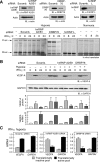
Figure 2. Specific HSR RNA requirements for DRBP76 interaction and RNA switch activity.
(A) Condition-dependent binding of VEGFA mRNA to GAIT and HILDA complexes. U937 cells were treated with IFN-γ for 24 h under Nmx. or Hpx. Lysates were subjected to IP with anti-hnRNP L or -EPRS antibodies (or IgG control) coupled with qRT-PCR using gene-specific primers. VEGFA mRNA was normalized to GAPDH mRNA, and results normalized to amount of VEGFA mRNA in EPRS IP of cells treated with IFN-γ under Nmx. (B) hnRNP A2/B1 and DRBP76 are essential for hnRNP L binding to VEGFA mRNA in vivo. U937 cells were transfected with hnRNP A2/B1- and DRBP76-specific (or scrambled) siRNA, and lysates immunoprecipitated with anti-hnRNP L antibody. Extracted RNA was subjected to RT-PCR using primers specific for VEGFA or β-actin mRNA. Efficiency of hnRNP L IP was shown by immunoblot. (C) Protein binding domains of VEGFA HSR. Recombinant hnRNP A2/B1, hnRNP L, and DRBP76 were incubated with [32P]UTP-labeled VEGFA HSR, CARE, AUSL, AUSL-A, and AUSL-D RNA and subjected to UV crosslinking. Crosslinked products were treated with RNase A and detected by SDS-PAGE and autoradiography. (D) HSR region required for RNA switch activity. Reporter constructs containing FLuc upstream of wild-type or mutant VEGFA HSR were transfected into U937 cells with a plasmid expressing RLuc driven by the SV40 promoter. Luciferase activity was measured after treatment with IFN-γ under Nmx. or Hpx. for 8 or 24 h. Relative luciferase activity (FLuc/RLuc) was determined from three independent experiments and reported as mean ± SD (n = 3). FLuc mRNA expression was determined by semi-quantitative RT-PCR (inset). (E) Permissible spacer length between GAIT element and CARE. Reporter constructs with FLuc upstream of wild-type or mutant VEGFA HSR containing poly(C) spacers were transfected into U937 cells together with a RLuc-bearing plasmid. Luciferase activity was measured after treatment with IFN-γ under Nmx. or Hpx. for 0, 8, and 24 h. Cells were treated as in (D) and luciferase activity determined in three independent experiments, and reported as mean ± SD (n = 3). FLuc mRNA expression was determined by semiquantitative RT-PCR (inset). (F) Schematic of heterotrimeric HILDA complex binding VEGFA HSR RNA in hypoxia.
Figure 3
Figure 3. HILDA complex is essential for VEGFA RNA switch activity.
(A) hnRNP A2/B1, DRBP76, and hnRNP L are required for hypoxia-inducible RNA switch activity in vitro. Effectiveness of knockdown by siRNA targeting hnRNP A2/B1, DRBP76, and hnRNP L, and scrambled (scramb.) control was determined by immunoblot analysis; β-actin was probed as loading control (top). FLuc reporter RNA bearing the VEGFA HSR and RLuc control transcripts were subjected to in vitro translation in RRL in presence of lysates from U937 cells transfected with scrambled or gene-specific siRNA and incubated with IFN-γ under hypoxia; lysates from IFN-γ-treated normoxic cells shown as control (bottom). (B) hnRNP A2/B1 and DRBP76 are required for robust in vivo expression of endogenous VEGF-A in hypoxia. Lysates from siRNA-treated cells as in (A) were probed with anti-VEGF-A and anti-GAPDH antibodies, and normalized VEGF-A expression was quantified by densitometry. Expression of VEGF-A mRNA was determined by qRT-PCR and normalized by GAPDH mRNA. Results are reported as mean ± SEM (n = 3). (C) hnRNP A2/B1 and DRBP76 are required for efficient VEGFA mRNA translation in presence of IFN-γ and Hpx. U937 cells were transfected with siRNA targeting hnRNP A2/B1 and DRBP76 (or scrambled siRNA), and then subjected to IFN-γ and Hpx. Cell lysates were fractionated on a sucrose gradient, and total RNA in translationally active and inactive pools subjected to qRT-PCR with VEGFA- and GAPDH-specific primers. Results are reported as mean ± SD (n = 3).
Figure 4
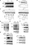
Figure 4. IFN-γ induces VHL-mediated polyubiquitination and degradation of prolylhydroxylated hnRNP L.
(A) Steady-state amount of hnRNP L mRNA is not regulated by IFN-γ. U937 cells were treated with IFN-γ under Nmx. or Hpx. for 0, 8, and 24 h. HnRNP L and β-actin mRNA were determined by semiquantitative RT-PCR. (B) IFN-γ induces hnRNP L degradation in normoxic cells. U937 cells were treated with CHX for up to 16 h under Nmx. and lysates subjected to immunoblot and quantitated by densitometry. (C) IFN-γ-inducible degradation of hnRNP L is proteasome-mediated. U937 cells were treated with CHX or CHX plus MG132 in presence of IFN-γ for up to 12 h under Nmx. (left panel) or Hpx. (right panel), and lysates subjected to immunoblot and quantitated by densitometry. (D) IFN-γ induces polyubiquitination of endogenous hnRNP L in vivo. U937 cells were treated with IFN-γ for up to 24 h in the absence or presence of MG132, and lysates subjected to IP with mouse-derived hnRNP L antibody followed by immunoblot with rabbit-derived hnRNP L antibody. (E) IFN-γ induces normoxia-dependent ubiquitination of hnRNP L. U937 cells were transfected with HA-ubiquitin, treated with MG132 in Nmx. or Hpx., immunoprecipitated with anti-hnRNP L antibody, and subjected to immunoblot with anti-HA antibody. (F) IFN-γ induces interaction of hnRNP L with VHL. Lysates from U937 cells treated with IFN-γ and MG132 for up to 24 h were immunoprecipitated with anti-VHL antibody and subjected to immunoblot with anti-hnRNP L, -VHL, -hnRNP A2/B1, and -DRBP76 antibodies. (G) IFN-γ-induced polyubiquitination and degradation of hnRNP L is mediated by VHL. U937 cells were transfected with VHL-specific (or scrambled) siRNA. After recovery, cells were treated with IFN-γ in the presence or absence of MG132 and lysates immunoblotted with anti-VHL, -hnRNP L, -ubiquitin, and -GAPDH antibodies.
Figure 5
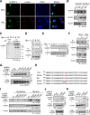
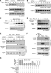
Figure 5. Hypoxia-inducible hnRNP L phosphorylation at Tyr359 suppresses nuclear translocation and cytoplasmic degradation.
(A) Hypoxia increases cytoplasmic localization of hnRNP L. U937 cells treated with IFN-γ for 24 h under Nmx. or Hpx. were immunostained using rabbit anti-hnRNP L and -β-actin antibodies. Cell nuclei were stained with DAPI. (B) Analysis of hypoxia-stimulated translocation of hnRNP L by cell fractionation. U937 cells treated with IFN-γ in Nmx. or Hpx. were fractionated and subjected to immunoblot. (C) Hypoxia induces hnRNP L phosphorylation in vivo. U937 cells were incubated under Hpx. and then with a 4-h pulse of 32P-orthophosphate between 6 and 10 h (denoted as 8 h) or between 22 and 26 h (denoted as 24 h). Lysates were immunoprecipitated with anti-hnRNP L antibody (or pre-immune IgG), and 32P-labeled protein detected by autoradiography. (D) Hypoxia induces tyrosine phosphorylation of hnRNP L. Lysates from cells treated with Hpx. for 0, 8, and 24 h were immunoprecipitated with anti-hnRNP L antibody and immunoblotted with antibodies targeting phosphoserine (P-Ser), phosphothreonine (P-Thr), or phosphotyrosine (P-Tyr). (E) Time course of hypoxia-inducible hnRNP L phosphorylation. U937 cells were treated with Hpx. for up to 24 h and lysates immunoprecipitated with anti-hnRNP L antibody and immunoblotted with antibodies targeting P-Tyr or hnRNP L. (F) Hypoxia induces cytoplasmic accumulation of P-Tyr-hnRNP L. Cytosolic and nuclear fractions from U937 cells treated with Hpx. for 24 h were immunprecipitated with anti-hnRNP L antibody and immunoblotted with anti-P-Tyr and -hnRNP L antibodies. Western blots were done using anti-HDAC1 and anti-tubulin antibodies. (G) Hypoxia induces Tyr359 phosphorylation of hnRNP L. pcDNA3-hnRNP L-Myc bearing selected Tyr-to-Ala mutations were transfected into U937 cells with endogenous hnRNP L knocked down by 3′UTR-targeting siRNA. After recovery, cells were treated with Hpx. for 24 h. Lysates were immunprecipitated with anti-hnRNP L antibody and immunoblotted with anti-P-Tyr and -hnRNP L antibodies. (H) Sequence conservation of hnRNP L phospho-site in vertebrate animals. Tyr phospho-site in aligned sequences is shown (red). (I) Cellular localization of phospho-mimetic and phospho-dead hnRNP L in normoxia. c-Myc-tagged, wild-type (WT), phospho-mimetic (Tyr-to-Asp, Y-D), and phospho-dead (Tyr-to-Ala, Y-A) mutant hnRNP L were transiently transfected into U937 cells and were determined in cytoplasmic and nuclear fractions by immunoblot analysis with anti-c-Myc, -HDAC1, and -tubulin antibodies. (J) Phospho-mimetic hnRNP L binds hnRNP A2/B1. c-Myc-tagged, wild-type (WT), phospho-dead (Y-A), and phospho-mimetic (Y-D) hnRNP L were expressed in U937 cells by transient transfection. Lysates were immunoprecipitated with anti-c-Myc antibody and immunoblotted with anti-hnRNP L and -hnRNP A2/B1 antibodies. Expression of c-Myc-tagged hnRNP L was determined with anti-c-Myc antibody of total lysate. (K) Phospho-mimetic hnRNP L inhibits VHL-mediated, proteasomal degradation of hnRNP L. c-Myc-tagged wild-type, phospho-dead, and phospho-mimetic hnRNP L was transiently transfected into U937 cells, and then treated with IFN-γ for 24 h. Lysates were immunoblotted with anti-c-Myc and -actin antibodies. C-Myc-tagged hnRNP L was determined by immunoblot with anti-c-Myc antibody of total lysate from cells not treated with IFN-γ as controls for transfection and expression of hnRNP L-bearing vectors.
Figure 6
Figure 6. hnRNP A2/B1 prevents IFN-γ-induced hnRNP L prolyl hydroxylation, blocks interaction with VHL, and stabilizes hnRNP L.
(A) Rapid degradation of hnRNP L in absence of hnRNP A2/B1. U937 cells were transfected with hnRNP A2/B1-specific (or scrambled) siRNA. After recovery, cells were treated with IFN-γ and Hpx. for 0, 8, and 24 h. Lysates were immunoblotted with anti-hnRNP A2/B1, -hnRNP L, and -GAPDH antibodies. (B) Time course of hnRNP L degradation in absence of hnRNP A2/B1. U937 cells were treated as in (A) for up to 24 h. Lysates were immunoblotted with anti-hnRNP L and -DRBP76 antibodies. (C) IFN-γ induces prolyl hydroxylation of hnRNP L. U937 cells were treated with IFN-γ and MG132 for up to 24 h. Lysates were immunoprecipitated with anti-hnRNP L antibody and immunoblotted with anti-hydroxyproline and -hnRNP L antibodies. (D) hnRNP L is not subject to prolyl hydroxylation in Hpx. U937 cells were treated with IFN-γ and Hpx. for up to 24 h. Lysates were immunoprecipitated with anti-hnRNP L antibody and immunoblotted with anti-hydroxyproline and -hnRNP L antibodies. (E) hnRNP A2/B1 inhibits prolyl hydroxylation of hnRNP L in hypoxia. U937 cells were transfected with hnRNP A2/B1-specific (or scrambled) siRNA. After recovery, cells were treated with IFN-γ, Hpx., and MG132 for up to 24 h. Lysates were immunoblotted with anti-hnRNP A2/B1 antibody. Lysates were also immunoprecipitated with anti-hnRNP L antibody and immunoblotted with anti-hydroxyproline and -hnRNP L antibodies. (F) Hypoxia induces binding of hnRNP A2/B1 to hnRNP L and prevents VHL binding. U937 cells were subjected to Nmx. or Hpx. for 24 h in the presence of IFN-γ stimulus. Lysates were immunoprecipitated with anti-hnRNP L antibody and immunoblotted with anti-hnRNP A2/B1 and -VHL antibodies. Total hnRNP L in cell lysates was determined by immunoblot. IP with pre-immune IgG of hypoxic lysate served as a control. (G) Reconstitution of RNA switch function of HILDA complex. Phospho-mimetic hnRNP L (Y359D) was pre-incubated with DRBP76 and hnRNP A2/B1 as indicated (5 pmol each) for 0.5 h on ice. In vitro translation of the FLuc reporter bearing the VEGFA HSR element (and RLuc control RNA) was determined in a wheat germ extract in the presence of 35S-Met, cytosolic extracts from IFN-γ-treated U937 cells, and HILDA components as indicated. In a control experiment, wild-type hnRNP L replaced phospho-mimetic hnRNP L. FLuc expression was normalized by RLuc and reported as mean ± SD, n = 3.
Figure 7
Figure 7. Regulation of hnRNP L expression by IFN-γ and hypoxia and the role of the HILDA complex in the VEGFA RNA switch.

References

    1. Tammela T, Enholm B, Alitalo K, Paavonen K (2005) The biology of vascular endothelial growth factors. Cardiovasc Res 65: 550–563. - PubMed
    1. Hua Z, Lv Q, Ye W, Wong CK, Cai G, et al. (2006) MiRNA-directed regulation of VEGF and other angiogenic factors under hypoxia. PLoS One 1: e116 doi:10.1371/journal.pone.0000116 - DOI - PMC - PubMed
    1. Pages G, Pouyssegur J (2005) Transcriptional regulation of the Vascular Endothelial Growth Factor gene—a concert of activating factors. Cardiovasc Res 65: 564–573. - PubMed
    1. Ferrara N, Davis-Smyth T (1997) The biology of vascular endothelial growth factor. Endocr Rev 18: 4–25. - PubMed
    1. Weis SM, Cheresh DA (2005) Pathophysiological consequences of VEGF-induced vascular permeability. Nature 437: 497–504. - PubMed

Publication types

MeSH terms

Substances