. 2013 Aug 19;8(8):e71149.
doi: 10.1371/journal.pone.0071149. eCollection 2013.

Deciphering the glycan preference of bacterial lectins by glycan array and molecular docking with validation by microcalorimetry and crystallography

Affiliations

Deciphering the glycan preference of bacterial lectins by glycan array and molecular docking with validation by microcalorimetry and crystallography

Jeremie Topin et al. PLoS One. .

Abstract

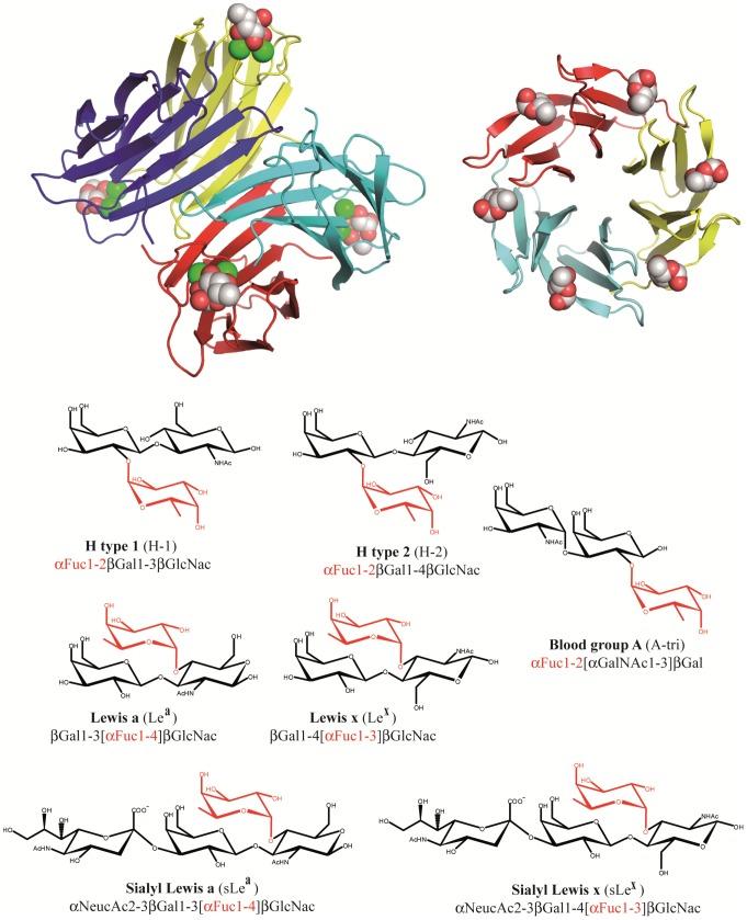
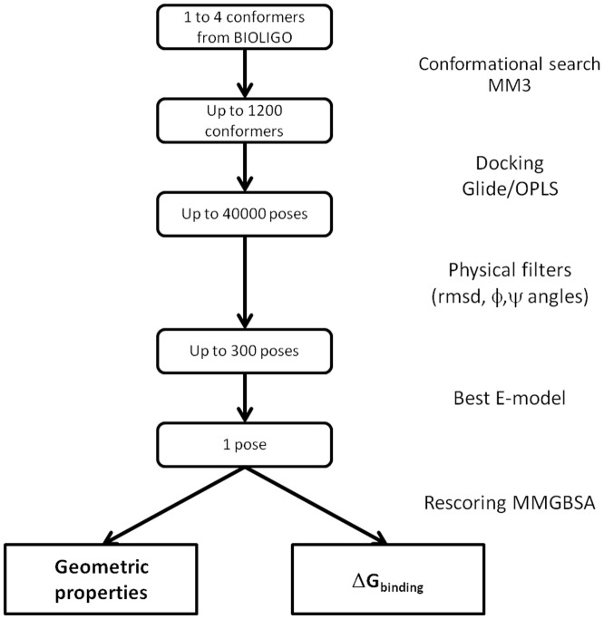
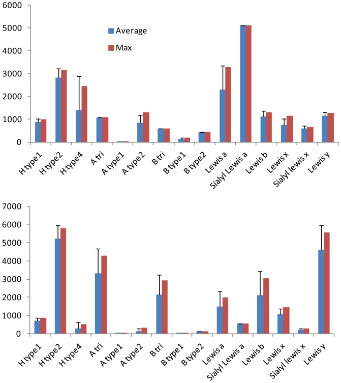
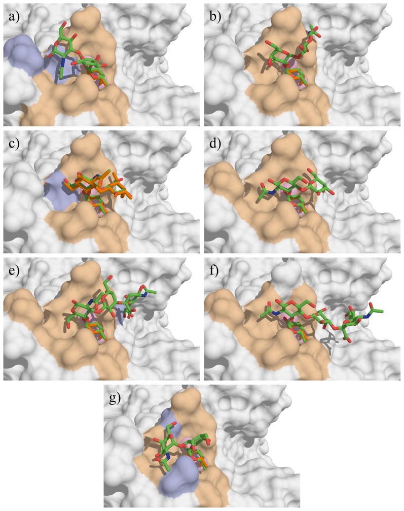
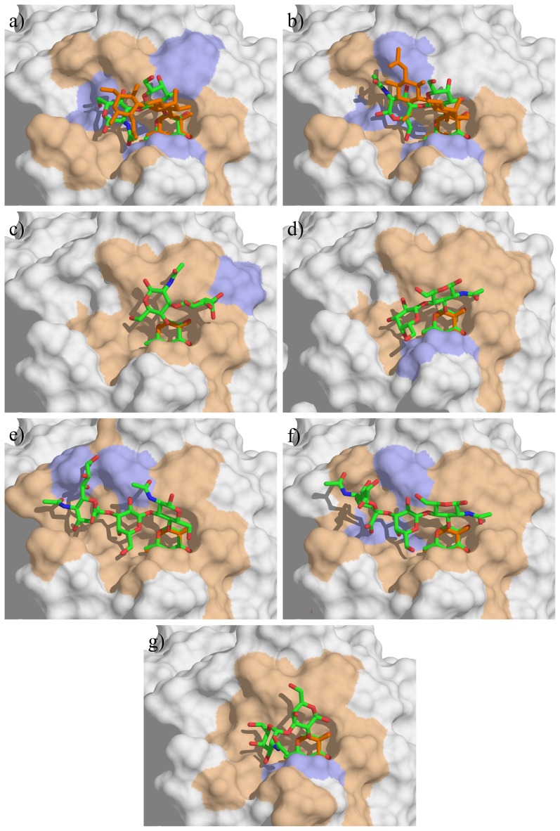
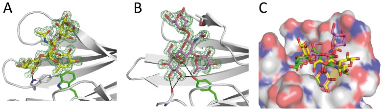
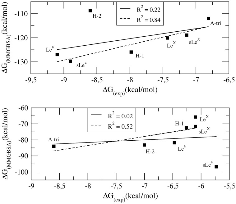
Recent advances in glycobiology revealed the essential role of lectins for deciphering the glycocode by specific recognition of carbohydrates. Integrated multiscale approaches are needed for characterizing lectin specificity: combining on one hand high-throughput analysis by glycan array experiments and systematic molecular docking of oligosaccharide libraries and on the other hand detailed analysis of the lectin/oligosaccharide interaction by x-ray crystallography, microcalorimetry and free energy calculations. The lectins LecB from Pseudomonas aeruginosa and BambL from Burkholderia ambifaria are part of the virulence factors used by the pathogenic bacteria to invade the targeted host. These two lectins are not related but both recognize fucosylated oligosaccharides such as the histo-blood group oligosaccharides of the ABH(O) and Lewis epitopes. The specificities were characterized using semi-quantitative data from glycan array and analyzed by molecular docking with the Glide software. Reliable prediction of protein/oligosaccharide structures could be obtained as validated by existing crystal structures of complexes. Additionally, the crystal structure of BambL/Lewis x was determined at 1.6 Å resolution, which confirms that Lewis x has to adopt a high-energy conformation so as to bind to this lectin. Free energies of binding were calculated using a procedure combining the Glide docking protocol followed by free energy rescoring with the Prime/Molecular Mechanics Generalized Born Surface Area (MM-GBSA) method. The calculated data were in reasonable agreement with experimental free energies of binding obtained by titration microcalorimetry. The established predictive protocol is proposed to rationalize large sets of data such as glycan arrays and to help in lead discovery projects based on such high throughput technology.


Conflict of interest statement

Competing Interests: The authors have declared that no competing interests exist.

Figures

Figure 1
Figure 1. Representation of the bacterial lectins and oligosaccharides used in the docking calculations.
Top: Graphical representation of the two bacterial lectins. From left to right LecB/fucose and BambL/fucose complexes have been adapted from PDB 1GZT and 3ZW2 respectively. The fucose ligand and calcium atoms are displayed as spheres. Bottom: Schematic representation and sequences of fucosylated oligosaccharides.
Figure 2
Figure 2. Flowchart of the docking/analysis protocol.
Figure 3
Figure 3. Selected data from the glycan array v4.1 performed on two bacterial lectins.
Binding is represented as fluorescent intensity obtained with LecB (top panel) and BambL (bottom panel) both at 0.2 µg.mL−1 and labeled with Alexa 488. Blue bar: average value with standard deviation over 5 experiments at same concentration, red bar: maximum response observed.
Figure 4
Figure 4. Docking of the oligosaccharides in LecB binding site.
a) H type 1, b) H type 2, c) Lea, d) Lex, e) sLea f) sLex and g) A-tri. The docked oligosaccharides are represented as sticks (carbon, oxygen and nitrogen atoms are colored green, red and blue respectively) and the ones from crystal structures are colored orange. Calcium ions are represented as pink spheres. The protein accessible surface is colored in beige for residues comprised within a sphere of 4 Å around the ligand and in blue for residues involved in hydrogen bond with ligand residues (except fucose).
Figure 5
Figure 5. Energy maps and docked conformations.
Isoenergy contrours (1 kcal/mole) are represented as a function of Φ (x-axis) and Ψ (y-axis)torsion angles for all glycosidic linkages of interest taken from Glyco3D (http://glyco3d.cermav.cnrs.fr/) with superimposition of oligosaccharide conformations derived from docking (circles and squares) or protein crystallography (stars). Torsion angles are defined as Φ = θ(O5-C1-O1-Cx) and Ψ = θ(C1-O1-C′x-C′x+1).
Figure 6
Figure 6. Docking of the oligosaccharides in BambL binding site.
a) H type 1, b) H type 2, c) Lea, d) Lex, e) sLea f) sLex and g) A-tri. The color codes are the same as in Figure 4.
Figure 7
Figure 7. Crystallographic structure of BambL in complex with Lex tetrasaccharide.
(A) Intramonomeric and (B) intermonomeric sites. Tetrasaccharide and key amino acids are represented as sticks, hydrogen bond to non-fucose carbohydrate residues as dotted lines; 2mF0-Dfc electron density map contoured at 1σ is shown ad green mesh. (C) Superimposition of Lex tetrasaccharide from crystal intramonomeric (yellow) and intermonomeric (pink) sites and Lex trisaccharide from docking (green).
Figure 8
Figure 8. Correlations between experimental binding free energies of binding measured from titration microcalorimetry and theoretical ones calculated using MM-GBSA.
Top: LecB, bottom: BambL (bottom). Calculations have been performed with all data (black lines) or with omission of H-2 for LecB or sLea for BambL, respectively.

Similar articles

Cited by

References

    1. Rillahan CD, Paulson JC (2011) Glycan microarrays for decoding the glycome. Annu Rev Biochem 80: 797–823. - PMC - PubMed
    1. Smith DF, Cummings RD (2013) Application of microarrays for deciphering the structure and function of the human glycome. Mol Cell Proteomics 12: 902–912. - PMC - PubMed
    1. Imberty A, Varrot A (2008) Microbial recognition of human cell surface glycoconjugates. Curr Opin Struct Biol 18: 567–576. - PubMed
    1. Sharon N (1996) Carbohydrate-lectin interactions in infectious disease. Adv Exp Med Biol 408: 1–8. - PubMed
    1. Springer SA, Gagneux P (2013) Glycan evolution in response to collaboration, conflict, and constraint. J Biol Chem 288: 6904–6911. - PMC - PubMed

Publication types