. 2013 Aug 19;8(8):e71349.
doi: 10.1371/journal.pone.0071349. eCollection 2013.

TBCK influences cell proliferation, cell size and mTOR signaling pathway

Affiliations

TBCK influences cell proliferation, cell size and mTOR signaling pathway

Yueli Liu et al. PLoS One. .

Abstract

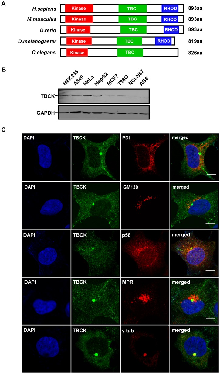
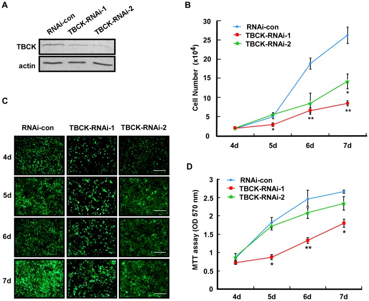
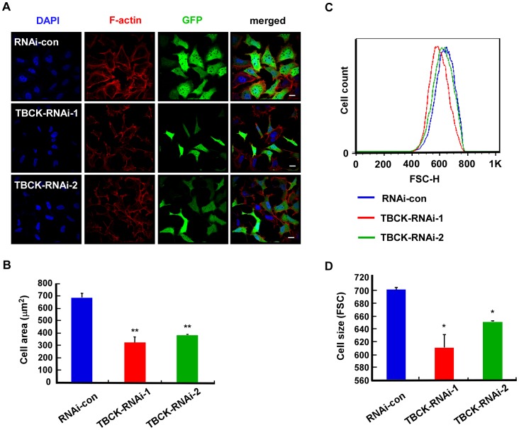
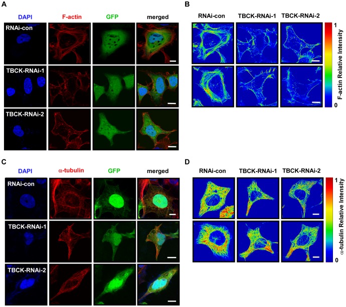
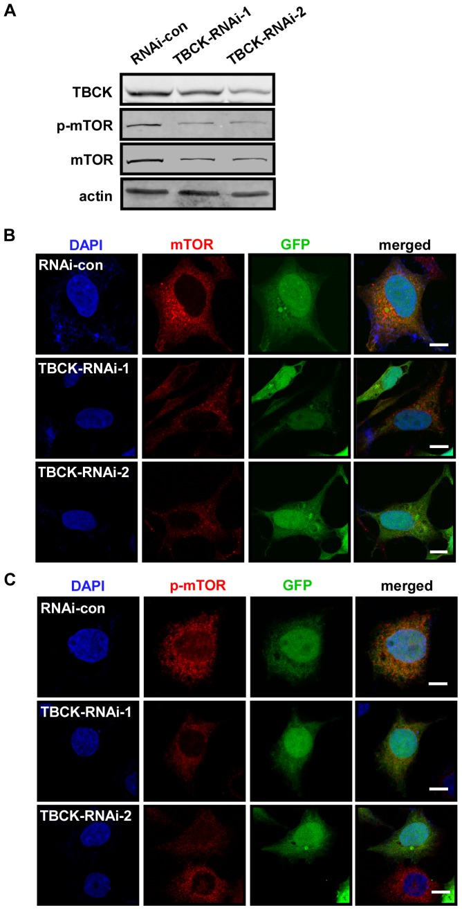
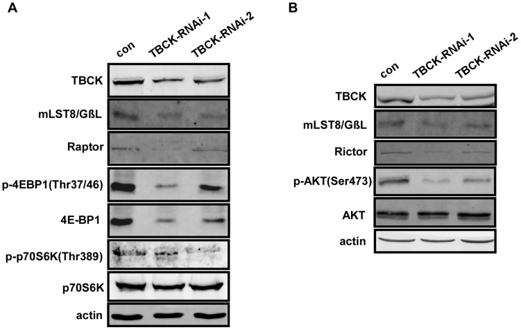
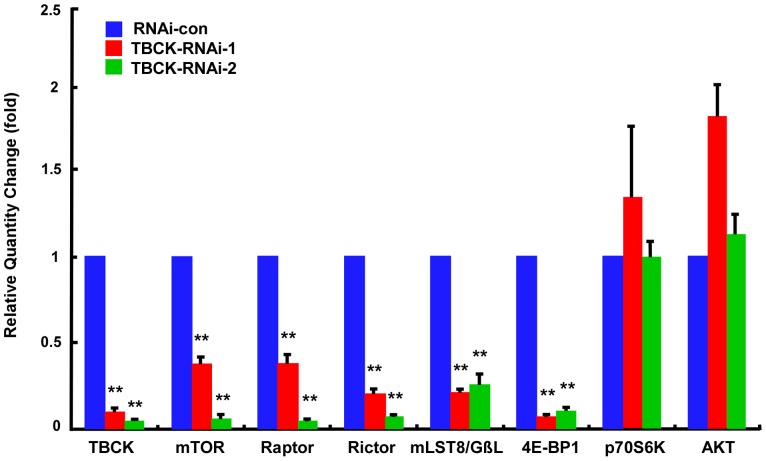
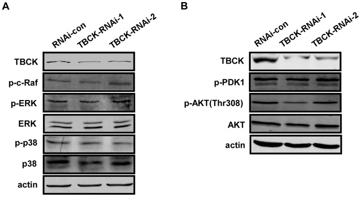
Mammalian target of rapamycin (mTOR) is a central regulator for both cell proliferation and cell growth; however, little is known about the regulation of mTOR expression at the transcriptional level. Here, we provide evidences that a conserved human protein TBCK (TBC1 domain containing kinase) is involved in the regulation of mTOR signaling pathway. Depletion of TBCK significantly inhibits cell proliferation, reduces cell size, and disrupts the organization of actin, but not microtubule. Knockdown of TBCK induces a significant decrease in the protein levels of components of mTOR complex (mTORC), and suppresses the activity of mTOR signaling, but not MAPK or PDK1/Akt pathway. Further results show that TBCK influences the expression of mTORC components at the transcriptional level. Thus, these data suggest that TBCK may play an important role in cell proliferation, cell growth and actin organization possibly by modulating mTOR pathway.


Conflict of interest statement

Competing Interests: The authors have declared that no competing interests exist.

Figures

Figure 1
Figure 1. Characterization of TBCK.
(A) Schematic comparison of TBCK homologs in H. sapiens, M. musculus, D. rerio, D. melanogaster, and C. elegans. Red, green and blue bars indicate possible kinase domain, TBC domain and rhodanese homology domain, respectively. (B) Expression of TBCK in mammalian cell lines. Total lysates from the indicated cells were resolved by SDS-PAGE and subjected to Western analysis. (C) Subcellular localization of TBCK. HEK293 cells grown on cover slides were immunostained with the antibodies as shown. DNA was visualized by DAPI. Bar, 10 μm.
Figure 2
Figure 2. Depletion of TBCK inhibits cell proliferation.
HEK293 cells infected with the lentiviruses targeting to the various regions of TBCK mRNA (TBCK-RNAi-1 or 2) were subjected to Western blotting (A), cell counts (B), fluorescence microscopy (C) and MTT analyses (D). Bar, 200 µm. Lentivirus-infected cells were GFP-positive. Data are shown as the mean of three independent experiments ± SE (* P<0.05, ** P<0.01).
Figure 3
Figure 3. TBCK depletion reduces cell size.
HEK293 cells were infected with the indicated lentivirus and subjected to fluorescence microscopy (A–B) and FACS analysis (C–D). (AB) GFP-positive signals indicate the cells infected by lentivirus. Bar, 20 µm. The area measurements of infected cells were quantified by Image-Pro Plus 6.0 software. Data are shown as the mean of three independent experiments ± SE (** P<0.01, n>200). (CD) Representative histogram of flow cytometry shows the size distribution (FSC-H) of GFP positive cells that were stained with propidium iodide. The mean of three independent experiments ± SE is shown (* P<0.05, n>100,000).
Figure 4
Figure 4. TBCK plays a role in actin organization.
HEK293 cells infected with the indicated lentivirus were subjected to immunofluorescence staining with phalloidin-TRITC (A) or anti-α-tubulin antibody (C). Lentivirus-infected cells were GFP-positive. DNA was visualized by DAPI. Bar, 10 µm. Single panel image of phalloidin-TRITC signal (B) or α-tubulin (D) is showed in rainbow palettes.
Figure 5
Figure 5. Depletion of TBCK influences mTOR signaling pathway.
HEK293 cells were infected with the indicated lentivirus and subjected to Western blotting (A) and immunofluorescence analysis (BC) with the antibodies as shown. GFP-positive signals indicate the cells infected by lentivirus. DNA was visualized by DAPI. Bar, 10 µm.
Figure 6
Figure 6. TBCK influences the protein level of mTOR components and the activities of mTOR complexes.
(AB) HEK293 cells infected with the indicated lentivirus were subjected to immunoblotting with the antibodies as shown.
Figure 7
Figure 7. Depletion of TBCK represses the mRNA level of mTOR complexes components.
Total RNAs from HEK293 cells infected with the indicated lentivirus were analyzed by quantitative RT-PCR with the targeted genes as shown. Error bars indicate SE (** P<0.01).
Figure 8
Figure 8. TBCK has no significant effect on MAPK and PDK1/AKT pathway.
(AB) HEK293 cells infected with the indicated lentivirus were subjected to Western blotting with the antibodies as shown.

References

    1. Schmelzle T, Hall MN (2000) TOR, a central controller of cell growth. Cell 103: 253–262. - PubMed
    1. Wullschleger S, Loewith R, Hall MN (2006) TOR signaling in growth and metabolism. Cell 124: 471–484. - PubMed
    1. Hay N, Sonenberg N (2004) Upstream and downstream of mTOR. Genes Dev 18: 1926–1945. - PubMed
    1. Loewith R, Hall MN (2011) Target of rapamycin (TOR) in nutrient signaling and growth control. Genetics 189: 1177–1201. - PMC - PubMed
    1. Kim J, Guan KL (2011) Amino acid signaling in TOR activation. Annu Rev Biochem 80: 1001–1032. - PubMed

Publication types

MeSH terms

Substances