. 2013 Aug 14;8(8):e71359.
doi: 10.1371/journal.pone.0071359. eCollection 2013.

Acute mechanical stretch promotes eNOS activation in venous endothelial cells mainly via PKA and Akt pathways

Affiliations

Acute mechanical stretch promotes eNOS activation in venous endothelial cells mainly via PKA and Akt pathways

Zhenqian Hu et al. PLoS One. .

Abstract

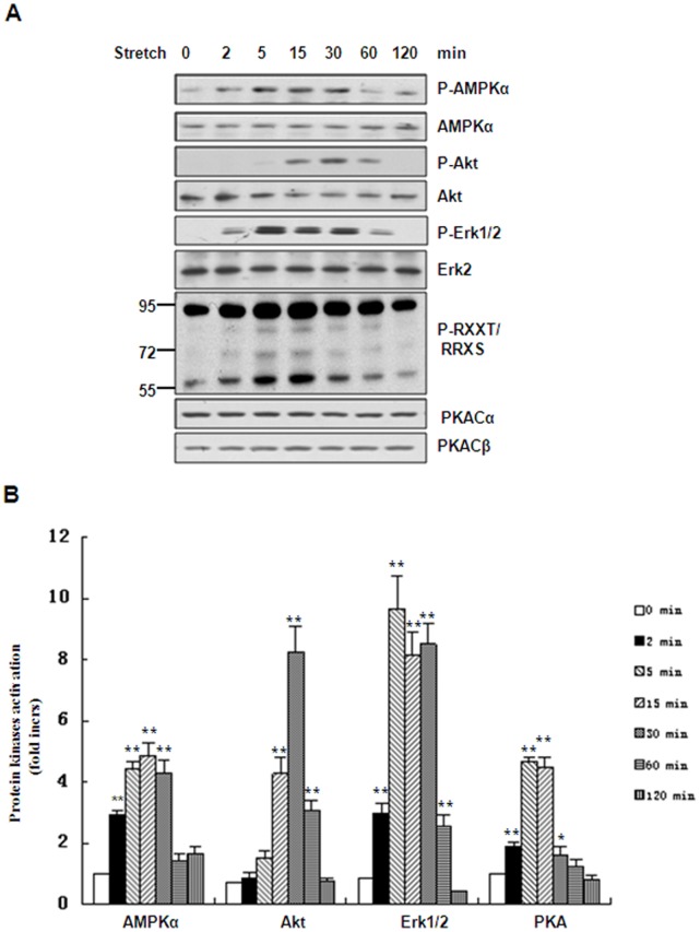
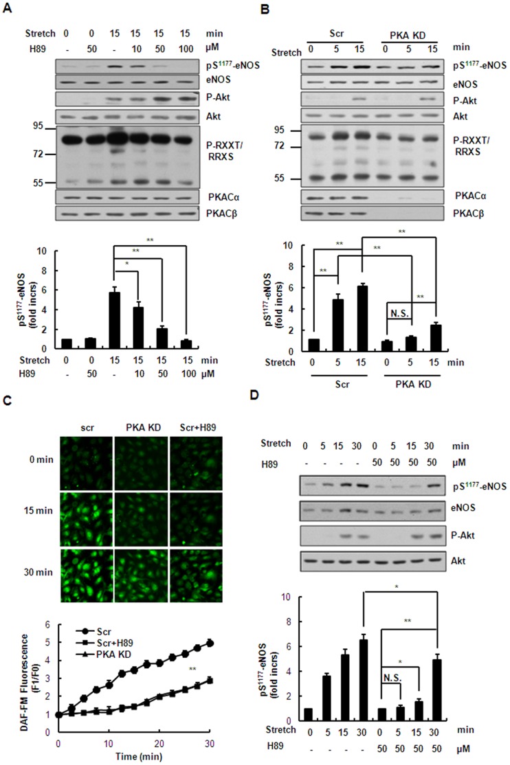
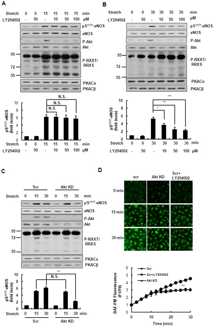
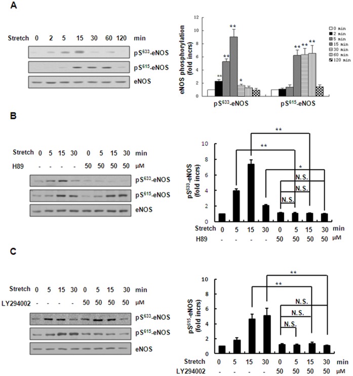
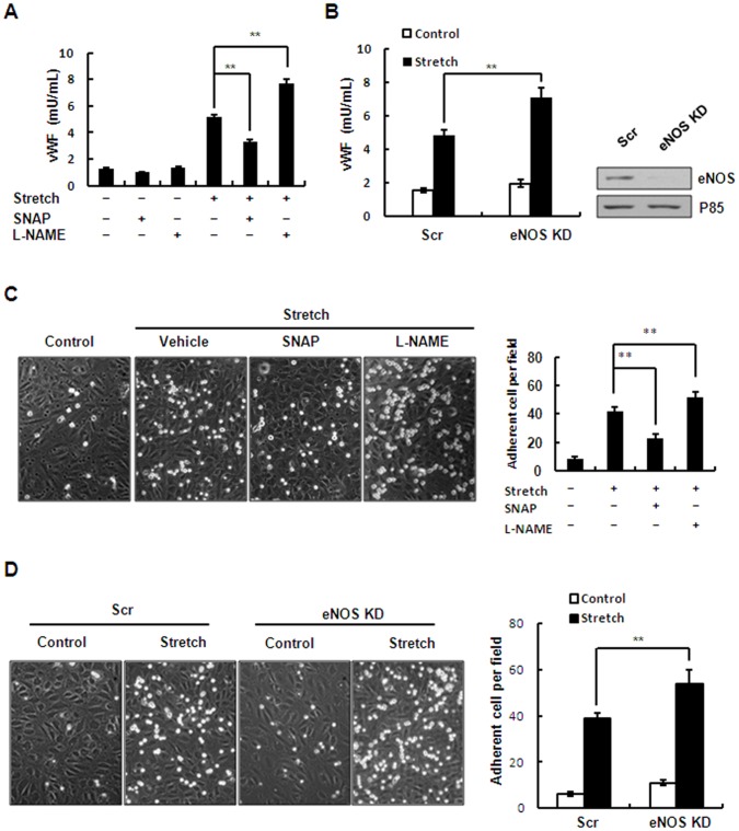
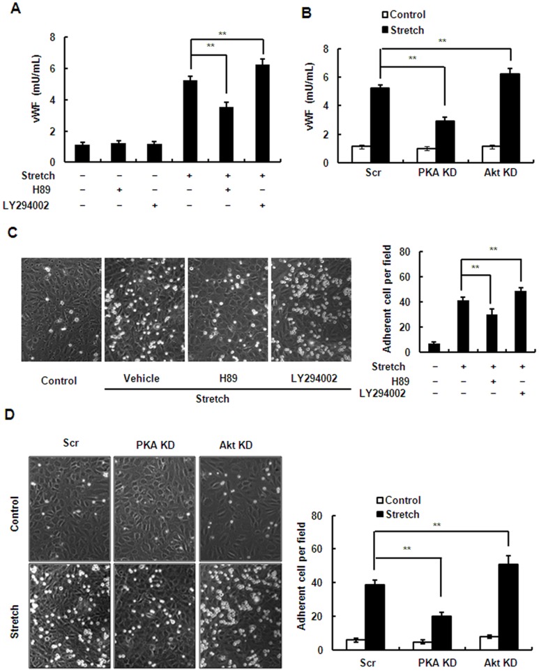
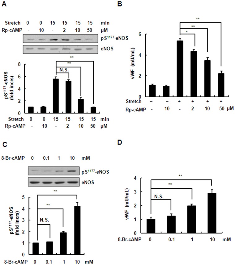
In the vasculature, physiological levels of nitric oxide (NO) protect against various stressors, including mechanical stretch. While endothelial NO production in response to various stimuli has been studied extensively, the precise mechanism underlying stretch-induced NO production in venous endothelial cells remains incompletely understood. Using a model of continuous cellular stretch, we found that stretch promoted phosphorylation of endothelial NO synthase (eNOS) at Ser¹¹⁷⁷, Ser⁶³³ and Ser⁶¹⁵ and NO production in human umbilical vein endothelial cells. Although stretch activated the kinases AMPKα, PKA, Akt, and ERK1/2, stretch-induced eNOS activation was only inhibited by kinase-specific inhibitors of PKA and PI3K/Akt, but not of AMPKα and Erk1/2. Similar results were obtained with knockdown by shRNAs targeting the PKA and Akt genes. Furthermore, inhibition of PKA preferentially attenuated eNOS activation in the early phase, while inhibition of the PI3K/Akt pathway reduced eNOS activation in the late phase, suggesting that the PKA and PI3K/Akt pathways play distinct roles in a time-dependent manner. Finally, we investigated the role of these pathways in stretch-induced endothelial exocytosis and leukocyte adhesion. Interestingly, we found that inhibition of the PI3K/Akt pathway increased stretch-induced Weibel-Palade body exocytosis and leukocyte adhesion, while inhibition of the PKA pathway had the opposite effects, suggesting that the exocytosis-promoting effect of PKA overwhelms the inhibitory effect of PKA-mediated NO production. Taken together, the results suggest that PKA and Akt are important regulators of eNOS activation in venous endothelial cells under mechanical stretch, while playing different roles in the regulation of stretch-induced endothelial exocytosis and leukocyte adhesion.


Conflict of interest statement

Competing Interests: The authors have declared that no competing interests exist.

Figures

Figure 1
Figure 1. Effects of stretch on eNOS activation and NO production.
(A) HUVECs were subjected to 50% stretch for the times indicated (0, 2, 5, 15, 30, 60 and 120 min). Phosphorylation of eNOS in cell lysates was analyzed by immunoblotting with phospho-eNOS (Ser1177). The same blot was stripped and re-probed with antibody detecting total eNOS to monitor the equal loading of samples (upper), and the quantitative analysis of Ser1177 phosphorylation for eNOS was normalized by arbitrarily setting the density of control cells (time = 0) to 1.0 (lower). (B) Upper: Western blots of phospho-eNOS (Ser1177) in HUVECs stretched to the indicated magnitudes for 15 min. Lower: quantitative analysis of Ser1177 phosphorylation of eNOS. (C) DAF-FM staining was performed to detect NO release under continuous stretch (time = 15 min) and 1 mM L-NAME (a NOS inhibitor) was used to pre-treat HUVECs for 1 h. (D) Quantitative analysis of NO release under continuous stretch. Results are representative of 3 individual experiments and expressed as mean ± S.D. (n = 4). *p<0.05; **p<0.01; N.S., not significant.
Figure 2
Figure 2. Effects of stretch on phosphorylation of AMPKα, Akt, Erk1/2 and activation of PKA.
(A) Western blots of phospho-AMPKα (Thr172), phospho-Akt (Ser473), phospho-Erk1/2 (Thr202/Tyr204) and PKA substrates in HUVECs stretched for the indicated times. The same blot was stripped and re-probed with antibodies detecting the total amount of each protein to monitor equal loading of samples. (B) Quantitative analysis of stretch-induced phosphorylation or activation of protein kinases for the times indicated. Results are representative of 3 individual experiments and expressed as mean ± S.D. (n = 4). *p<0.05; **p<0.01.
Figure 3
Figure 3. AMPKα and ERK pathways are not involved in stretch-induced phosphorylation of eNOS.
(A) Upper: Western blots of phospho-eNOS (Ser1177) and phospho-AMPKα (Thr172) in HUVECs under continuous stretch, pretreated with Compound C (5–50 µM). Lower: Quantitative analysis of Ser1177 phosphorylation of eNOS. (B) Upper: Western blots of phospho-AMPKα (Thr172) and phospho-eNOS (Ser1177) in HUVECs expressing scrambled (Scr) or AMPKα1-targeting (AMPKα1 KD) shRNAs under continuous stretch. Lower: Quantitative analysis of Ser1177 phosphorylation of eNOS. (C) Upper: Western blots of phospho-Erk1/2 (Thr202/Tyr204) and phospho-eNOS (Ser1177) in HUVECs after stretch, pretreated with PD98059 (5–50 µM). Lower: Quantitative analysis of Ser1177 phosphorylation of eNOS. (D) Quantitative analysis of NO release in HUVECspretreated with 10 µM Compound C or PD980598, under continuous stretch. Results are representative of 3 individual experiments and expressed as mean ± S.D. (n = 4). N.S., not significant.
Figure 4
Figure 4. PKA pathway mediates stretch-induced phosphorylation of eNOS and NO production in the early phase.
(A) Upper: Western blots of phospho-Akt (Ser473), phospho-eNOS (Ser1177) and PKA substrates in HUVECs under continuous stretch (15 min), pretreated with H89 (10–100 µM). Lower: quantitative analysis of Ser1177 phosphorylation of eNOS. (B) Upper: Western blots of phospho-Akt (Ser473), phospho-eNOS (Ser1177) and PKA substrates in HUVECs expressing scrambled (Scr) or PKA-targeting (PKA KD) shRNAs under continuous stretch. Lower: quantitative analysis of Ser1177 phosphorylation of eNOS. (C) Upper: DAF-FM staining of HUVECs expressing scrambled (Scr) or PKA-targeting (PKA KD) shRNAs with or without 50 µM H89 pretreatment, under continuous stretch. Lower: quantitative analysis of NO release. (D) Upper: Western blots of phospho-Akt (Ser473) and phospho-eNOS (Ser1177) in HUVECs pretreated with or without 50 µM H89 under continuous stretch for the indicated time periods. Lower: quantitative analysis of Ser1177 phosphorylation of eNOS. Results are representative of 3 individual experiments and expressed as mean ± S.D. (n = 4). *p<0.05; **p<0.01.
Figure 5
Figure 5. PI3K/Akt pathway mediates stretch-induced phosphorylation of eNOS and NO production in the late phase.
(A) Upper: Western blots of phospho-Akt (Ser473), phospho-eNOS (Ser1177), and phospho-PKA substrates in HUVECs under continuous stretch (15 min), pretreated with LY294002 (10–100 µM). Lower: quantitative analysis of Ser1177 phosphorylation of eNOS. (B) Western blots of phospho-Akt (Ser473), phospho-eNOS (Ser1177), and PKA substrates in HUVECs under continuous stretch (30 min), pretreated with LY294002 (10–100 µM). Lower: quantitative analysis of Ser1177 phosphorylation of eNOS. (C) Upper: Western blots of phospho-Akt (Ser473), phospho-eNOS (Ser1177), and PKA substrates in HUVECs expressing scrambled (Scr) or Akt1/2-targeting (Akt KD) shRNAs under continuous stretch. Lower: quantitative analysis of Ser1177 phosphorylation of eNOS. (D) Upper: DAF-FM staining of HUVECs expressing scrambled (Scr) or Akt1/2-targeting (Akt KD) shRNAs with or without 50 µM LY294002 pretreatment, under continuous stretch Lower: quantitative analysis of NO release. Results are representative of 3 individual experiments and expressed as mean ± S.D. (n = 4). *p<0.05; **p<0.01; N.S., not significant.
Figure 6
Figure 6. PKA and PI3K/Akt pathways respectively mediate stretch-induced eNOS-Ser633 and Ser615 phosphorylation.
(A) Left: Western blots of Ser633 and Ser615 phosphorylation of eNOS in HUVECs under continuous stretch (50%) for the times indicated. Right: quantitative analysis of Ser633and Ser615phosphorylation of eNOS. (B) Left: Western blots of Ser633 and Ser615 phosphorylation of eNOS in HUVECs under continuous stretch (50%) for the times indicated, pretreated with or without 50 µM H89. Right: quantitative analysis of Ser633 and Ser615 phosphorylation of eNOS. (C) Left: Western blots of Ser633 and Ser615 phosphorylation of eNOS in HUVECs under continuous stretch (50%) for the times indicated, pretreated with or without 50 µMLY294002. Right: quantitative analysis of Ser633 and Ser615 phosphorylation of eNOS. Results are representative of 3 individual experiments and expressed as mean ± S.D. (n = 4). *p<0.05; **p<0.01; N.S., not significant.
Figure 7
Figure 7. Effect of stretch-induced eNOS activation and NO production on WPB exocytosis and leukocyte adhesion.
(A) Quantitative analysis of stretch-induced vWF release from HUVECs pretreated with SNAP (50 µM), L-NAME (1 mM) or vehicle (DMSO). (B) Left: quantitative analysis of stretch-induced vWF release from HUVECs expressing scrambled (Scr) or eNOS-targeting (eNOS KD) shRNAs. Right: Western blots of the knockdown efficiency of shRNAs targeting eNOS in HUVECs. (C) Left: HL-60 cell adhesion to HUVEC monolayers after stretch, pretreated with SNAP (50 µM), L-NANE (1 mM) or vehicle (DMSO). Right: quantitative analysis of HL-60 adhesion. (D) Left: stretch-induced HL-60 cell adhesion to HUVECs expressing scrambled (Scr) or eNOS-targeting (eNOS KD) shRNAs. Right: quantitative analysis of HL-60 adhesion. Results are representative of 3 individual experiments and expressed as mean ± S.D. (n = 4). **P<0.01.
Figure 8
Figure 8. Inhibition of the PI3K/Akt pathway increases stretch-induced WPB exocytosis and leukocyte adhesion while inhibition of PKA has the opposite effect.
(A) vWF release from HUVECs pretreated with H89 (50 µM), LY294002 (50 µM) or vehicle (DMSO) in response to stretch. (B) vWF release from HUVECs expressing scrambled (Scr), PKA-targeting (PKA KD), or Akt1/2-targeting (Akt KD) shRNA under continuous stretch. (C) Left: HL-60 cell adhesion to HUVEC monolayers after stretch, pretreated with H89 (50 µM), LY294002 (50 µM) or vehicle (DMSO). Right: quantitative analysis of HL-60 adhesion. (D) Left: HL-60 cell adhesion to HUVECs expressing scrambled (Scr), PKA-targeting (PKA KD), or Akt1/2-targeting (Akt KD) shRNA after stretch. Right: quantitative analysis of HL-60 adhesion. Results are representative of 3 individual experiments and expressed as mean ± S.D. (n = 4), **p<0.01.
Figure 9
Figure 9. Effects of antagonist and agonist on phosphorylation of eNOS and vWF release.
(A) Upper: Western blots of phospho-eNOS (Ser1177) in HUVECs under continuous stretch (15 min), pretreated with 5–50 µM Rp-cAMP (cAMP inhibitor). Lower: quantitative analysis of Ser1177 phosphorylation of eNOS. (B) Stretch-induced vWF release from HUVECs pretreated with 5–50 µM Rp-cAMP as indicated for 1 h. (C) Upper: Western blots of phospho-eNOS (Ser1177) in HUVECs in response to 0.1–10 mM 8-Br-cAMP for 5 min. Lower: quantitative analysis of Ser1177 phosphorylation of eNOS. (D) vWF release from HUVECs in response to 0.1–10 mM 8-Br-cAMP. Results are representative of 3 individual experiments and expressed as mean ± SD (n = 4). *P<0.05, **P<0.01; N.S., not significant.

References

    1. Joannides R, Haefeli WE, Linder L, Richard V, Bakkali EH, et al. (1995) Nitric oxide is responsible for flow-dependent dilatation of human peripheral conduit arteries in vivo . Circulation 91: 1314–1319. - PubMed
    1. Murohara T, Asahara T, Silver M, Bauters C, Masuda H, et al. (1998) Nitric oxide synthase modulates angiogenesisin response to tissue ischemia. J Clin Invest 101: 2567–2578. - PMC - PubMed
    1. Wang GR, Zhu Y, Halushka PV, Halushka PV, Lincoln TM, et al. (1998) Mechanism of platelet inhibition by nitric oxide: in vivo phosphorylation of thromboxane receptor by cyclic GMP-dependent protein kinase. Proc Natl Acad Sci U S A 95: 4888–4893. - PMC - PubMed
    1. Kubes P, Suzuki M, Granger DN (1991) Nitric oxide: An endogenous modulator of leukocyte adhesion. Proc Natl Acad Sci U S A 88, 4651–4655. - PMC - PubMed
    1. Feliers D, Chen X, Akis N, Choudhury GG, Madaio M, et al. (2005) VEGF regulation of endothelial nitric oxide synthase in glomerular endothelial cells. Kidney Int 68: 1648–59. - PubMed

Publication types

MeSH terms

Substances

LinkOut - more resources