. 2013 Aug 16;8(8):e71661.
doi: 10.1371/journal.pone.0071661. eCollection 2013.

High fat diet accelerates pathogenesis of murine Crohn's disease-like ileitis independently of obesity

Affiliations

High fat diet accelerates pathogenesis of murine Crohn's disease-like ileitis independently of obesity

Lisa Gruber et al. PLoS One. .

Abstract

Background: Obesity has been associated with a more severe disease course in inflammatory bowel disease (IBD) and epidemiological data identified dietary fats but not obesity as risk factors for the development of IBD. Crohn's disease is one of the two major IBD phenotypes and mostly affects the terminal ileum. Despite recent observations that high fat diets (HFD) impair intestinal barrier functions and drive pathobiont selection relevant for chronic inflammation in the colon, mechanisms of high fat diets in the pathogenesis of Crohn's disease are not known. The aim of this study was to characterize the effect of HFD on the development of chronic ileal inflammation in a murine model of Crohn's disease-like ileitis.

Methods: TNF(ΔARE/WT) mice and wildtype C57BL/6 littermates were fed a HFD compared to control diet for different durations. Intestinal pathology and metabolic parameters (glucose tolerance, mesenteric tissue characteristics) were assessed. Intestinal barrier integrity was characterized at different levels including polyethylene glycol (PEG) translocation, endotoxin in portal vein plasma and cellular markers of barrier function. Inflammatory activation of epithelial cells as well as immune cell infiltration into ileal tissue were determined and related to luminal factors.

Results: HFD aggravated ileal inflammation but did not induce significant overweight or typical metabolic disorders in TNF(ΔARE/WT). Expression of the tight junction protein Occludin was markedly reduced in the ileal epithelium of HFD mice independently of inflammation, and translocation of endotoxin was increased. Epithelial cells showed enhanced expression of inflammation-related activation markers, along with enhanced luminal factors-driven recruitment of dendritic cells and Th17-biased lymphocyte infiltration into the lamina propria.

Conclusions: HFD feeding, independently of obesity, accelerated disease onset of small intestinal inflammation in Crohn's disease-relevant mouse model through mechanisms that involve increased intestinal permeability and altered luminal factors, leading to enhanced dendritic cell recruitment and promoted Th17 immune responses.


Conflict of interest statement

Competing Interests: FPM is an employee of Nestlé SA, part of the Nestlé Food and Beverage Company. This does not alter the authors’ adherence to all PLOS ONE policies on sharing data and material.

Figures

Figure 1
Figure 1. HFD accelerates the development of intestinal inflammation.
WT and TNFΔARE/WT mice were fed control diet or HFD from the age of 4 weeks until the age of 8, 12 or 16 weeks. H&E stained sections of distal ileum (A, B) and proximal colon (C) were blindly assessed for the degree of histopathology. Data sets with different superscript letters differ significantly from each other. Disease activity index (DAI) was assessed as a combined score of weight loss, stool consistency and blood in stool (D). n = 6 per group for 8 weeks and 12 weeks, n = 9 per group for 16 weeks timepoint; Data sets with different superscript letters differ significantly from each other according to two-way ANOVA: a,b within 8 weeks, a’, b’ within 12 weeks, a”, b” within 16 weeks timepoint. *p<0.05 **p<0.01 ***p<0.001 between dietary groups within the same genotype according to Student’s t-test.
Figure 2
Figure 2. HFD fails to induce obesity or impaired glucose tolerance in TNFΔARE/WT mice.
Body weight of WT and TNFΔARE/WT at the ages of 8, 12 and 16 weeks was assessed (A). Glucose tolerance was investigated in vivo by an oral glucose tolerance test (B). Tissue weights and (C) mean adipocyte size of mesenteric adipose depot (D) were determined. n = 6 per group for 8 weeks and 12 weeks, n = 9 per group for 16 weeks timepoint; Data sets with different superscript letters differ significantly from each other according to two-way ANOVA: a,b within 8 weeks, a’, b’ within 12 weeks, a”, b”; c”, d” within 16 weeks timepoint.
Figure 3
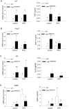
Figure 3. Lack of induction of inflammatory gene expression in adipose tissues of TNFΔARE/WT mice under HFD.
Expression in adipose tissue and corresponding plasma levels of tnf (A), mcp1 (B) and il6 (C) and leptin (D) were analyzed in 12-week-old animals using qPCR and ELISA respectively. Cytokine/adipokine mRNA expression was normalized to gapdh expression and expressed as fold change relative to WT on control diet. n = 6 per group; data sets with different superscript letters differ significantly from each other according to two-way ANOVA.
Figure 4
Figure 4. Metabolic composition of the plasma but not distal ileum is affected by the HFD.
Metabolites in Plasma (A) and distal ileum tissue (B) of 12-week-old mice (n = 6 per group) were analyzed using the Biocrates Life Sciences AbsoluteIDQ kit. The metabolomics data are represented as PCA plots. Statistical analyses for dietary groups and genotype differences were then performed using O-PLS-DA.
Figure 5
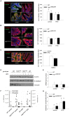
Figure 5. Loss of ileal occludin expression and increased endotoxin translocation but not PEG translocation upon HFD feeding.
Protein expression of Occludin and E-Cadherin in ileal IEC was assessed by immunefluorescence analysis in distal ileum (A) and proximal colon (B) of 12-week-old TNFΔARE/WT mice and WT littermates (n = 4 per group), as well as ileum of SWR/J mice (C) (n = 4–5 per group) with subsequent quantification. Findings were confirmed by Western Blotting using β-actin as loading control (D). Gene expression of occludin was analyzed by qPCR (n = 6 per group) (E). Translocation of orally applied PEG of the average molecular sizes 1500 and 3000 Da were determined by LC/MS, determining the sum of sodium adducts as area units in counts per second in the respective SIM traces (F), and Plasma endotoxin concentrations in hepatic portal vein EDTA-plasma (G) were analyzed using an LAL Chromogenic Endpoint Assay (n = 6 per group). Data sets with different superscript letters differ significantly from each other according to two-way ANOVA; *p<0.05 according to Student’s t-test.
Figure 6
Figure 6. HFD triggers activation of inflammatory responses in IEC.
Expression levels of tnf (A), icam1 (B), ccl20 (C) and cxcl2 (D) mRNA were determined in isolated ileal IEC of 12-weeks-old animals using qPCR. Relative mRNA expression was normalized to gapdh and expressed as fold change relative to WT on control diet. n = 6 per group; Data sets with different superscript letters differ significantly from each other according to two-way ANOVA.
Figure 7
Figure 7. Cecal lysates trigger chemokine production and DC recruitment.
Plasma CCL20 concentration was measured by ELISA (A), and CD11c+ dendritic cells in the LP were quantified by cell cytometry (B) (n = 6 per group). Mode K cells were stimulated in technical triplicates with cecal lysates (50 µg protein/ml) or cecal water (50 µg protein/ml) of mice fed the different diets for 24 h. TNF (20 ng/ml) was used as a positive control. CCL20 concentration was measured in the supernatants using ELISA (C). With these conditioned supernatants, the chemotaxis of DCs generated from bone-marrow was assessed in triplicates using a transwell migration assay (D). Data are representative for three independent experiments. Data sets with different superscript letters differ significantly from each other according to two-way ANOVA; *p<0.05 according to Student’s t-test.
Figure 8
Figure 8. Diet-induced luminal changes are associated with Th17 biased immune response.
Il23p19 (A) and il1235p35 (B) mRNA expression of BM-DCs was quantified by qPCR after 24 h stimulation with CM from Mode K cells treated with cecal lysate (50 µg protein/ml) of mice fed the different diets or unstimulated Mode K cells. Experiments were performed in triplicates. Expression of the cytokines il23p19 (C) and il12p35 (D), the transcription factors rorgt (E) and tbet (F), and the effector cytokines il17a (G) and ifng (H) were quantified in isolated LPLs using qPCR. mRNA expression was normalized to gapdh expression and expressed as –fold change relative to WT on control diet (n = 6 per group). Data sets with different superscript letters differ significantly from each other; *p<0.05 according to Student’s t-test.

References

    1. Hass DJ, Brensinger CM, Lewis JD, Lichtenstein GR (2006) The impact of increased body mass index on the clinical course of Crohn’s disease. Clin Gastroenterol Hepatol 4: 482–488. - PubMed
    1. Long MD, Crandall WV, Leibowitz IH, Duffy L, del Rosario F, et al. (2011) Prevalence and epidemiology of overweight and obesity in children with inflammatory bowel disease. Inflamm Bowel Dis 17: 2162–2168. - PMC - PubMed
    1. Blain A, Cattan S, Beaugerie L, Carbonnel F, Gendre JP, et al. (2002) Crohn’s disease clinical course and severity in obese patients. Clin Nutr 21: 51–57. - PubMed
    1. Hou JK, Abraham B, El-Serag H (2011) Dietary intake and risk of developing inflammatory bowel disease: a systematic review of the literature. Am J Gastroenterol 106: 563–573. - PubMed
    1. Ding S, Chi MM, Scull BP, Rigby R, Schwerbrock NM, et al. (2010) High-fat diet: bacteria interactions promote intestinal inflammation which precedes and correlates with obesity and insulin resistance in mouse. PLoS One 5: e12191. - PMC - PubMed

Publication types

MeSH terms