. 2013 Aug 16;8(8):e71697.
doi: 10.1371/journal.pone.0071697. eCollection 2013.

Cell cycle regulated phosphorylation of the telomere-associated protein TIN2

Affiliations

Cell cycle regulated phosphorylation of the telomere-associated protein TIN2

Shuqun Yang et al. PLoS One. .

Abstract

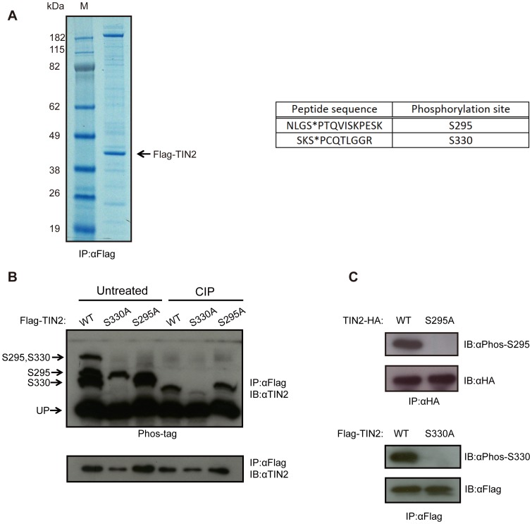
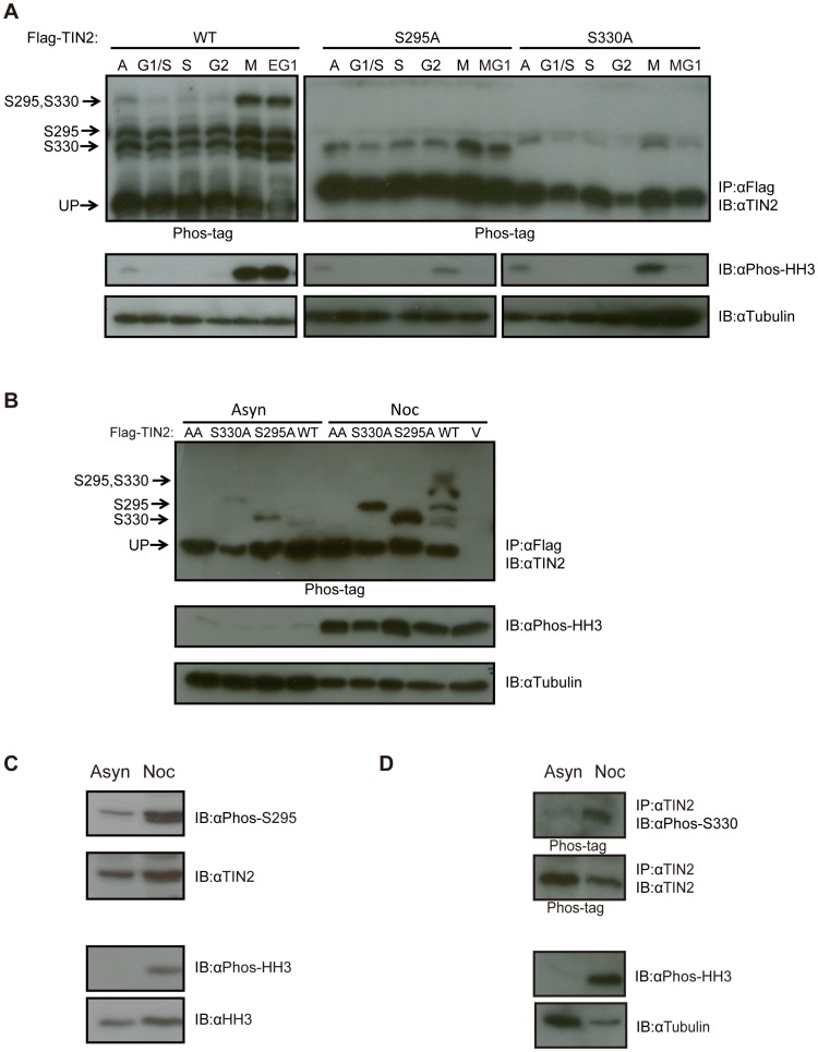
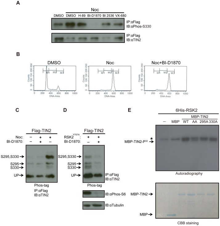
The protein TIN2 is a member of telomere-binding protein complex that serves to cap and protect mammalian chromosome ends. As a number of proteins in this complex are phosphorylated in a cell cycle-dependent manner, we investigated whether TIN2 is modified by phosphorylation as well. We performed phospho-proteomic analysis of human TIN2, and identified two phosphorylated residues, serines 295 and 330. We demonstrated that both these sites were phosphorylated during mitosis in human cells, as detected by Phos-tag reagent and phosphorylation-specific antibodies. Phosphorylation of serines 295 and 330 appeared to be mediated, at least in part, by the mitotic kinase RSK2. Specifically, phosphorylation of TIN2 at both these residues was increased upon expression of RSK2 and reduced by an inhibitor of the RSK family of kinases. Moreover, RSK2 phosphorylated TIN2 in vitro. The identification of these specifically timed post-translational events during the cell cycle suggests a potential mitotic regulation of TIN2 by phosphorylation.


Conflict of interest statement

Competing Interests: The authors have declared that no competing interests exist.

Figures

Figure 1
Figure 1. TIN2 is phosphorylated on Serine 295 and Serine 330.
(A) Identification of phosphorylation sites on TIN2 by mass spectrometry. Left, A lysate from HeLa cells stably infected with a retrovirus encoding N-terminal Flag epitope-tagged TIN2 (Flag-TIN2) was subjected to immunoprecipitation (IP) with an anti-Flag antibody, resolved by SDS-PAGE, and detected by Coomassie Brilliant Blue staining. M: marker lane. Right, The purified protein was then recovered and digested by trypsin, followed by TiO2 enrichment and mass spectrometry analysis, revealing two peptides with phosphorylated serine residues (denoted with an *). Representative of one experiment. (B) Detection of phosphorylation of TIN2 at S295 and S330 by the Phos-tag reagent. Lysates from HeLa cells stably infected with a retrovirus encoding Flag-TIN2 in the wild-type (WT), S330A, or S295A configuration were subjected to immunoprecipitation (IP) with an anti-Flag antibody and then either left untreated or treated with calf intestine phosphatase (CIP), followed by SDS-PAGE either in the presence (top) or the absence (bottom) of the Phos-tag reagent and immunoblotted (IB) with an anti-TIN2 antibody. The supershifted bands corresponding to S295, S330, or S295 and S330 phosphorylation, as well as the unphosphorylated TIN2 (UP), are denoted on the left. Representative of three experiments. (C) Detection of S295 and S330 phosphorylation of TIN2 with phosphorylation-specific antibodies. Lysates from HeLa cells stably infected with a retrovirus encoding C-terminal HA epitope-tagged TIN2 (TIN2-HA) or Flag-TIN2 in WT, S295A, or S330A configuration were subjected to immunoprecipitation (IP) with either an anti-HA or anti-Flag antibody, resolved by SDS-PAGE, and immunoblotted (IB) with either an anti-Phos-S295 or anti-Phos-S330 antibody to detected the phosphorylated TIN2, and either an anti-HA or anti-Flag antibody to detect total ectopic TIN2 as a loading control. Representative of two experiments.
Figure 2
Figure 2. TIN2 is phosphorylated on S295 and S330 during mitosis.
(A) Detection of S295 and S330 phosphorylation of TIN2 during mitosis by the Phos-tag reagent after release from a double thymidine block. HeLa cells stably infected with a retrovirus encoding Flag-TIN2 in the WT, S330A, or S295A configuration were collected from asynchronous populations (A), populations arrested with a double thymidine block corresponding to the G1/S phase of the cell cycle, or populations at the points corresponding to S, G2, M and early or middle G1 (EG1 or MG1) after release from the double thymidine block. Derived lysates were then either subjected to (top) immunoprecipitation (IP) with an anti-Flag antibody and resolved by SDS-PAGE in the presence of the Phos-tag reagent and immunoblotted (IB) with an anti-TIN2 antibody or (bottom) resolved by normal SDS-PAGE and immunoblotted with either an anti-Phos-HH3 antibody to monitor cell cycle progression or an anti-Tubulin antibody as a loading control. The supershifted bands corresponding to S295, S330, or S295 and S330 phosphorylation, as well as the unphosphorylated TIN2 (UP) are denoted on the left of the upper panels. Left and right panels are different exposures. Representative of two experiments. (B) Detection of S295 and S330 phosphorylation of TIN2 by the Phos-tag reagent in cells arrested with nocodazole. HeLa cells stably infected with a retrovirus encoding no transgene (vector, V) or Flag-TIN2 in the WT, S330A, S295A, or AA configuration were collected from asynchronous populations (Asyn) or populations arrested in G2/M by treatment with nocodazole (Noc). Derived lysates were then subjected to either (top) immunoprecipitation (IP) with αFlag and resolved by SDS-PAGE in the presence of the Phos-tag reagent and immunoblotted (IB) with an anti-TIN2 antibody or (bottom) resolved by normal SDS-PAGE and immunoblotted with either an anti-Phos-HH3 antibody to monitor cell cycle progression or an anti-Tubulin antibody as a loading control. The supershifted bands corresponding to S295, S330, or S295 and S330 phosphorylation, as well as the unphosphorylated TIN2 (UP), are denoted on the left of the upper panel. Representative of three experiments. (C) Detection of S295 phosphorylation of endogenous TIN2 with a phosphorylation-specific antibody in cells arrested with nocodazole. Lysates from HeLa cells were collected from asynchronous populations (Asyn) or populations arrested in G2/M by treatment with nocodazole (Noc), resolved by SDS-PAGE and immunoblotted (IB) with an anti-Phos-S295, anti-TIN2, anti-Phos-HH3, or anti-HH3 (loading control) antibody. Representative of two experiments. (D) Detection of S330 phosphorylation of endogenous TIN2 with a phosphorylation-specific antibody in cells arrested with nocodazole. HeLa cells were collected from asynchronous populations (Asyn) or populations arrested in G2/M by treatment with nocodazole (Noc). Derived lysates were then either subjected to (top) immunoprecipitation (IP) with an anti-TIN2 antibody, resolved by SDS-PAGE in the presence of the Phos-tag reagent, and immunoblotted (IB) with either an anti-Phos-S330 or anti-TIN2 antibody, or (bottom) resolved by normal SDS-PAGE and immunoblotted with an anti-Phos-HH3 antibody, to monitor cell cycle progression, or an anti-Tubulin antibody as a loading control. Representative of one experiment.
Figure 3
Figure 3. TIN2 is phosphorylated by the mitotic kinase RSK2.
(A) Detection of S330 phosphorylation of TIN2 with a phosphorylation-specific antibody in cells arrested with nocodazole and treated with kinase inhibitors. HeLa cells stably expressing wild-type Flag-TIN2 were treated with DMSO, H-89, BI-D1870, BI 2536 or VX-680 in the presence of either nocodazole (Noc) or vehicle (DMSO). Derived lysates were immunoprecipitated (IP) with an anti-Flag antibody, resolved by SDS-PAGE, and immunoblotted (IB) with an anti-Phos-S330 antibody or, as a loading control, an anti-TIN2 antibody. Representative of two experiments. (B) DNA profiles of HeLa cells treated with BI-D1870. HeLa cells treated with DMSO, nocodazole (Noc), or nocodazole+ BI-D1870 were harvested, stained with propidium iodide, and subjected to fluorescence-activated cell sorting (FACS) analysis. Representative of two experiments. (C) Detection of S295 and S330 phosphorylation of TIN2 by the Phos-tag reagent in asynchronous or nocodazole arrested cells with or without the RSK2 inhibitor BI-D1870. 293T cells were either untreated or treated with nocodazole (Noc), BI-D1870, or both compounds. Derived lysates were then subjected to immunoprecipitation (IP) with an anti-Flag antibody and resolved by SDS-PAGE in the presence of the Phos-tag reagent and immunoblotted (IB) with an anti-TIN2 antibody. The supershifted bands corresponding to S295, S330, or S295 and S330 phosphorylation, as well as the unphosphorylated TIN2 (UP), are denoted on the left. Representative of two experiments. (D) Detection of S295 and S330 phosphorylation of TIN2 by the Phos-tag reagent in asynchronous cells with ectopic RSK2 and/or the RSK2 inhibitor BI-D1870. 293T cells transiently transfected with Flag-TIN2 and the Y707A constitutively active mutant form of RSK2 (Flag-RSK2Y707A) were either left untreated or treated with RSK kinase inhibitor BI-D1870. Derived lysates were split into two portions. The first portions were subjected to immunoprecipitation (IP) with an anti-Flag antibody, resolved by SDS-PAGE in the presence of the Phos-tag reagent, and immunoblotted (IB) with an anti-TIN2 antibody. The supershifted bands corresponding to S295, S330, or S295 and S330 phosphorylation, as well as the unphosphorylated TIN2 (UP), are denoted on the left (top). The second portions were resolved by normal SDS-PAGE and immunoblotted with either an anti-Phospho-S6 antibody to monitor RSK2 kinase activity, or an anti-Tubulin antibody as a loading control (bottom). Representative of two experiments. (E) Detection of TIN2 phosphorylation by RSK2 in vitro. Recombinant maltose-binding protein (MBP) or N-terminal MBP-tagged TIN2 (MBP-TIN2) in the WT, S295A, S330A, or AA mutant configuration were captured with amylose resin and eluted with maltose. No protein (-) or equal amounts of the aforementioned purified MBP-TIN2 proteins were incubated with recombinant N-terminal 6His-tagged RSK2 (6His-RSK2) in the presence of ATP32, after which the reaction products were resolved by SDS-PAGE and either (top) exposed to autographic film or (bottom) stained with Coomassie Brilliant Blue (CBB staining). Phosphorylated (P32) MBP-TIN2 and a non-specific band (*) are denoted on the left top panel. MBP-TIN2 and MBP are denoted on the left bottom panel. Representative of two experiments.

Similar articles

Cited by

References

    1. Kim SH, Kaminker P, Campisi J (1999) TIN2, a new regulator of telomere length in human cells. Nat Genet 23: 405–412. - PMC - PubMed
    1. Loayza D, De Lange T (2003) POT1 as a terminal transducer of TRF1 telomere length control. Nature 423: 1013–1018. - PubMed
    1. Houghtaling BR, Cuttonaro L, Chang W, Smith S (2004) A dynamic molecular link between the telomere length regulator TRF1 and the chromosome end protector TRF2. Curr Biol 14: 1621–1631. - PubMed
    1. Kim SH, Beausejour C, Davalos AR, Kaminker P, Heo SJ, et al. (2004) TIN2 mediates functions of TRF2 at human telomeres. J Biol Chem 279: 43799–43804. - PubMed
    1. Ye JZ, Donigian JR, van Overbeek M, Loayza D, Luo Y, et al. (2004) TIN2 binds TRF1 and TRF2 simultaneously and stabilizes the TRF2 complex on telomeres. J Biol Chem 279: 47264–47271. - PubMed

Publication types

MeSH terms