. 2013 Aug 20;8(8):e71851.
doi: 10.1371/journal.pone.0071851. eCollection 2013.

Accurate detection and quantification of the fish viral hemorrhagic Septicemia virus (VHSv) with a two-color fluorometric real-time PCR assay

Affiliations

Accurate detection and quantification of the fish viral hemorrhagic Septicemia virus (VHSv) with a two-color fluorometric real-time PCR assay

Lindsey R Pierce et al. PLoS One. .

Abstract

Viral Hemorrhagic Septicemia virus (VHSv) is one of the world's most serious fish pathogens, infecting >80 marine, freshwater, and estuarine fish species from Eurasia and North America. A novel and especially virulent strain - IVb - appeared in the Great Lakes in 2003, has killed many game fish species in a series of outbreaks in subsequent years, and shut down interstate transport of baitfish. Cell culture is the diagnostic method approved by the USDA-APHIS, which takes a month or longer, lacks sensitivity, and does not quantify the amount of virus. We thus present a novel, easy, rapid, and highly sensitive real-time quantitative reverse transcription PCR (qRT-PCR) assay that incorporates synthetic competitive template internal standards for quality control to circumvent false negative results. Results demonstrate high signal-to-analyte response (slope = 1.00±0.02) and a linear dynamic range that spans seven orders of magnitude (R(2) = 0.99), ranging from 6 to 6,000,000 molecules. Infected fishes are found to harbor levels of virus that range to 1,200,000 VHSv molecules/10(6) actb1 molecules with 1,000 being a rough cut-off for clinical signs of disease. This new assay is rapid, inexpensive, and has significantly greater accuracy than other published qRT-PCR tests and traditional cell culture diagnostics.


Conflict of interest statement

Competing Interests: The authors have declared that no competing interests exist.

Figures

Figure 1
Figure 1. Real-time PCR amplification plots for various experiments.
ABI 7500 real-time PCR results for (a) a true VHSv positive fish sample, (b) the relationship between the VHSv Native Template (NT) and Internal Standard (IS) with the NT held constant and the IS varied, (c) the relationship between VHSv NT and IS with the IS held constant and the NT varied, and (d) the relationship between VHSv NT:IS with concentrations held constant for dilutions of 1∶1–1∶20. Green = NT, red = IS, y = fluorescence of the reporter dye minus the baseline (Δ fluorescence), x = cycle threshold.
Figure 2
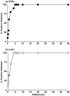
Figure 2. True accuracy of the 2-color fluorometric test.
Results are based on % positives from 10 separate runs of 12 dilutions using a known Internal Standard Mixture (ISM). Dilutions are: 40, 20, 10, 7, 6, 5, 4, 2, 1, 0.7, 0.4, and 0.1 molecules. The 2-color fluorometric test yields 100% positives for (a) ≥ 5 molecules of VHSv and (b) ≥ 4 molecules for actb1.
Figure 3
Figure 3. Relationship between the number of observed and expected molecules for NT:IS concentrations of 1∶1–1∶20.
The concentration of Native Template (NT) is held constant and the Internal Standard (IS) varied for dilutions of: 1∶1 (6×10⁴ molecules), 1∶2, 1∶3, 1∶4, 1∶5, 1∶6, 1∶7, 1∶8, 1∶9, 1∶10, 1∶12, 1∶14, 1∶16, 1∶18, and 1∶20 (3×10³molecules). The 2-color fluorometric assay yields a linear relationship for (a) VHSv (R 2 = 0.99, F = 1514.00, df = 1, 13, p<0.001) with a mean CV of 5% for dilutions 1∶1–1∶10 and 7% for concentrations down to 1∶20, and for (b) actb1 (R 2 = 0.99, F = 1283.00, df = 1, 13, p<0.001), CV = 5% and 7%. The same linear pattern is observed when the IS was held constant and NT varied for (c) VHSv (R 2 = 0.99, F = 5124.00, df = 1, 13, p<0.001), CV = 5% for 1∶1–1∶10 and 7% for dilutions down to 1∶20, and (d) actb1 (R 2 = 0.99, F = 2434.00, df = 1, 13, p<0.001), CV = 3% and 6%. Error bars = standard error of results for triplicate runs. Dotted line = partition of dilutions from 1∶1–1∶10 (right) and 1∶12–1∶20 (left).
Figure 4
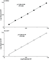
Figure 4. Relationship between the numbers of observed versus expected molecules when NT:IS concentrations are 1∶1.
Results are based on dilutions of the Native Template (NT) and Internal Standard (IS) of 6×10⁶, 6×10⁵, 6×10⁴, 6×10³, 6×10², 60, 6, and 0.6 molecules for VHSv and actb1. The 2-color fluorometric assay yields a linear relationship for (a) VHSv over seven orders of magnitude (from 6×10⁶ to 6×100 VHSv molecules) with a slope of 1.00 (R 2 = 0.99, F = 9404.00, df = 1, 5, p<0.001), and mean CV of 9%. A linear trend also is obtained for (b) actb1 (R 2 = 0.99, F = 1347.00, df = 1, 5, p<0.001). Slope = 1.04, mean CV = 10%. Error bars = standard error of triplicate runs.
Figure 5
Figure 5. Relative numbers of VHSv positives and negatives from our 2-color fluorometric and capillary electrophoresis StaRT-PCR assays, which indicates identical numbers of positives and negatives.
Compared to these tests, for 43 fishes (25 positives, 18 negatives (including 5 laboratory controls)), (a) SYBR® green has 44% false negative error (χ 2 = 5.67, df = 1, p = 0.02), and cell culture has 56% error (χ 2 = 9.36, df = 1, p = 0.002). For 63 fish samples (39 positives, 24 negatives (including 11 laboratory controls)), (b) SYBR® green qRT-PCR has 33% false negative error (χ 2 = 5.37, df = 1, p = 0.02), whereas the 2-color fluorometric and capillary electrophoresis tests show zero false negatives.
Figure 6
Figure 6. Mean log numbers of VHSv molecules/106 actb1 molecules from our new 2-color fluorometric assay versus the prior Agilent capillary electrophoresis approach.
Results indicate a linear relationship between the two tests (R 2 = 0.91, df = 1, 38, F = 396.40, p<0.001) and do not significantly differ (t = 1.42, df = 78, NS). Arrow = Estimated threshold concentration of VHSv for fish with clinical signs of infection using our new assay, from a χ 2 test of nine symptomatic fish (1×10³ VHSv molecules/106 actb1 molecules = 3.6×10² VHSv molecules). Triangle = false negative range for SYBR® green qRT-PCR (1.0×100–1.6×10² VHSv/106 actb1 molecules = 0.6×100–2.5×10² VHSv molecules). Square = false negative range for cell culture (1.0×100–2.2×10³ VHSv/106 actb1 molecules = 0.6×100–6.1×10³ VHSv molecules.

Similar articles

Cited by

References

    1. Rao JR, Fleming CC, Moore JE (2006) Molecular Diagnostics Current Technology and Applications. Horizon Bioscience, Norfolk, Norwich, United Kingdom.
    1. Park S, Zhang Y, Lin S, Wang TH, Yang S (2011) Advances in microfluidic PCR for point-of-care infectious disease diagnostics. Biotech Adv 29: 830–839. - PMC - PubMed
    1. Coutlée F, Viscidi RP, Saint-Antoine P, Kessous A, Yolken RH (1991) The polymerase chain reaction: a new tool for the understanding and diagnosis of HIV-1 infection at the molecular level. Mol Cell Probe 5: 241–259. - PubMed
    1. Ellis JS, Zambon MC (2002) Molecular diagnosis of influenza. Rev Med Virol 12: 375–389. - PubMed
    1. Chai Z, Ma W, Fu F, Lang Y, Wang W, et al. (2013) A SYBR Green-based real-time RT-PCR assay for simple and rapid detection and differentiation of highly pathogenic and classical type 2 porcine reproductive and respiratory syndrome virus circulating in China. Arch Virol 158: 407–415. - PubMed

Publication types

MeSH terms