. 2013 Aug 20;8(8):e71980.
doi: 10.1371/journal.pone.0071980. eCollection 2013.

The β-blocker Nebivolol Is a GRK/β-arrestin biased agonist

Affiliations

The β-blocker Nebivolol Is a GRK/β-arrestin biased agonist

Catherine E Erickson et al. PLoS One. .

Abstract

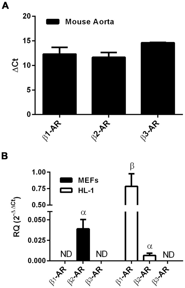
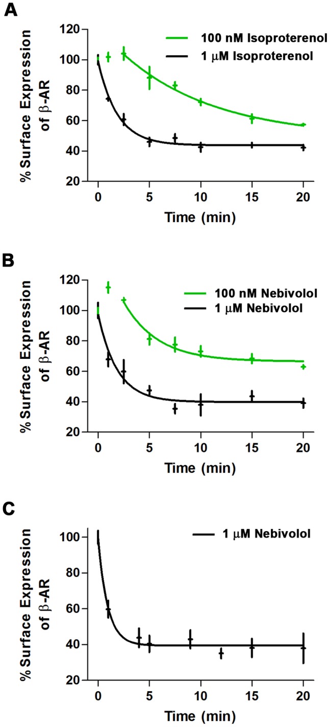
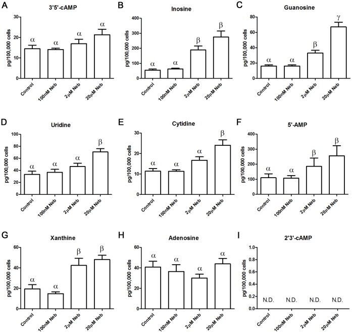
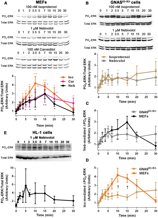
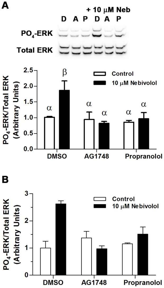
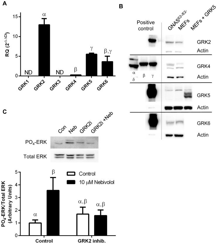
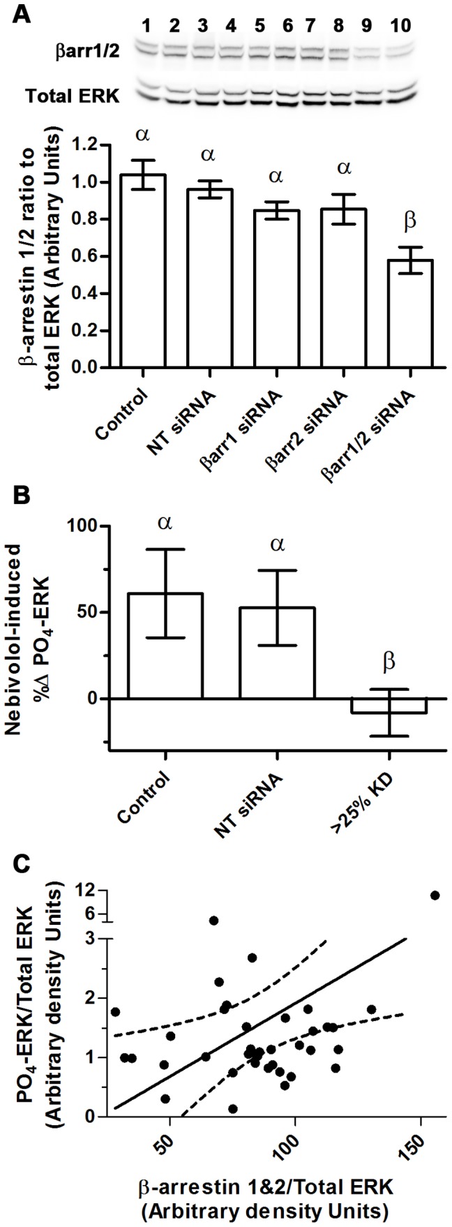
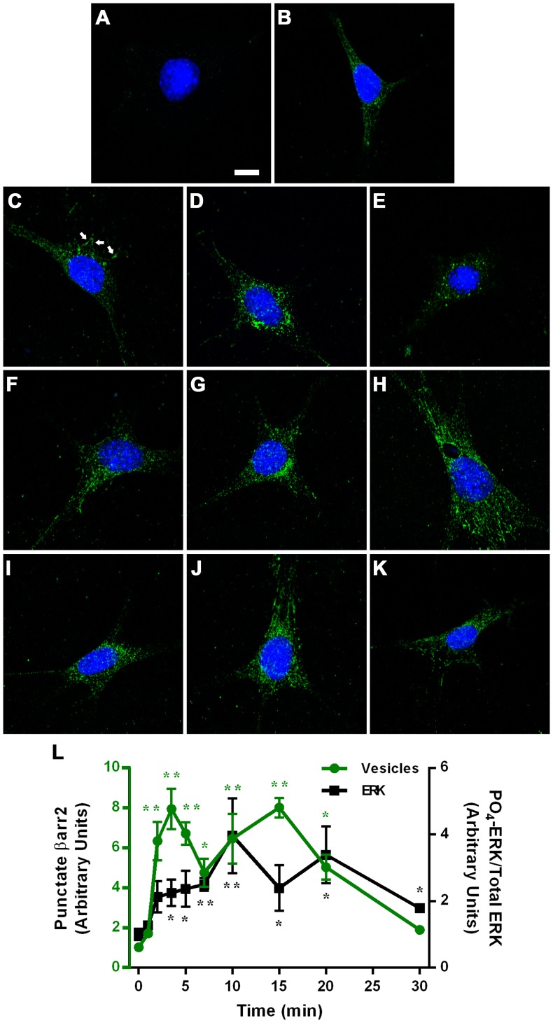
Nebivolol, a third generation β-adrenoceptor (β-AR) antagonist (β-blocker), causes vasodilation by inducing nitric oxide (NO) production. The mechanism via which nebivolol induces NO production remains unknown, resulting in the genesis of much of the controversy regarding the pharmacological action of nebivolol. Carvedilol is another β-blocker that induces NO production. A prominent pharmacological mechanism of carvedilol is biased agonism that is independent of Gαs and involves G protein-coupled receptor kinase (GRK)/β-arrestin signaling with downstream activation of the epidermal growth factor receptor (EGFR) and extracellular signal-regulated kinase (ERK). Due to the pharmacological similarities between nebivolol and carvedilol, we hypothesized that nebivolol is also a GRK/β-arrestin biased agonist. We tested this hypothesis utilizing mouse embryonic fibroblasts (MEFs) that solely express β2-ARs, and HL-1 cardiac myocytes that express β1- and β2-ARs and no detectable β3-ARs. We confirmed previous reports that nebivolol does not significantly alter cAMP levels and thus is not a classical agonist. Moreover, in both cell types, nebivolol induced rapid internalization of β-ARs indicating that nebivolol is also not a classical β-blocker. Furthermore, nebivolol treatment resulted in a time-dependent phosphorylation of ERK that was indistinguishable from carvedilol and similar in duration, but not amplitude, to isoproterenol. Nebivolol-mediated phosphorylation of ERK was sensitive to propranolol (non-selective β-AR-blocker), AG1478 (EGFR inhibitor), indicating that the signaling emanates from β-ARs and involves the EGFR. Furthermore, in MEFs, nebivolol-mediated phosphorylation of ERK was sensitive to pharmacological inhibition of GRK2 as well as siRNA knockdown of β-arrestin 1/2. Additionally, nebivolol induced redistribution of β-arrestin 2 from a diffuse staining pattern into more intense punctate spots. We conclude that nebivolol is a β2-AR, and likely β1-AR, GRK/β-arrestin biased agonist, which suggests that some of the unique clinically beneficial effects of nebivolol may be due to biased agonism at β1- and/or β2-ARs.


Conflict of interest statement

Competing Interests: The authors have declared that no competing interests exist.

Figures

Figure 1
Figure 1. qPCR detection of β1-, β2-, and β3-ARs in mouse cells and tissue.
Total RNA was isolated from mouse aorta (A), MEFs, and HL-1 cells (B) and subjected to 40 rounds of Taq-Man qPCR. Bars with different Greek letters are statistically different (p<0.05) from each other according to a one-way ANOVA followed by Kruskal-Wallis posthoc test. ND indicates that the transcript was not detected; data are expressed as mean ± SEM, n = 3 for cells and n = 2 for aorta.
Figure 2
Figure 2. Ligand-mediated β-AR internalization.
MEFs (A and B) and HL-1 cells (C) were stimulated for the indicated amount of time with isoproterenol (A) or nebivolol (B and C), and surface receptor levels were determined as described in the methods. Two doses of isoproterenol (A) and nebivolol (B) were used in the MEFs: 100 nM (green) and 1 µM (black). Data are expressed as mean ± SEM (in both the x and y axis); n = 3 to 4.
Figure 3
Figure 3. Nebivolol-mediated changes in nucleoside and related compounds concentrations.
MEFs were treated with the indicated amount of nebivolol, or vehicle control, for 30 minutes in the presence of IBMX. Each compound was measured from a single sample by LC-MS/MS; the compound examined is listed above each histogram. Bars with different Greek letters are statistically different (p<0.05) from each other according to a one-way ANOVA followed by Tukey-Kramer post hoc test. Data are expressed as mean ± SEM; n = 6.
Figure 4
Figure 4. Nebivolol time-dependently and Gαs-independently signals to ERK.
MEFs were treated with 100 nM of isoproterenol, 1 µM of nebivolol, or 100 nM of carvedilol for the indicated time points (A); representative blots are shown above the graph. Data are compared by one-way ANOVA with Kruskal-Wallis Multiple comparison post-hoc test; two asterisk (**) indicates significantly different than control by Bonferroni method (more stringent) and one asterisk (*) indicates significantly different by the regular test (less stringent). Gnas E2−/E2− cells were treated with 100 nM of isoproterenol and 1 µM of nebivolol for the indicated time points (B); representative blots are shown above the graph. These data were compared as in panel A. The data obtained from the MEFs and Gnas E2−/E2− cells were compared directly for nebivolol- (C) and isoproterenol-mediated (D) phosphorylation of ERK. Data are compared by two-way ANOVA with Sidak’s multiple comparisons post-hoc test; the cross (†) indicates that the MEFs and Gnas E2−/E2− cells are significantly different from each other. HL-1 cardiomyocytes were treated with 10 µM of nebivolol for the indicated time points (E); representative blots are shown above the graph. These data were compared as in panel A. All data are expressed as mean ± SEM; n = 3 to 6.
Figure 5
Figure 5. Nebivolol signals to ERK through β-ARs and the EGFR.
Cells were pretreated with inhibitors for 30 minutes then stimulated with 10 µM of nebivolol for 7 minutes. Representative blots from MEFs (A) are presented above the histogram of the data; D = DMSO, A = 1 µM of AG1748, and P = 30 µM of propranolol. Open bars are controls, and black bars are nebivolol treated. (B) Identical confirmatory experiments were conducted in two sets of HL-1 cells. All data were analyzed via a two-way ANOVA with Tukey-Kramer post hoc analysis, and significance is denoted by Greek letters. Any data set with a different Greek letter is statistically different p<0.05. Data are expressed as mean ± SEM; n = 4 for MEFs and n = 2 for HL-1 cells.
Figure 6
Figure 6. GRK expression and role in nebivolol-mediated phosphorylation of ERK.
qPCR was used to identify the GRKs present in MEFs (A); data are expressed as mean ± SEM, n = 3. Western blots for GRK2, 4, 5, and 6 as well as actin, as a control, from GNASE2−/E2− cells, MEFs, and heterologously expressed GRKs from 293T cells (positive controls) were used to confirm the qPCR data via identifying the protein expression of GRKs (B). MEFs were pretreated with the GRK inhibitor for 30 minutes then stimulated with 10 µM of nebivolol for 7 minutes (C). Representative blots are presented above the histogram of the data. Open bars are controls, and black bars are nebivolol treated. Data are expressed as mean ± SEM; n = 4. All data were analyzed by a one-way ANOVA with Tukey-Kramer post hoc analysis, and significance is denoted by Greek letters. Any data set with a different Greek letter is statistically different p<0.05.
Figure 7
Figure 7. β-arrestins are required for nebivolol-mediated phosphorylation of ERK.
siRNA directed towards β-arrestin 1 and 2 were transfected into MEFs; 72 hours later the cells were stimulated with 10 µM of nebivolol for 7 minutes. A representative blot for β-arrestins and total ERK is depicted in panel A above the histogram of the data: even numbers were treated with nebivolol; 1 & 2– non targeting siRNA, 3 & 4– mock transfection, 5 & 6– βarr1 siRNA, 7 & 8– βarr2 siRNA, 9 & 10– βarr1+ βarr2 siRNA. The efficacy of the siRNA was analyzed via a one-way ANOVA with Tukey-Kramer post hoc analysis; statistical difference is denoted by Greek letters; any data set with a different Greek letter is statistically different p<0.05. Data are expressed as mean ± SEM; n = 16. The ERK phosphorylation data (B), collected only from samples where β-arrestins were knocked down by 25% or more, are expressed as the percent change induced by nebivolol. These data were analyzed via a one-way ANOVA with a nonparametric Kruskal-Wallis post hoc test. Statistical difference (p<0.05) is denoted by Greek letters; any data set with a different Greek letter is statistically different. Data are expressed as mean ± SEM; n = 5 to 9. Due to the poor knockdown of β-arrestins, a scatter plot of nebivolol-induced phosphorylation of ERK and β-arrestin level was generated (C) and a Pearson’s correlation was run to determine the relationship between β-arrestin level and nebivolol-mediated phosphorylation of ERK (n = 38); the dotted lines represent the 95% confidence interval of the line.
Figure 8
Figure 8. Subcellular distribution of β-arrestin 2 following stimulation with nebivolol.
MEFs were treated with vehicle (A and B) or 10 µM of nebivolol for 1 (C), 2, (D), 3.5 (E), 5 (F), 7 (G), 10 (H), 15 (I), 20 (J), or 30 (K) minutes, and stained as described in the methods. Only Alexa 647 secondary antibody and Hoechst 33342 were used in panel A; addition of primary βarr2 antibody (H-9), shown in green, occurred in B through K. All settings remained identical for each image; the scale bar in panel A represents 10 µm. Panel L represents vesicular βarr2 in green on the left axis (n = 8) and 10 µM nebivolol-mediated phosphorylation of ERK in black on the right axis (n = 3 to 7). For both data sets a Kruskal-Wallis Multiple comparison test was run and two asterisk (**) indicates significantly different from control by the Bonferroni method (more stringent) and one asterisk (*) indicates significantly different by the regular test (less stringent).

References

    1. Van de Water A, Janssens W, Van Neuten J, Xhonneux R, De Cree J, et al. (1988) Pharmacological and hemodynamic profile of nebivolol, a chemically novel, potent, and selective beta 1-adrenergic antagonist. J Cardiovasc Pharmacol 11: 552–563. - PubMed
    1. Brixius K, Bundkirchen A, Bolck B, Mehlhorn U, Schwinger RH (2001) Nebivolol, bucindolol, metoprolol and carvedilol are devoid of intrinsic sympathomimetic activity in human myocardium. Br J Pharmacol 133: 1330–1338. - PMC - PubMed
    1. Bowman AJ, Chen CP, Ford GA (1994) Nitric oxide mediated venodilator effects of nebivolol. Br J Clin Pharmacol 38: 199–204. - PMC - PubMed
    1. Cockcroft JR, Chowienczyk PJ, Brett SE, Chen CP, Dupont AG, et al. (1995) Nebivolol vasodilates human forearm vasculature: evidence for an L-arginine/NO-dependent mechanism. J Pharmacol Exp Ther 274: 1067–1071. - PubMed
    1. Feng MG, Prieto MC, Navar LG (2012) Nebivolol induced vasodilation of renal afferent arterioles involves beta3 adrenergic receptor and nitric oxide synthase activation. Am J Physiol Renal Physiol. - PubMed

Publication types

MeSH terms