. 2013 Aug 19;8(8):e72023.
doi: 10.1371/journal.pone.0072023. eCollection 2013.

Directed in vitro myogenesis of human embryonic stem cells and their in vivo engraftment

Affiliations

Directed in vitro myogenesis of human embryonic stem cells and their in vivo engraftment

Yongsung Hwang et al. PLoS One. .

Erratum in

Abstract

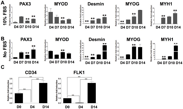
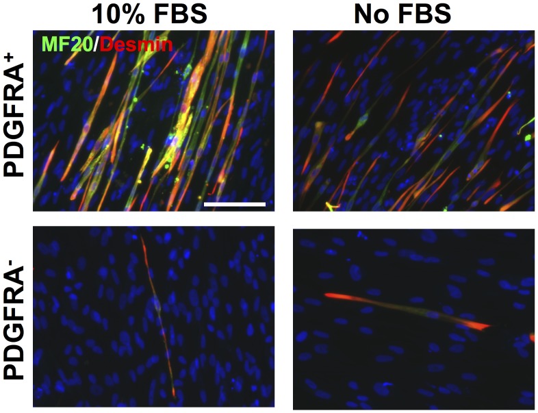
Development of human embryonic stem cell (hESC)-based therapy requires derivation of in vitro expandable cell populations that can readily differentiate to specified cell types and engraft upon transplantation. Here, we report that hESCs can differentiate into skeletal muscle cells without genetic manipulation. This is achieved through the isolation of cells expressing a mesodermal marker, platelet-derived growth factor receptor-α (PDGFRA), following embryoid body (EB) formation. The ESC-derived cells differentiated into myoblasts in vitro as evident by upregulation of various myogenic genes, irrespective of the presence of serum in the medium. This result is further corroborated by the presence of sarcomeric myosin and desmin, markers for terminally differentiated cells. When transplanted in vivo, these pre-myogenically committed cells were viable in tibialis anterior muscles 14 days post-implantation. These hESC-derived cells, which readily undergo myogenic differentiation in culture medium containing serum, could be a viable cell source for skeletal muscle repair and tissue engineering to ameliorate various muscle wasting diseases.


Conflict of interest statement

Competing Interests: The authors have declared that no competing interests exist.

Figures

Figure 1
Figure 1. Derivation of PDGFRA+ myogenic progenitors from hESCs.
(A) Schematic depicting the isolation protocol for PDGFRA+ cells. (B) Undifferentiated hESC colony showing OCT4-GFP expression. (C) EB formation. (D) EB attached to the Matrigel-coated tissue culture plates. (E) Migration of cells from EBs. (F) FACS demonstrating isolation of PDGFRA+/OCT4-GFP population. (G) Cells attached to 0.1% gelatin-coated tissue culture plates sorted against PDGFRA+ (left) and PDGFRA (right). Scale bar = 200 µm.
Figure 2
Figure 2. Characterization of the proliferative potential of PDGFRA+ cells.
(A) Phase contrast images of PDGFRA+ cells undergoing myogenic differentiation at different time points (top: 10% FBS, bottom: No FBS). (B) Proliferation profiles of cells grown in serum-containing and serum-free media. (C) Population doubling time. Scale bar = 200 µm. *p<0.05 and **p<0.01.
Figure 3
Figure 3. In vitro myogenic differentiation potential demonstrated by quantitative PCR.
Gene expression profiles of PDGFRA+ cells as a function of culture in serum-containing (A) and serum-free (B) media. Statistical analysis was performed between serum-containing (A) and serum-free (B) myogenic media for each corresponding time point. **p<0.01. (C) PDGFRA+ cells undergoing myogenic differentiation show upregulation of endothelial markers. **p<0.01.
Figure 4
Figure 4. Terminal myogenic differentiation characterized by immunofluorescent staining.
Immunofluorescent staining for MF20 (green) and desmin (red) of PDGFRA+ (top) PDGFRA (bottom) cells grown in serum-containing (left) and serum-free (right) media. Scale bar = 100 µm.
Figure 5
Figure 5. Cell shape analyses of PDGFRA+ cells undergoing myogenic differentiation.
(A) Alignment and orientation of PDGFRA+ cells grown in serum-containing (left) and serum-free (right) media. (B, C) Shape indices for PDGFRA+ cells while undergoing terminal myogenic differentiation in serum-containing (left bar) and serum-free (right bar) media. n = 794, 385, 425, and 210, respectively. (D) Estimated differentiation indices of PDGFRA+ in serum and serum-free media. (E) Estimated fusion indices of differentiated PDGFRA+ (MF20 positive cells) in serum and serum -free media. n = 722 and 245, respectively. **p<0.01.
Figure 6
Figure 6. In vitro mesodermal differentiation of PDGFRA+ cells.
Osteogenic differentiation as characterized by staining for Alizarin Red S (A), alkaline phosphatase (B), and gene expression (E). Adipogenic differentiation as characterized by Oil Red O staining (C) and gene expression (F). Chondrogenic differentiation as characterized by Safranin O staining (D) and gene expression (G). UN, undifferentiated PDGFRA+ cells cultured in growth medium; DIFF, PDGFRA+ cells cultured in differentiation inducing medium. Scale bar = 200 µm. **p<0.01.
Figure 7
Figure 7. Engraftment of myogenic progenitors in NOD/SCID mice.
(A) Immunofluorescent staining of TA muscle sections from NOD/SCID mice injected with cells grown for 4 days (left) and 14 days with (CTX+) and without cardiotoxin injury (CTX-) for mouse laminin (red), human lamin A/C (green), and nuclei (blue). Corresponding high magnification images for muscles treated with cells cultured for 14 day without (B) and with (C) cardiotoxin injury. Scale bar = 50 µm, 20 µm, and 20 µm, repectively.

References

    1. Thomson JA, Itskovitz-Eldor J, Shapiro SS, Waknitz MA, Swiergiel JJ, et al. (1998) Embryonic stem cell lines derived from human blastocysts. Science 282: 1145–1147. - PubMed
    1. Barberi T, Willis LM, Socci ND, Studer L (2005) Derivation of multipotent mesenchymal precursors from human embryonic stem cells. PLoS Med 2: e161. - PMC - PubMed
    1. Crook JM, Peura TT, Kravets L, Bosman AG, Buzzard JJ, et al. (2007) The Generation of Six Clinical-Grade Human Embryonic Stem Cell Lines. Cell Stem Cell 1: 490–494. - PubMed
    1. Gunaseeli I, Doss MX, Antzelevitch C, Hescheler J, Sachinidis A (2010) Induced pluripotent stem cells as a model for accelerated patient- and disease-specific drug discovery. Curr Med Chem 17: 759–766. - PMC - PubMed
    1. Jang J, Yoo JE, Lee JA, Lee DR, Kim JY, et al. (2012) Disease-specific induced pluripotent stem cells: a platform for human disease modeling and drug discovery. Exp Mol Med 44: 202–213. - PMC - PubMed

Publication types

MeSH terms

Substances