

Effects of androgen deprivation on cerebral morphometry in prostate cancer patients--an exploratory study
- PMID: 23977199
- PMCID: PMC3747074
- DOI: 10.1371/journal.pone.0072032
Effects of androgen deprivation on cerebral morphometry in prostate cancer patients--an exploratory study
Abstract
Background: Androgen deprivation therapy (ADT) is a common treatment for non-metastatic, low-risk prostate cancer, but a potential side effect of ADT is impaired brain functioning. Previous work with functional magnetic resonance imaging (MRI) demonstrated altered prefrontal cortical activations in cognitive control, with undetectable changes in behavioral performance. Given the utility of brain imaging in identifying the potentially deleterious effects of ADT on brain functions, the current study examined the effects of ADT on cerebral structures using high resolution MRI and voxel-based morphometry (VBM).
Methods: High resolution T1 weighted image of the whole brain were acquired at baseline and six months after ADT for 12 prostate cancer patients and 12 demographically matched non-exposed control participants imaged at the same time points. Brain images were segmented into gray matter, white matter and cerebral ventricles using the VBM toolbox as implemented in Statistical Parametric Mapping 8.
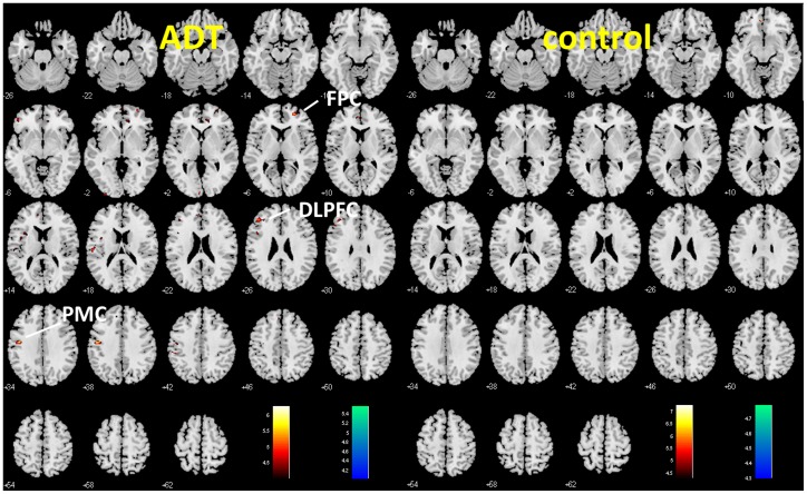
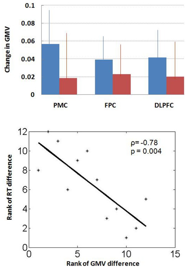
Results: Compared to baseline scan, prostate cancer patients undergoing ADT showed decreased gray matter volume in frontopolar cortex, dorsolateral prefrontal cortex and primary motor cortex, whereas the non-exposed control participants did not show such changes. In addition, the decrease in gray matter volume of the primary motor cortex showed a significant correlation with longer reaction time to target detection in a working memory task.
Conclusions: ADT can affect cerebral gray matter volumes in prostate cancer patients. If replicated, these results may facilitate future studies of cognitive function and quality of life in men receiving ADT, and can also help clinicians weigh the benefits and risks of hormonal therapy in the treatment of prostate cancer.
Conflict of interest statement
Figures
References
-
- Meng MV, Grossfeld GD, Sadetsky N, Mehta SS, Lubeck DP, et al. (2002) Contemporary patterns of androgen deprivation therapy use for newly diagnosed prostate cancer. Urology 60 (Suppl 3A)7–12. - PubMed
-
- Shahinian VB, Kuo Y-F, Freeman JL, Orihuela E, Goodwin JS (2005) Increasing use of gonadotropin-releasing hormone agonists for localized prostate cancer. Cancer 103: 1615–24. - PubMed
-
- Zhang Y, Skolarus TA, Miller DC, Wei JT, Hollenbeck BK (2011) Understanding prostate cancer spending growth among medicare beneficiaries. Urology 77: 326–331. - PubMed
-
- Alibhai SM, Gogov S, Allibhai Z (2006) Long-term side effects of androgen deprivation therapy in men with non-metastatic prostate cancer: a systematic literature review. Crit Rev Oncol Hematol 60: 201–215. - PubMed
Publication types
MeSH terms
Substances
Grants and funding
LinkOut - more resources
Full Text Sources
Other Literature Sources
Medical
