. 2013 Aug 20;8(8):e72594.
doi: 10.1371/journal.pone.0072594. eCollection 2013.

Intestinal-specific TNFα overexpression induces Crohn's-like ileitis in mice

Affiliations

Intestinal-specific TNFα overexpression induces Crohn's-like ileitis in mice

Giorgos Bamias et al. PLoS One. .

Abstract

Background and aim: Human and animal studies have clearly established tumor necrosis factor (TNF)α as an important mediator of Crohn's disease pathogenesis. However, whether systemic or only local TNFα overproduction is required for the development of chronic intestinal inflammation and Crohn's disease remains unclear. The aim of this study was to assess the contribution of intestinal epithelial-derived TNFα to the development of murine Crohn's-like ileitis.

Methods: We adapted the well-established TNF(∆ARE/+) mouse model of Crohn's disease (which systemically overexpresses TNFα) to generate a homozygous mutant strain that overexpress TNFα only within the intestinal epithelium. Intestinal-specific TNF(i∆ARE/i∆ARE) mice were examined for histopathological signs of gut inflammation and extraintestinal manifestations of Crohn's disease. The mucosal immune phenotype was characterized, and the contribution of specific lymphocyte populations to the pathogenesis of TNF(i∆ARE/i∆ARE) ileitis was assessed.

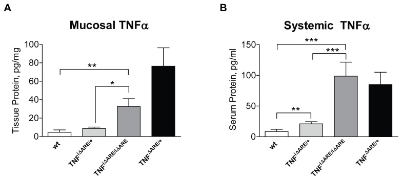
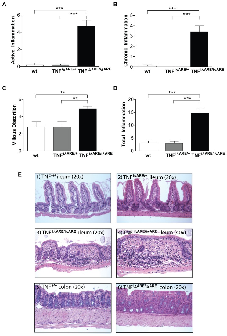
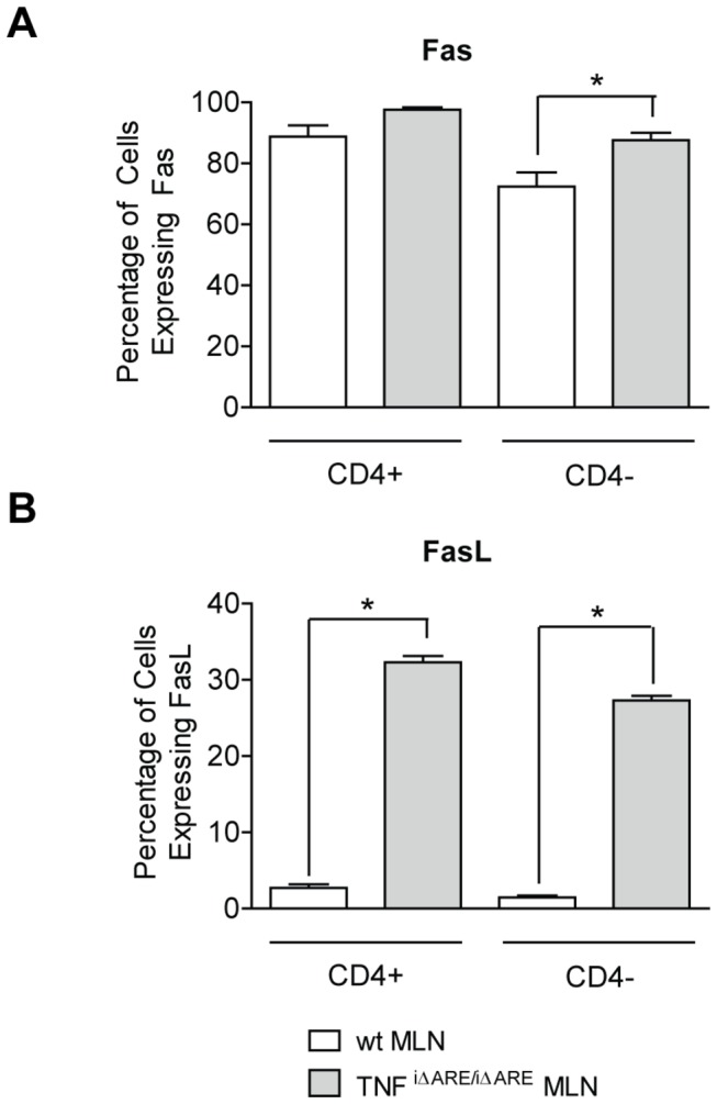
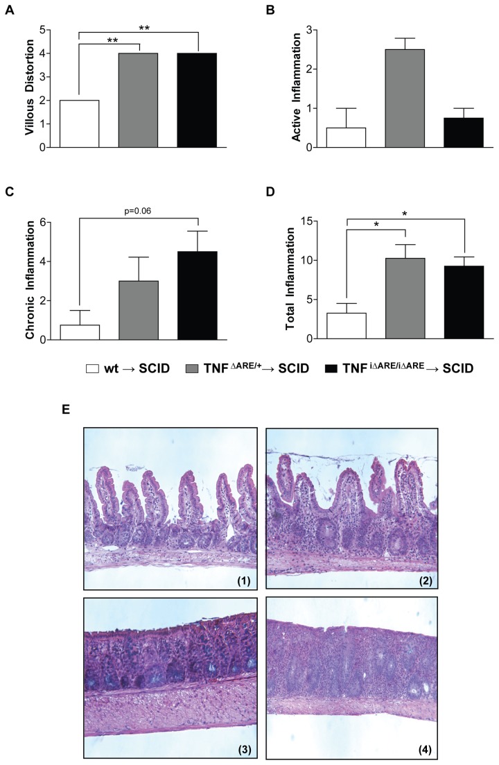
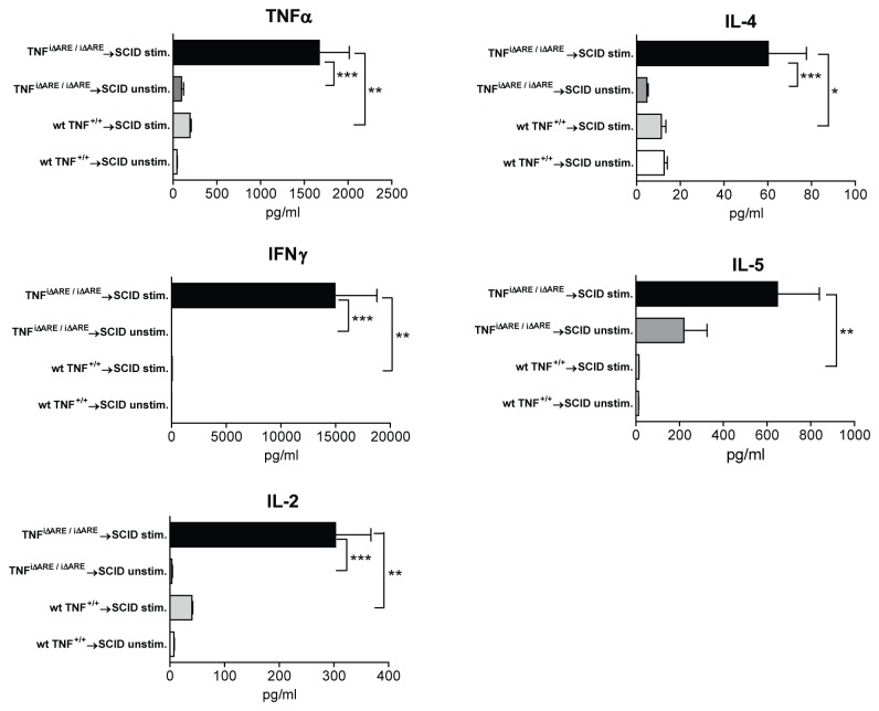
Results: TNF(i∆ARE/i∆ARE) mice had increased mucosal and systemic TNFα levels compared to wild-type controls (P<0.001), as well as severe chronic ileitis with increased neutrophil infiltration and villous distortion, but no extraintestinal manifestations (P<0.001 vs. wild-type controls). The gut mucosal lymphocytic compartment was also expanded in TNF(i∆ARE/i∆ARE) mice (P<0.05), consisting of activated CD69(+) and CD4(+)CD62L(-) lymphocytes (P<0.05). FasL expression was significantly elevated in the mesenteric lymph nodes of TNF(i∆ARE/i∆ARE) mice (P<0.05). Adoptive transfer of mucosal TNF(i∆ARE/i∆ARE) lymphocytes resulted in ileitis in immunologically naïve severe combined immunodeficiency recipients (P<0.05 vs. wild-type controls), indicating an effector phenotype that was associated with increased production of both Th1 (IFNγ) and Th2 (IL-5, IL-13) cytokines.

Conclusion: Intestinal epithelial-derived TNFα is sufficient for the induction of Crohn's-like ileitis, but not for the occurrence of extraintestinal manifestations, in TNF(i∆ARE/i∆ARE) mice. These effects were associated with generation of effector lymphocytes within the intestinal mucosa and dysregulated apoptosis. Thus, targeted intestinal blockade of TNFα may provide an effective means to neutralize gut-derived TNFα with reduced side effects.


Conflict of interest statement

Competing Interests: The authors have declared that no competing interests exist

Figures

Figure 1
Figure 1. Mucosal and systemic expression of TNFα protein is elevated in TNFi∆ARE/i∆ARE mice.
Intestinal epithelial-specific overexpression of TNFα was induced by expressing the tnf ΔARE mutation under the promoter of the intestinal-specific I-FABP gene, which is expressed primarily by IECs. (A) Pieces of small intestinal tissue were homogenized at the time of animal sacrifice, and the concentration of TNFα protein was measured by ELISA. Values are expressed as pg of TNFα per mg of total protein content, which was measured by a Bradford Protein Assay. (B) Blood was drawn from mice by cardiac puncture at the time of sacrifice. Serum was collected and stored until use. The concentration of TNFα protein was measured by ELISA. Murine ileal tissue and sera were analyzed individually. A significant increase in the total protein content in both small intestinal tissue homogenates and murine sera was seen in homozygous TNFi∆ARE/i∆ARE mice (n=11), as compared with either heterozygous TNFi∆ARE/+, (n=10) or wt mice (combined wt for TNFi∆ARE/i∆ARE and systemic TNFΔARE/+, n=16). Mice with systemic overexpression of TNFα were included in the analysis, serving as positive controls (TNFΔARE/+ mice, n=6). Graphs represent mean values ± SEM for each experimental group of mice. *P<0.05, ** < P<0.01, *P<0.001.
Figure 2
Figure 2. Histopathological features of TNFi∆ARE/i∆ARE mice.
Histological assessment of inflammation in the terminal ileum was done using a validated scoring system. Indices were calculated for (A) villous distortion, (B) active inflammation (neutrophil infiltration), and (C) chronic inflammation (mononuclear cell infiltration). (D) The total inflammatory score represents the sum of all 3 individual indices. Values for all indices were significantly elevated in homozygous TNFi∆ARE/i∆ARE mice (n=11), whereas no inflammatory changes were detected in heterozygous TNFi∆ARE/+ mice (n=10) or wt mice (n=10); all mice were evaluated at 16-20 weeks of age. Graphs represent mean values ± SEM for each group of mice. *P<0.05, ** < P<0.01, *P<0.001. (E) Representative photomicrographs of H&E stained sections from wt TNF+/+, TNFi∆ARE/+, and TNFi∆ARE/i∆ARE mice. 1) wt TNF+/+ ileum showing normal villous architecture with no blunting or evidence of acute or chronic inflammatory infiltration, 20x. (2) TNFi∆ARE/+ ileum showing mild villous blunting with no significant inflammation, 20x. (3) TNFi∆ARE/i∆ARE ileum showing evidence of chronic ileitis with villous blunting; acute and chronic inflammatory infiltration is seen within the LP and extending into the submucosa, 20x. (4) Higher magnification of ileum histology from a TNFi∆ARE/i∆ARE mouse showing evidence of marked inflammatory infiltration and distortion of normal villous architecture and crypts, 40x. (5) Colon histology of wt TNF+/+ mice showing no evidence of colonic inflammation, 20x. (6) Colon histology of a TNFi∆ARE/i∆ARE mouse showing normal colonic histology with only a few lymphocytes infiltrating the LP.
Figure 3
Figure 3. Increased apoptosis of lymphocytes in the MLNs of TNFi∆ARE/i∆ARE mice.
Single cell suspensions were obtained from MLNs, and the expression of various surface markers was analyzed by flow cytometry. Fluorochrome-tagged monoclonal antibodies against CD4, Fas, and FasL were utilized for identification of the respective populations. Multiple color flow cytometry was performed on a FACS calibur system to determine the percentage of cells expressing surface markers and the intensity of expression. The vast majority of MLN cells stained positive for Fas and no significant differences detected between wt and TNFi∆ARE/i∆ARE mice. Conversely, expression of FasL was significantly elevated in MLN cells from TNFi∆ARE/i∆ARE mice as compared to wt controls. This difference remained significant when expression of FasL was examined separately in the CD4+ or CD4- populations. Mice were processed individually. Three separate experiments with 3-4 mice per group gave similar results, and one representative experiment is shown. Graphs represent mean values ± SEM for each group of experimental mice. *P<0.05, ** < P<0.01, *P<0.001.
Figure 4
Figure 4. Adoptive transfer of CD4+ MLN cells from TNFi∆ARE/i∆ARE mice into SCID recipients.
CD4+ enriched (purity >95%) populations were obtained from single cell suspensions of MLNs from TNFi∆ARE/i∆ARE via positive selection by use of a magnetic cell-sorting system. Donor cells (1x10⁶) were injected intraperitoneally into MHC-matched 6-8-wk-old SCID mice. Recipient mice were euthanized 8 weeks after the transfer. Histological assessment of inflammation in the terminal ileum was done by a validated scoring system. Indices were calculated for (A) villous distortion, (B) active inflammation (neutrophil infiltration), and (C) chronic inflammation (mononuclear cell infiltration). (D) The total inflammatory score represents the sum of all 3 individual indices. Total inflammation was significantly elevated in TNFi∆ARE/i∆ARE → SCID mice (n=4) and TNF∆ARE/+ → SCID mice (n=8), compared to wt TNF+/+ → SCID (n=4). Graphs represent mean values ± SEM for each group of mice. *P<0.05, ** < P<0.01, *P<0.001. (E) Representative photomicrographs of H&E stained sections from recipient SCID mice displaying the characteristics of adoptively transferred TNFi∆ARE/i∆ARE → SCID ileitis. (1). Representative wt → SCID mouse showing normal histological appearance of the terminal ileum. (2). Representative TNFi∆ARE/i∆ARE → SCID mouse showing a mixed acute and chronic inflammatory infiltrate with moderate ileitis. Three separate experiments using 4-8 mice per group were performed and gave similar results, and one representative experiment is shown.
Figure 5
Figure 5. Mucosal effector responses in the adoptive transfer model of TNFi∆ARE/i∆ARE→SCID ileitis.
CD4+ cells from the MLNs of TNFi∆ARE/i∆ARE or wt mice were injected into MHC-matched SCID mice. Recipient mice were euthanized 8 weeks after the transfer. Single cell suspensions from MLNs were cultured in complete medium (106 cells/mL), either with no stimulation or stimulated with immobilized anti-CD3 monoclonal antibody for 48 hours. Concentration of IFNγ, IL-4, IL-5, IL-2, and TNFα were concomitantly determined in culture supernatants by a cytometric bead array. In the absence of stimulation, no substantial cytokine secretion was detected by MLN cells from either strain of mice. Upon stimulation with anti-CD3, there was a significant increase in the secretion of all cytokines in MLN cells from TNFi∆ARE/i∆ARE → SCID mice (n=7), but not from wt → SCID recipients (n=4), indicating the presence of effector lymphocytes in the former, but not the latter, group. Individual mice were processed separately. Three separate experiments were performed, similar to Figure 4. Graphs represent mean values ± SEM for each condition.

References

    1. Bamias G, Nyce MR, De La Rue SA, Cominelli F, American College of P, et al. (2005) New concepts in the pathophysiology of inflammatory bowel disease. Ann Intern Med 143: 895-904. - PubMed
    1. Papadakis KA, Targan SR (2000) Role of cytokines in the pathogenesis of inflammatory bowel disease. Annu Rev Med 51: 289-298. doi:10.1146/annurev.med.51.1.289. PubMed: 10774465. - DOI - PubMed
    1. Locksley RM, Killeen N, Lenardo MJ (2001) The TNF and TNF receptor superfamilies: integrating mammalian biology. Cell 104: 487-501. doi:10.1016/S0092-8674(01)00237-9. PubMed: 11239407. - DOI - PubMed
    1. Matsuda R, Koide T, Tokoro C, Yamamoto T, Godai T et al. (2009) Quantitative cytokine mRNA expression profiles in the colonic mucosa of patients with steroid naive ulcerative colitis during active and quiescent disease. Inflamm Bowel Dis 15: 328-334. doi:10.1002/ibd.20759. PubMed: 18942752. - DOI - PubMed
    1. Raddatz D, Bockemühl M, Ramadori G (2005) Quantitative measurement of cytokine mRNA in inflammatory bowel disease: relation to clinical and endoscopic activity and outcome. Eur J Gastroenterol Hepatol 17: 547-557. doi:10.1097/00042737-200505000-00012. PubMed: 15827446. - DOI - PubMed

Publication types

MeSH terms