. 2013 Aug 19;8(8):e72610.
doi: 10.1371/journal.pone.0072610. eCollection 2013.

Sustained release of BMP-2 in bioprinted alginate for osteogenicity in mice and rats

Affiliations

Sustained release of BMP-2 in bioprinted alginate for osteogenicity in mice and rats

Michelle T Poldervaart et al. PLoS One. .

Abstract

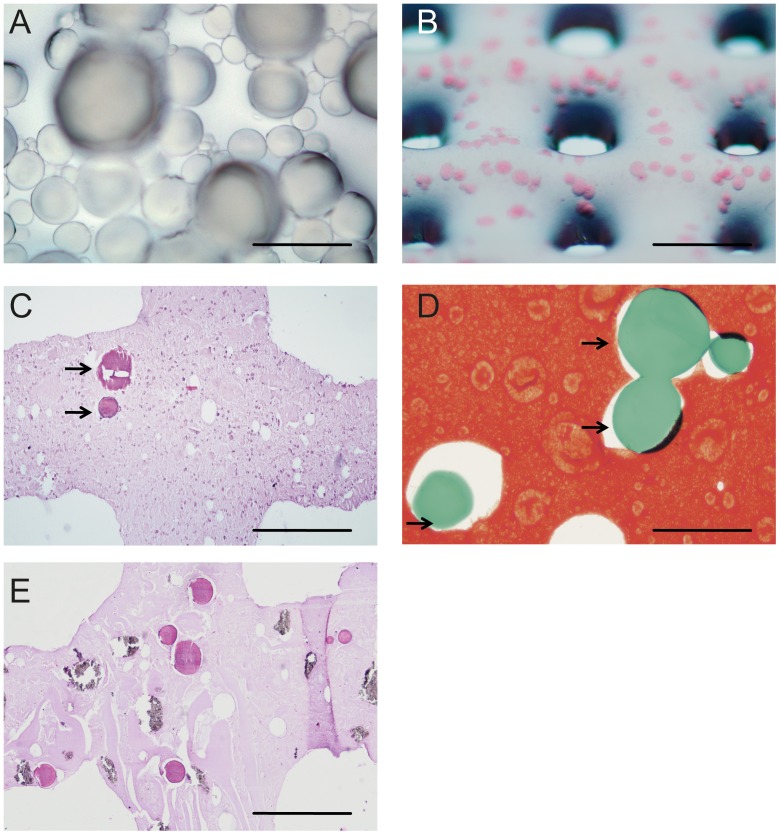
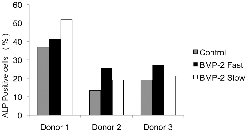
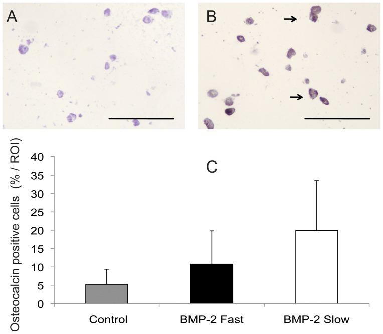
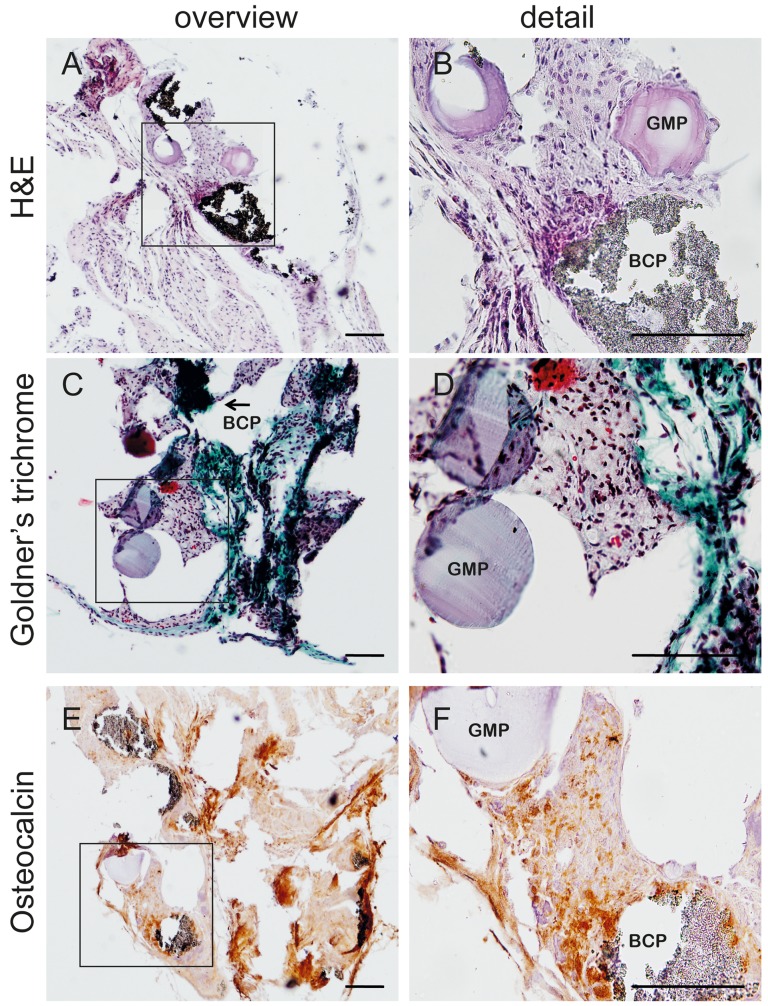
The design of bioactive three-dimensional (3D) scaffolds is a major focus in bone tissue engineering. Incorporation of growth factors into bioprinted scaffolds offers many new possibilities regarding both biological and architectural properties of the scaffolds. This study investigates whether the sustained release of bone morphogenetic protein 2 (BMP-2) influences osteogenicity of tissue engineered bioprinted constructs. BMP-2 loaded on gelatin microparticles (GMPs) was used as a sustained release system, which was dispersed in hydrogel-based constructs and compared to direct inclusion of BMP-2 in alginate or control GMPs. The constructs were supplemented with goat multipotent stromal cells (gMSCs) and biphasic calcium phosphate to study osteogenic differentiation and bone formation respectively. BMP-2 release kinetics and bioactivity showed continuous release for three weeks coinciding with osteogenicity. Osteogenic differentiation and bone formation of bioprinted GMP containing constructs were investigated after subcutaneous implantation in mice or rats. BMP-2 significantly increased bone formation, which was not influenced by the release timing. We showed that 3D printing of controlled release particles is feasible and that the released BMP-2 directs osteogenic differentiation in vitro and in vivo.


Conflict of interest statement

Competing Interests: The authors have declared that no competing interests exist.

Figures

Figure 1
Figure 1. Bioprinted gelatin microparticles.
A. Gelatin microparticles; B. GMPs in alginate, directly after printing, GMPs in pink; C. Section of bioprinted alginate construct, directly after printing (H/E staining). Arrows indicate GMPs; D. Safranin O staining of bioprinted alginate construct, directly after printing. Arrows indicate GMPs; E. Section of bioprinted alginate construct containing BCP, after 3 weeks of culturing (H/E staining). Experiments were performed in triplicate, representative pictures are shown. Scale bars indicate in A: 200 µm, B: 2 mm, C,E: 500 µm, D: 100 µm.
Figure 2
Figure 2. Cumulative release profiles of BMP-2 from gelatin microspheres.
A. Detection of 125I-BMP-2 in PBS supplemented with collagenase, performed in triplicate. Results are presented as mean±SD (small SDs hardly visible in figure); B. BMP-2 concentrations in PBS without collagenase, as determined by ELISA.
Figure 3
Figure 3. Bioactivity of released BMP-2.
Frequency of ALP positive MSCs after culturing for three weeks without BMP-2 (control), with BMP-2 in the hydrogel (fast release), and with BMP-2 laden GMPs (slow release), determined for 3 individual donors. Statistical analysis performed on mean values per donor.
Figure 4
Figure 4. Osteocalcin staining of bioprinted scaffolds after 3 weeks of culturing.
A. Control staining (isotype-matched control antibody); B. Osteocalcin positive cells from a scaffold with slow release of BMP-2. Arrows indicate some of the positively stained cells (brown). Scale bars represent 50 µm; C. Percentage of osteocalcin positive cells per region of interest, no statistically significant differences were found. Results are presented as mean±SD.
Figure 5
Figure 5. Osteogenic differentiation in vivo.
Presence of gelatin microparticles (GMP) and biphasic calcium phosphate granules (BCP) after 6 weeks of subcutaneous implantation in mice as indicated in the pictures. An overview and detail of HE, Goldner's trichrome and osteocalcin stainings are shown. Scale bars represent 100 µm.
Figure 6
Figure 6. Bone formation after 12 weeks of subcutaneous implantation in rats.
A. Control group; B. Fast release of BMP-2 group; C. Sustained BMP-2 released from GMP group. Section were stained with methylene blue and basic fuchsin. Biphasic calcium phosphate (BCP) is indicated in the pictures. D. Quantification of bone content by histomorphometry, n = 10. E. 3D reconstruction of micro-CT showing bone formation (orange) in a scaffold of the BMP-2 fast group. F. Quantification of bone content by micro-CT, n = 10. Representative pictures from each group are given. Scale bars represent 100 µm in A-C, 1 mm in E; Results are presented as mean ± SD, * p<0.01 compared to control.

References

    1. Giannoudis PV, Dinopoulos H, Tsiridis E (2005) Bone substitutes: an update. Injury 36 Suppl 3S20–27. - PubMed
    1. Silber JS, Anderson DG, Daffner SD, Brislin BT, Leland JM, et al. (2003) Donor site morbidity after anterior iliac crest bone harvest for single-level anterior cervical discectomy and fusion. Spine (Phila Pa 1976) 28: 134–139. - PubMed
    1. Cross M, Dexter TM (1991) Growth factors in development, transformation, and tumorigenesis. Cell 64: 271–280. - PubMed
    1. Lee K, Silva EA, Mooney DJ (2011) Growth factor delivery-based tissue engineering: general approaches and a review of recent developments. J R Soc Interface 8: 153–170. - PMC - PubMed
    1. Rickard DJ, Sullivan TA, Shenker BJ, Leboy PS, Kazhdan I (1994) Induction of rapid osteoblast differentiation in rat bone marrow stromal cell cultures by dexamethasone and BMP-2. Dev Biol 161: 218–228. - PubMed

Publication types

MeSH terms