

doi: 10.3978/j.issn.2225-319X.2012.04.15.
Video-assisted thoracoscopic lobectomy using a standardized three-port anterior approach - The Copenhagen experience
Affiliations
- PMID: 23977470
- PMCID: PMC3741716
- DOI: 10.3978/j.issn.2225-319X.2012.04.15
Item in Clipboard
Video-assisted thoracoscopic lobectomy using a standardized three-port anterior approach - The Copenhagen experience
Ann Cardiothorac Surg.
2012 May.
No abstract available
Figures
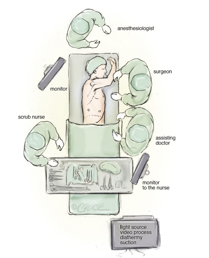
Operating room set-up for the anterior approach of video-assisted thorascopic lobectomy
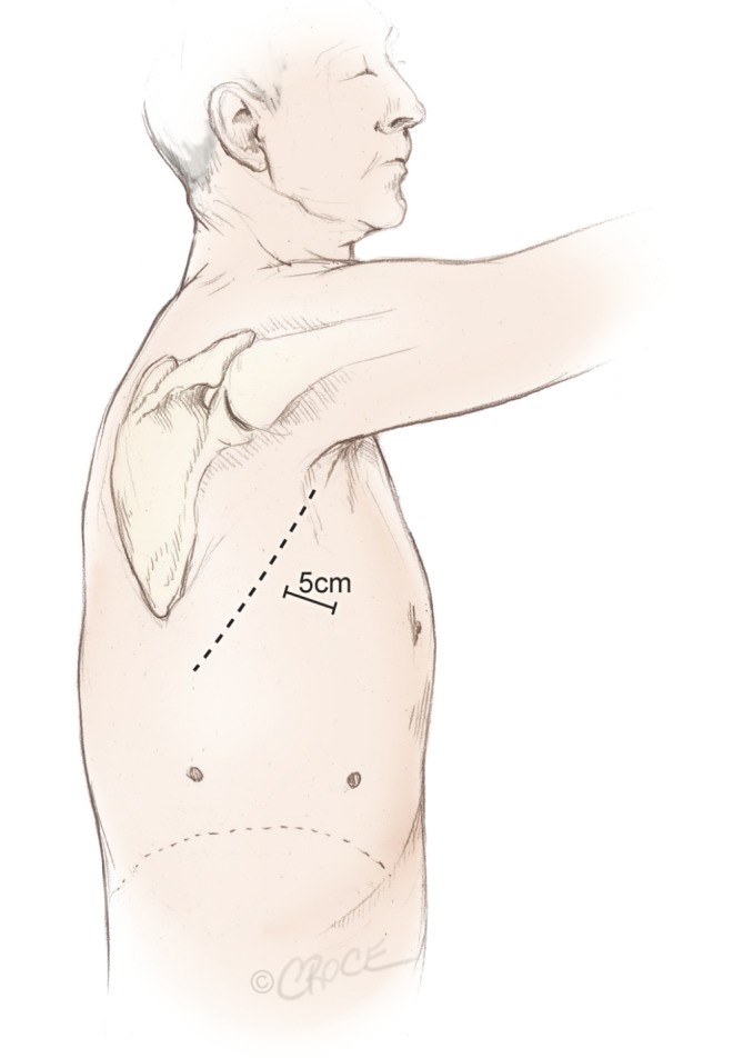
Three incisions made for the anterior approach forming a triangular configuration, with the utility incision at the apex of the triangle, measuring 5 cm in length
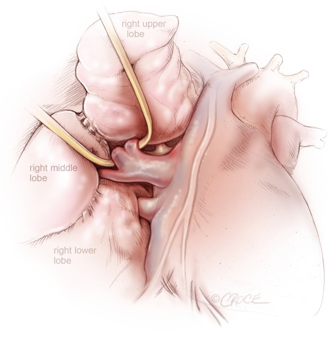
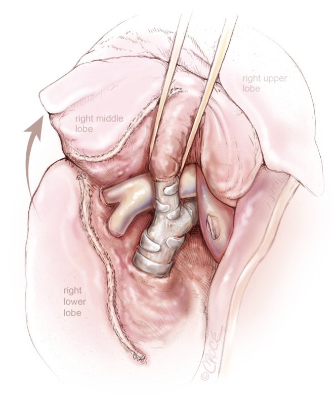
VATS right upper lobectomy: right superior pulmonary vein from right upper lobe is encircled by a vascular loop, while the pulmonary venous drainage from the right middle lobe is clearly seen
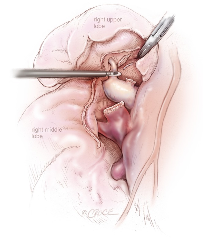
VATS right upper lobectomy: after division of right upper lobe pulmonary vein and truncus anterior, posterior ascending segmental artery to the right upper lobe is being divided by a Ligasure
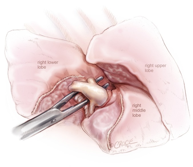
VATS right middle lobectomy: bronchus of the right middle lobe is presented through the oblique fissure after division of the right middle lobe vein
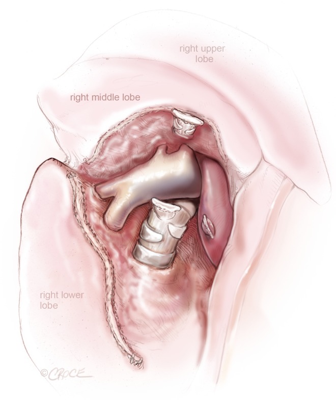
VATS right middle lobectomy: after the division of the right middle lobe vein and bronchus, pulmonary artery branches to the right middle lobe and lower lobe is exposed
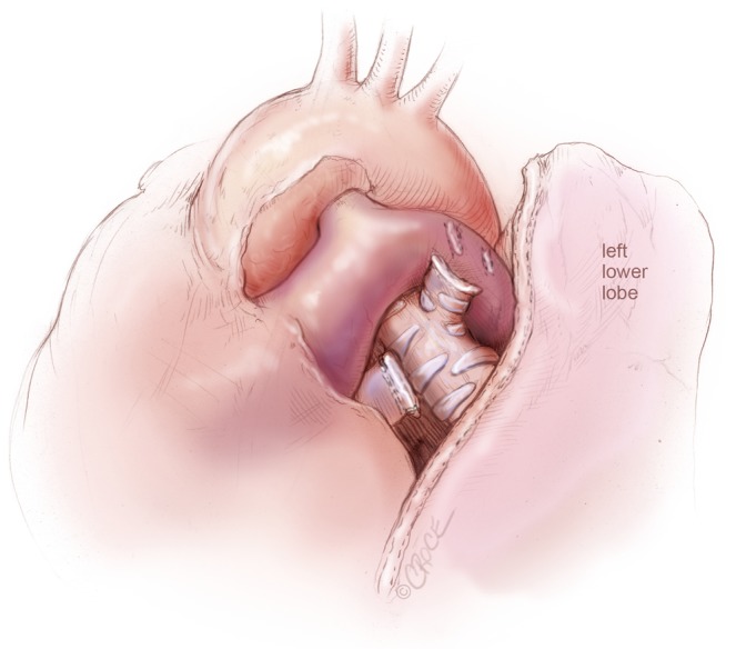
VATS left upper lobectomy: left upper lobe bronchial and vascular stumps are visualized, after left upper lobectomy and en bloc removal of Station 5 and Station 6 lymph nodes
VATS right lower lobectomy: pulmonary artery to right lower lobe, including superior segmental artery is isolated, after dividing the oblique fissure anteriorly

VATS lymph node dissection: superior mediastinal lymph node dissection on the right side
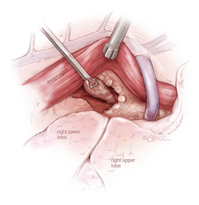
VATS lymph node dissection: subcarinal lymph node dissection from the right side, by retracting the esophagus posteriorly and the lung anteriorly to expose the membranous trachea and the subcarinal region
References
-
- Downey RJ, Cheng D, Kernstine K, et al. Video-Assisted Thoracic Surgery for Lung Cancer Resection. A Consensus Statement of the International Society of Minimally Invasive Cardiothoracic Surgery (ISMICS) 2007. Innovations (Phila) 2007;2:293-302 - PubMed
-
- Whitson BA, Groth SS, Duval SJ, et al. Surgery for early-stage non-small cell lung cancer: a systematic review of the video-assisted thoracoscopic surgery versus thoracotomy approaches to lobectomy. Ann Thorac Surg 2008;86:2008-16; discussion 2016-8. - PubMed
-
- Yan TD, Black D, Bannon PG, et al. Systematic review and meta-analysis of randomized and nonrandomized trials on safety and efficacy of video-assisted thoracic surgery lobectomy for early-stage non-small-cell lung cancer. J Clin Oncol 200927:2553-62 - PubMed
-
- Lewis RJ, Caccavale RJ, Sisler GE, et al. One hundred video-assisted thoracic surgical simultaneously stapled lobectomies without rib spreading. Ann Thorac Surg 1997;63:1415-21; discussion 1421-2 - PubMed
-
- Okada M, Sakamoto T, Yuki T, et al. Hybrid surgical approach of video-assisted minithoracotomy for lung cancer: significance of direct visualization on quality of surgery. Chest 2005;128:2696-701 - PubMed
LinkOut - more resources
Full Text Sources
Medical