. 2012 Sep;1(3):381-93.
doi: 10.3978/j.issn.2225-319X.2012.08.05.

Thoracoabdominal aortic aneurysm repair with a branched graft

Affiliations

Thoracoabdominal aortic aneurysm repair with a branched graft

Kim I de la Cruz et al. Ann Cardiothorac Surg. 2012 Sep.
No abstract available


Figures

Figure 1
Figure 1
Preoperative anatomy: a typical patient undergoing a Crawford extent II thoracoabdominal aortic aneurysm repair will have an aneurysm extending from the left subclavian artery down to the aortic bifurcation. Note that in this case, the celiac axis, superior mesenteric artery, and right and left renal arteries are displaced away from each other, making their reattachment as a single patch undesirable
Figure 2
Figure 2
Incision and exposure: the patient is positioned such that the upper body is at 60 degrees from horizontal and the hips are at 30 degrees from horizontal. A sigmoid-shaped skin incision is made from behind the left scapula, along the 7th rib, across the costal margin, and toward the left periumbilical region. The chest is entered through the 6th intercostal space. Left medial visceral rotation and circumferential division of the diaphragm enable exposure of the entire thoracoabdominal aorta. The use of table-mounted self-retaining retractors maintains stable exposure throughout the procedure
Figure 3
Figure 3
Left heart bypass, placement of aortic clamps, and opening the proximal descending thoracic aorta: a cannula is placed in the left atrium via left inferior pulmonary venotomy and connected to the drainage line of the left heart bypass circuit. Another cannula is placed in the distal descending thoracic aorta and connected to the inflow line of the circuit. A Y-line from the inflow tubing is connected to two balloon perfusion catheters; these are used to provide selective perfusion to the celiac and superior mesenteric arteries later during the repair. A separate cold renal perfusion system is set up to enable intermittent administration of 4 °C lactated Ringer¡¯s solution to the renal arteries through two balloon perfusion catheters. After left heart bypass flow is initiated, the proximal aortic clamp is placed just distal to the left subclavian artery, and the distal aortic clamp is applied across the mid-descending thoracic aorta. The aortic segment between the two clamps is opened longitudinally by using electrocautery
Figure 4
Figure 4
Tailoring the graft and constructing the proximal anastomosis: after back-bleeding intercostal arteries are ligated, the aorta is transected 2 to 3 cm distal to the aortic clamp and separated from the underlying esophagus. A four-branched aortic graft is tailored by stretching the graft so that it is taut, lining up the origin of the celiac graft with the origin of the left renal artery, and cutting the proximal end of the aortic graft at the point where it reaches the proximal anastomosis site (inset). This ensures that the origin of each branch graft is positioned slightly inferior to the origin of its paired artery, and thereby facilitates the formation of gentle curves in the branch grafts that help prevent the grafts from becoming kinked. The anastomosis between the aortic graft and the proximal aortic cuff is sewn
Figure 5
Figure 5
Visceral perfusion and the intercostal patch anastomosis: after the proximal anastomosis is completed, left heart bypass is stopped, the distal aortic clamp and cannula are removed, and the aorta is opened longitudinally down to the aortic bifurcation. Briskly back-bleeding intercostal and lumbar arteries are suture ligated. The celiac and superior mesenteric arteries are cannulated with balloon perfusion catheters, and selective perfusion is initiated. The renal arteries are likewise cannulated with balloon perfusion catheters to enable intermittent perfusion with cold crystalloid solution. Two pairs of intercostal arteries have been selected for reattachment; these are sutured to an opening in the side of the graft
Figure 6
Figure 6
Distal anastomosis: after the patch reimplantation of the intercostal arteries, the aortic cross-clamp is moved down to the aortic graft distal to the intercostal patch, thereby allowing reperfusion of the reimplanted intercostal arteries. The distal end of the aortic graft is trimmed to the appropriate length, and the distal anastomosis is performed
Figure 7
Figure 7
Right renal artery anastomosis: after the four branch grafts are clamped, the aortic clamp is removed so that flow is restored to the iliac arteries. The right-sided 8-mm branch graft is trimmed to the appropriate length and anastomosed to the origin of the right renal artery
Figure 8
Figure 8
Superior mesenteric artery anastomosis: the inferior 10-mm branch graft is used to reattach the superior mesenteric artery. The perfusion catheter is removed from the superior mesenteric artery, and flow to the celiac axis is stopped; this prevents visceral back-bleeding from impairing visualization of the anastomosis. The branch graft is cut to appropriate length and sutured to the origin of the superior mesenteric artery
Figure 9
Figure 9
Celiac axis anastomosis: a hemostat clamp remains in place on the superior mesenteric artery graft to ensure a dry field during the celiac anastomosis. After the celiac axis catheter is removed, the superior 10-mm branch graft is trimmed to appropriate length and sutured to the origin of the celiac axis
Figure 10
Figure 10
Left renal artery mobilization and anastomosis: the origin of the left renal artery is separated from the aortic wall as a button, and its proximal portion is mobilized. The remaining 8-mm branch graft is cut to appropriate length—with care taken to ensure that the graft will not kink once the abdominal organs are returned to their anatomic positions—and sewn to the left renal artery in an end-to-end configuration
Figure 11
Figure 11
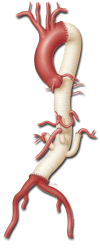
Completed repair: the use of the branched graft essentially eliminates residual aortic tissue in the visceral segment, thereby preventing future visceral patch aneurysm formation

References

    1. De Rango P, Estrera AL, Miller C, 3rd, et al. Operative outcomes using a side-branched thoracoabdominal aortic graft (STAG) for thoraco-abdominal aortic repair. Eur J Vasc Endovasc Surg 2011;41:41-7 - PubMed
    1. Kokotsakis J, Lazopoulos G, Ashrafian H, et al. Thoracoabdominal aneurysm repair using a four-branched thoracoabdominal graft: a case series. Cases J 2009;2:7144. - PMC - PubMed
    1. Kouchoukos NT, Masetti P, Castner CF. Use of presewn multiple branched graft in thoracoabdominal aortic aneurysm repair. J Am Coll Surg 2005;201:646-9 - PubMed
    1. Kulik A, Castner CF, Kouchoukos NT. Patency and durability of presewn multiple branched graft for thoracoabdominal aortic aneurysm repair. J Vasc Surg 2010;51:1367-72 - PubMed
    1. Dardik A, Perler BA, Roseborough GS, et al. Aneurysmal expansion of the visceral patch after thoracoabdominal aortic replacement: an argument for limiting patch size? J Vasc Surg 2001;34:405-9; discussion 410 - PubMed