. 2013 Nov-Dec;16(6):405-14.
doi: 10.2350/13-08-1359-OA.1. Epub 2013 Aug 26.

Microtomographic analysis of lower urinary tract obstruction

Affiliations

Microtomographic analysis of lower urinary tract obstruction

Joseph R Siebert et al. Pediatr Dev Pathol. 2013 Nov-Dec.

Abstract

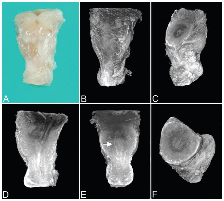
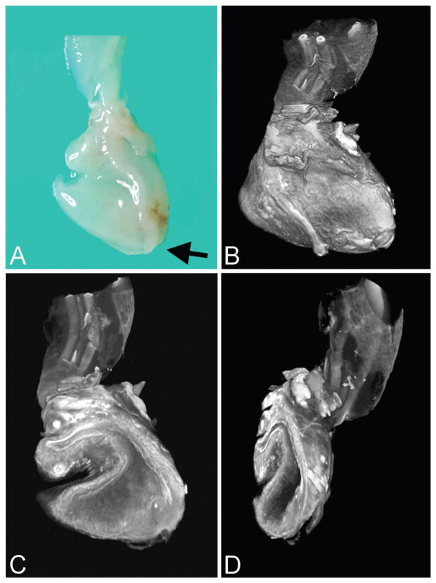
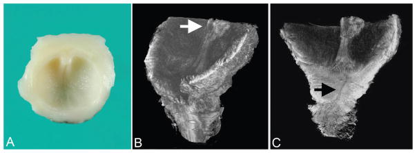
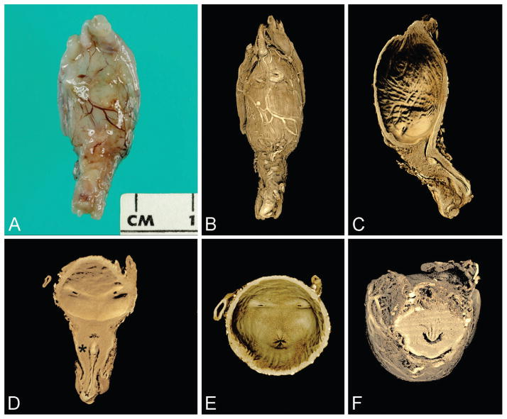
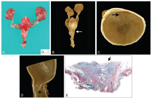
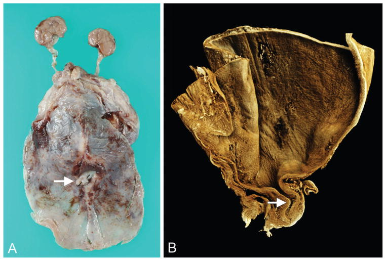
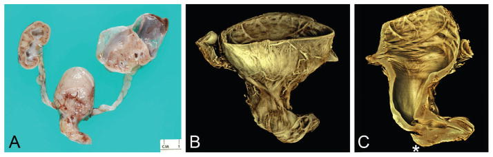
Prenatal obstruction of the lower urinary tract may result in megacystis, with subsequent development of hydroureter, hydronephrosis, and renal damage. Oligo- or anhydramnios, pulmonary hypoplasia, and prune belly syndrome are lethal consequences. Causes and mechanisms responsible for obstruction remain unclear but might be clarified by anatomic study at autopsy. To this end, we employed 2 methods of tomographic imaging-optical projection tomography and contrast-enhanced microCT scanning-to elucidate the anatomy of the intact urinary bladder and urethra in 10 male fetuses with lower urinary tract obstruction. Images were compared with those from 9 age-matched controls. Three-dimensional images, rotated and sectioned digitally in multiple planes, permitted thorough examination while preserving specimens for later study. Both external and internal features of the bladder and urethra were demonstrated; small structures (ie, urethral crest, verumontanum, prostatic utricle, ejaculatory ducts) were seen in detail. Types of obstruction consisted of urethral atresia (n = 5), severe urethral stenosis (n = 2), urethral diaphragm (n = 2), or physical kinking (n = 1); classic (Young type I) posterior urethral valves were not encountered. Traditional light microscopy was then used to verify tomographic findings. The prostate gland was hypoplastic or absent in all cases; in 1, prostatic tissue was displaced inferior to the verumontanum. Findings support previous views that dissection may produce valve-like artifacts (eg, bisection of an obstructing diaphragm) and that deformation of an otherwise normal urethra may result in megacystis. The designation "posterior urethral valves" should not be used as a generic expression of urethral obstruction unless actual valves are demonstrated.


Figures

Figure 1
Figure 1
Control specimen (27 weeks) imaged by optical projection tomography. Note: All images are presented in the anatomic position unless otherwise specified. A. Anterior surface of urinary bladder outlet and proximal-most penile shaft (gross specimen). B. Same orientation as A. C. Virtual dissection with superior-oblique view of bladder outlet. D. Lateral view of bladder outlet and lumen of posterior urethra. E. Anterior view of posterior urethra with verumontanum (arrow). F. Superior-oblique view of verumontanum showing prostatic utricle centrally and U-shaped urethral lumen (normal at this level). A color version of this figure is available online.
Figure 2
Figure 2
Urethral atresia imaged by optical projection tomography (case 1). A. External view of disrupted urinary bladder without urethral orifice (arrow marks atretic region) and portion of umbilical cord and umbilical arteries at top of image (gross specimen). B. Same orientation as A. C. Virtual section of bladder interior with patent urachus (left) and cord vessels at top of image. D. Oblique view of bladder interior showing bladder wall without urethral orifice. A color version of this figure is available online.
Figure 3
Figure 3
Type III posterior urethral valve (urethral diaphragm) imaged by optical projection tomography (case 3). A. Gross specimen consisting of transected posterior urethra, portion of verumontanum, and inferior urethral crests extending to obstructive diaphragm. B. Superior-oblique view of posterior urethra showing inferior portion of verumontanum (arrow) and inferior urethral crests that lead to obstructive diaphragm. C. Coronal section of urethra showing same structures as A plus stenotic, discontinuous urethra (arrow). A color version of this figure is available online.
Figure 4
Figure 4
Control specimen imaged by microCT (23 weeks). A. External view of anterior bladder with umbilical arteries present laterally and proximal penile shaft inferiorly (gross specimen). B. Rendered external view (same orientation as A). C. Virtual section of bladder and urethra in median plane. D. Virtual section of bladder and urethra in coronal plane; note insertion of ureters and verumontanum (asterisk) with inferior urethral crest. E. Base of urinary bladder showing urethral orifice centrally and 2 ureteral orifices laterally. F. Virtual section of posterior urethra in transverse plane showing verumontanum, ejaculatory ducts, and prostatic utricle (U-shaped urethral lumen is normal configuration at this level). A color version of this figure is available online. Color and shadowing are computer-generated.
Figure 5
Figure 5
Type III posterior urethral valve (urethral diaphragm) imaged by microCT (case 6). A. Anterior view of gross specimen showing megacystis, dilated (“keyhole”) posterior urethra with penis, dilated ureters and renal pelves, and cystic kidneys. B. Rendered external view (cystic kidneys were bisected at autopsy) showing close correspondence to A (arrow marks plane of virtual section shown in C). C. Interior view of transversely sectioned dilated posterior urethra showing inferior portion of verumontanum and urethral crest (arrow) extending to obstructive diaphragm. D. Virtual section in median plane providing lateral view of obstructive diaphragm and stenotic distal urethra (arrow). E. Photomicrograph of trichrome-stained urethra showing obstructing diaphragm (arrow) and patent urethra distally (×15). A color version of this figure is available online. Color and shadowing are computer-generated.
Figure 6
Figure 6
Tortuous, stenotic posterior urethra imaged by microCT (case 7). A. Gross specimen showing massive distention of urinary bladder and mildly dilated ureters (arrow marks base of penis). B. Medial virtual section on rendered volume showing dilated urinary bladder and tortuous, stenotic posterior urethra (arrow). A color version of this figure is available online. Color and shadowing are computer-generated.
Figure 7
Figure 7
Atypical kinking of posterior urethra with poststenotic dilatation and more distal obstructing diaphragm (case 10). A. Gross specimen showing hydronephrosis, with large perirenal cyst (urinoma), hydroureter, megacystis, dilated posterior urethra, and proximal penis. B. Rendered volume after microCT imaging of sectioned bladder, posterior urethra, and proximal penis. C. Virtual section in median plane showing dilated urinary bladder and posterior urethra; from proximal to distal, note regions of urethral stenosis followed by poststenotic dilatation, obstructing diaphragm, and patent urethra. (This distal diaphragm is consistent with a “congenital obstructive posterior urethral membrane,” or COPUM, as described by others [20].) When scanned, the lumen of the bladder/proximal urethra and lumen of the more distal urethra did not lie in the same plane; this was overcome by manually defining separate planes for the tissue with the software, the joining of which resulted in the faint oblique line visible across the stenotic urethral lumen (asterisk). A color version of this figure is available online. Color and shadowing are computer-generated.

References

    1. Young HH, Frontz WA, Baldwin JC. Congenital obstruction of the posterior urethra. J Urol. 1919;3:289–365. - PubMed
    1. Stevens FD. Anus and Genito-Urinary Tracts. Edinburgh: E. & S. Livingstone Ltd; 1963. Congenital Malformations of the Rectum.
    1. Schreuder MF, van der Horst HJ, Bökenkamp A, et al. Posterior urethral valves in three siblings: a case report and review of the literature. Birth Defects Res A Clin Mol Teratol. 2008;82:232–235. - PubMed
    1. Siebert JR, Walker MPR. Familial recurrence of urethral stenosis/atresia. Birth Defects Res A. 2009;85:715–719. - PubMed
    1. Kajbafzadeh A. Congenital urethral anomalies in boys. Part I: posterior urethral valves. Urol J. 2005;2:59–78. - PubMed

Publication types

LinkOut - more resources