. 2013 Aug;46(8):398-403.
doi: 10.5483/bmbrep.2013.46.8.237.

Floridoside suppresses pro-inflammatory responses by blocking MAPK signaling in activated microglia

Affiliations

Floridoside suppresses pro-inflammatory responses by blocking MAPK signaling in activated microglia

MinJeong Kim et al. BMB Rep. 2013 Aug.

Abstract

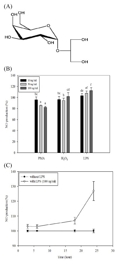
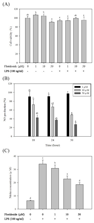
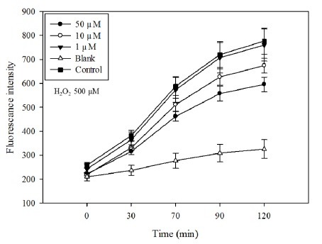
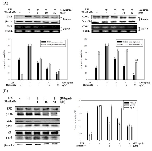
Inflammatory conditions mediated by activated microglia lead to chronic neuro-degenerative diseases such as Alzheimer's, Parkinson's, and Huntington's diseases. This study was conducted to determine the effect of floridoside isolated from marine red algae Laurencia undulata on LPS (100 ng/ml) activated inflammatory responses in BV-2 microglia cells. The results show that floridoside has the ability to suppress pro-inflammatory responses in microglia by markedly inhibiting the production of nitric oxide (NO) and reactive oxygen species (ROS). Moreover, floridoside down-regulated the protein and gene expression levels of iNOS and COX-2 by significantly blocking the phosphorylation of p38 and ERK in BV-2 cells. Collectively, these results indicate that floridoside has the potential to be developed as an active agent for the treatment of neuro-inflammation.


Figures

Fig. 1.
Fig. 1.. The structure of floridoside and the effect of different stimulators on NO production of BV-2 cells. (A) Chemical structure of floridoside. (B) Effect of different stimulators on the NO production of BV-2 cells. Cells were maintained in serum-free medium for 1h and treated with different concentrations (10, 50, 100 ng/ml) of the stimulators for 24 h. Conditioned media was collected and NO production level was assessed by using Griess reagent. (C) Time dependent effect of LPS (100 ng/ml) on BV-2 cells. LPS was treated to BV-2 cells cultured in serum free media. Conditioned media was collected at different times (3, 6, 18, 24 and 50 h) and NO concentration was measured. Data are presented as means ± standard deviation (SD) of three independent experiments (n = 3 total). a-fMeans with different letters are significantly different (P < 0.05) according to Tukey b multiple range test.
Fig. 2.
Fig. 2.. Effect of floridoside on the viability and activation of BV-2 cells. (A) Effect of floridoside on viability of BV-2 cells. Cells were serum starved for 1 h and treated with LPS (100 ng/ml) followed by the treatment with floridoside (1, 10 and 50 μM) 24 h. Cell viability was assessed by MTT assay. (B) Time dependent NO inhibitory effect of floridoside. Cells were cultured in serum-free media and treated with LPS (100 ng/ml) for 1 h and then incubated with floridoside (1, 10 and 50 μM) for 50 h. Conditioned media was collected at different times (18, 24 and 50 h) and NO production levels were compared. (C) Inhibitory effects of different concentrations (1, 10 and 50 μM) of floridoside on LPS-induced NO production in BV-2 cells. Conditioned medium was collected after 24 h and NO concentration was measured using the Griess reaction. Standard nitrite curve was used to quantify the NO production level. Data are presented as means ± standard deviation (SD) of three independent experiments (n = 3 total). a-eMeans with different letters are significantly different (P < 0.05) according toTukey b multiple range test.
Fig. 3.
Fig. 3.. ROS scavenging effect of floridoside in H2O2 (500 μM) activated BV-2 cells. Intracellular ROS levels were measured by detecting the fluorescence intensity of the oxidant-sensitive florescent probe DCFH-DA. Florescence intensity was recorded at different time intervals (0-120 min) in the presence of floridoside (1, 10 and 50 μM).
Fig. 4.
Fig. 4.. Effect of floridoside on the expression of signaling molecules in LPS activated BV-2 cells. (A) Floridoside mediated inhibition of protein and mRNA expression of COX-2 and iNOS. Protein and mRNA levels of COX-2 and iNOS were analyzed by Western blot and RT-PCR, respectively. β-actin and β-tubulin expressions were used as an internal controls. (B) Effect of floridoside on inhibition of MAPK molecules, p38, JNK and ERK1/2. Protein expression levels were analyzed by Western blot. The expression levels were quantified and presented in graphical form. Data are presented as means ± standard deviation (SD) of three independent experiments (n = 3 total). a-fMeans withdifferent letters are significantly different (P < 0.05) according to Tukey b multiple range test.

References

    1. Yokoyama A., Yang L., Itoh S., Mori K., Tanaka J. Microglia, a potential source of neurons, astrocytes, and oligodendrocytes. Glia. (2004);45:96–104. doi: 10.1002/glia.10306. - DOI - PubMed
    1. Vilhardt F. Microglia: phagocyte and glia cell. Int. J. Biochem. Cell Biol. (2005);37:17–21. doi: 10.1016/j.biocel.2004.06.010. - DOI - PubMed
    1. Matyszak M. K. Inflammation in the CNS: balance between immunological privilege and immune responses. Prog. Neurobiol. (1998);56:19–35. doi: 10.1016/S0301-0082(98)00014-8. - DOI - PubMed
    1. Lull M. E., Block M. L. Microglial activation and chronic neurodegeneration. Neurotherapeutics. (2010);7:354–365. doi: 10.1016/j.nurt.2010.05.014. - DOI - PMC - PubMed
    1. Lehnardt S., Massillon L., Follett P., Jensen F. E., Ratan R., Rosenberg P. A., Volpe J. J., Vartanian T. Activation of innate immunity in the CNS triggers neurodegeneration through a Toll-like receptor 4-dependent pathway. Proc. Natl. Acad. Sci. U. S. A. (2003);100:8514–8519. doi: 10.1073/pnas.1432609100. - DOI - PMC - PubMed

Publication types

MeSH terms