

Nanoparticle-nanoparticle interactions in biological media by atomic force microscopy
- PMID: 23978039
- PMCID: PMC4438084
- DOI: 10.1021/la4019585
Nanoparticle-nanoparticle interactions in biological media by atomic force microscopy
Abstract
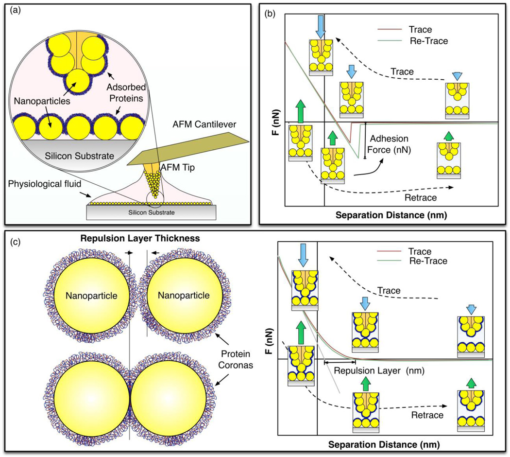
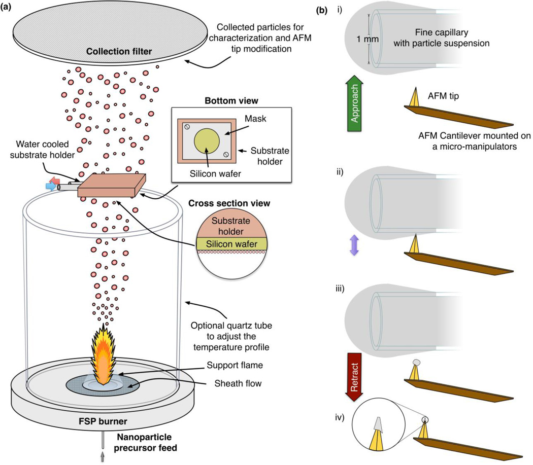
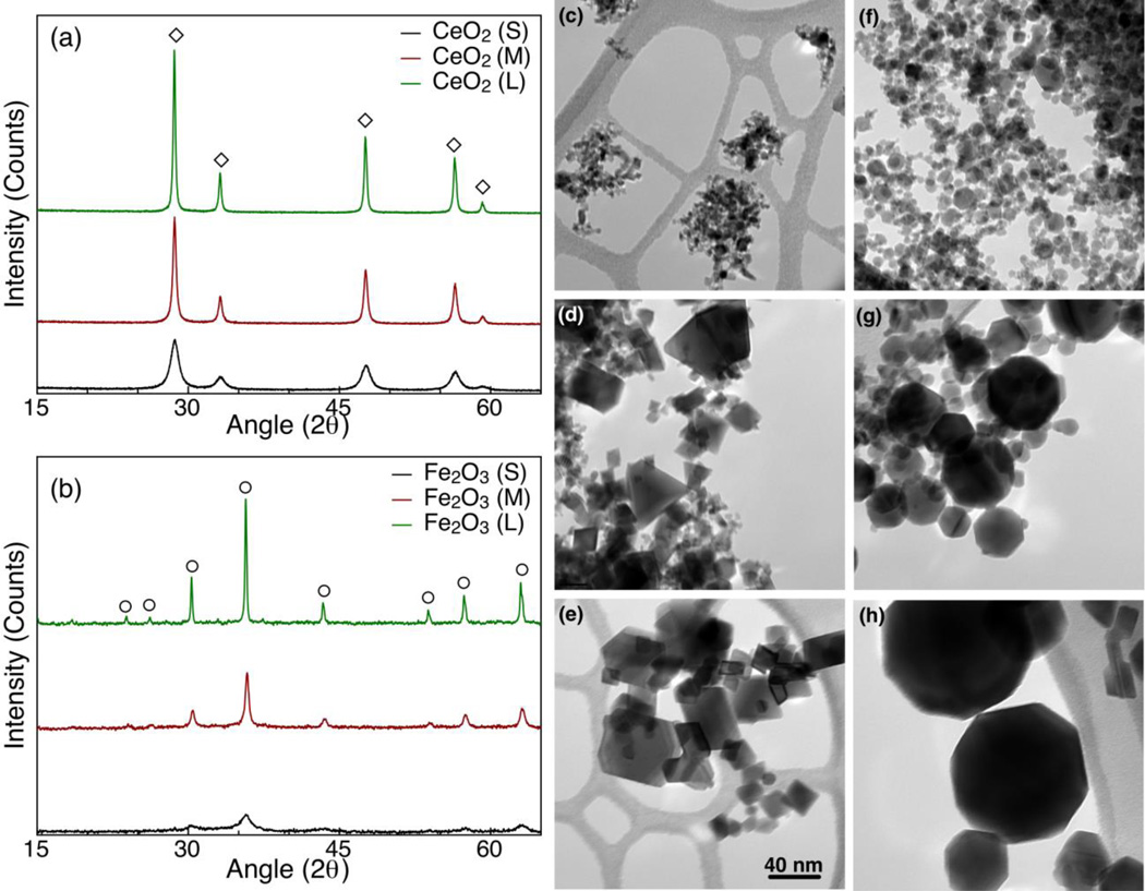
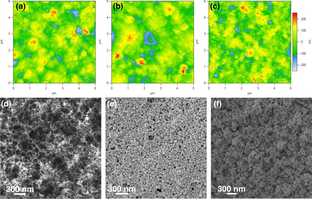
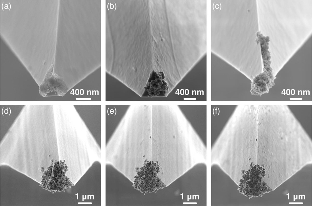
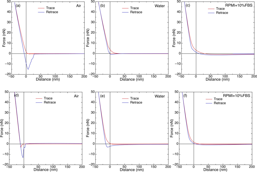
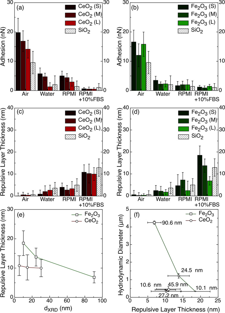
Particle-particle interactions in physiological media are important determinants for nanoparticle fate and transport. Herein, such interactions are assessed by a novel atomic force microscopy (AFM)-based platform. Industry-relevant CeO2, Fe2O3, and SiO2 nanoparticles of various diameters were made by the flame spray pyrolysis (FSP)-based Harvard Versatile Engineering Nanomaterials Generation System (Harvard VENGES). The nanoparticles were fully characterized structurally and morphologically, and their properties in water and biological media were also assessed. The nanoparticles were attached on AFM tips and deposited on Si substrates to measure particle-particle interactions. The corresponding force was measured in air, water, and biological media that are widely used in toxicological studies. The presented AFM-based approach can be used to assess the agglomeration potential of nanoparticles in physiological fluids. The agglomeration potential of CeO2 nanoparticles in water and RPMI 1640 (Roswell Park Memorial Institute formulation 1640) was inversely proportional to their primary particle (PP) diameter, but for Fe2O3 nanoparticles, that potential is independent of PP diameter in these media. Moreover, in RPMI+10% Fetal Bovine Serum (FBS), the corona thickness and dispersibility of the CeO2 are independent of PP diameter, while for Fe2O3, the corona thickness and dispersibility were inversely proportional to PP diameter. The present method can be combined with dynamic light scattering (DLS), proteomics, and computer simulations to understand the nanobio interactions, with emphasis on the agglomeration potential of nanoparticles and their transport in physiological media.
Figures
Similar articles
-
Real-Time Nanoparticle-Cell Interactions in Physiological Media by Atomic Force Microscopy.ACS Sustain Chem Eng. 2014 Jul 7;2(7):1681-1690. doi: 10.1021/sc500152g. Epub 2014 Jun 10. ACS Sustain Chem Eng. 2014. PMID: 25068097 Free PMC article.
-
Imaging and size measurement of nanoparticles in aqueous medium by use of atomic force microscopy.Anal Bioanal Chem. 2018 Feb;410(5):1525-1531. doi: 10.1007/s00216-017-0799-3. Epub 2017 Dec 18. Anal Bioanal Chem. 2018. PMID: 29256078
-
Effects of cell culture media on the dynamic formation of protein-nanoparticle complexes and influence on the cellular response.ACS Nano. 2010 Dec 28;4(12):7481-91. doi: 10.1021/nn101557e. Epub 2010 Nov 17. ACS Nano. 2010. PMID: 21082814
-
Size Measurement of Nanoparticles Using Atomic Force Microscopy: Version 1.1.2009 Oct. In: National Cancer Institute’s Nanotechnology Characterization Laboratory Assay Cascade Protocols [Internet]. Bethesda (MD): National Cancer Institute (US); 2005 May 1–. NIST - NCL Joint Assay Protocol, PCC-6. 2009 Oct. In: National Cancer Institute’s Nanotechnology Characterization Laboratory Assay Cascade Protocols [Internet]. Bethesda (MD): National Cancer Institute (US); 2005 May 1–. NIST - NCL Joint Assay Protocol, PCC-6. PMID: 39013048 Free Books & Documents. Review.
-
How Corona Formation Impacts Nanomaterials as Drug Carriers.Mol Pharm. 2020 Mar 2;17(3):725-737. doi: 10.1021/acs.molpharmaceut.9b01111. Epub 2020 Jan 24. Mol Pharm. 2020. PMID: 31939673 Review.
Cited by
-
Redox-Responsive Polysulfide-Based Biodegradable Organosilica Nanoparticles for Delivery of Bioactive Agents.ACS Appl Mater Interfaces. 2017 Jun 28;9(25):21133-21146. doi: 10.1021/acsami.7b04351. Epub 2017 Jun 13. ACS Appl Mater Interfaces. 2017. PMID: 28609092 Free PMC article.
-
Decreased Uptake and Enhanced Mitochondrial Protection Underlie Reduced Toxicity of Nanoceria in Human Monocyte-Derived Macrophages.J Biomed Nanotechnol. 2016 Dec;12(12):2139-50. doi: 10.1166/jbn.2016.2320. J Biomed Nanotechnol. 2016. PMID: 29368911 Free PMC article.
-
Development of reference metal and metal oxide engineered nanomaterials for nanotoxicology research using high throughput and precision flame spray synthesis approaches.NanoImpact. 2018 Apr;10:26-37. doi: 10.1016/j.impact.2017.11.007. Epub 2017 Dec 2. NanoImpact. 2018. PMID: 30035243 Free PMC article.
-
Silica coating influences the corona and biokinetics of cerium oxide nanoparticles.Part Fibre Toxicol. 2015 Oct 12;12:31. doi: 10.1186/s12989-015-0106-4. Part Fibre Toxicol. 2015. PMID: 26458946 Free PMC article.
-
Insights into Characterization Methods and Biomedical Applications of Nanoparticle-Protein Corona.Materials (Basel). 2020 Jul 10;13(14):3093. doi: 10.3390/ma13143093. Materials (Basel). 2020. PMID: 32664362 Free PMC article. Review.
References
-
- Scheringer M. Nanoecotoxicology: Environmental Risks of Nanomaterials. Nature Nanotech. 2008;3:322–323. - PubMed
-
- Bello D, Martin J, Santeufemio C, Sun Q, Lee Bunker K, Shafer M, Demokritou P. Physicochemical Morphological Characterisation of Nanoparticles From Photocopiers: Implications for Environmental Health. Nanotoxicology. 2012 - PubMed
-
- Philbert MA, Alexeeff GV, Bahadori T, Balbus JM, Bawendi MG, Biswas P, Colvin V, Klaine SJ, Maynard AD, Monteiro-Riviere NA, Oberdörster G, Ratner MA, Teeguarden JG, Weisner M. Review of Federal Strategy for Nanotechnology-Related Enviromental, Health, and Safety Research. Washington, DC: The National Academic Press; 2008. pp. 1–131.
-
- Nel AE, Mädler L, Velegol D, Xia T, Hoek EMV, Somasundaran P, Klaessig F, Castranova V, Thompson M. Understanding Biophysicochemical Interactions at the Nano-Bio Interface. Nat. Mater. 2009;8:543–557. - PubMed
Publication types
MeSH terms
Substances
Grants and funding
LinkOut - more resources
Full Text Sources
Other Literature Sources
Miscellaneous
