. 2013 Oct 1;85(19):9173-80.
doi: 10.1021/ac401868b. Epub 2013 Sep 12.

A new approach to measuring protein backbone protection with high spatial resolution using H/D exchange and electron capture dissociation

Affiliations

A new approach to measuring protein backbone protection with high spatial resolution using H/D exchange and electron capture dissociation

Rinat R Abzalimov et al. Anal Chem. .

Abstract

Inadequate spatial resolution remains one of the most serious limitations of hydrogen/deuterium exchange-mass spectrometry (HDX-MS), especially when applied to larger proteins (over 30 kDa). Supplementing proteolytic fragmentation of the protein in solution with ion dissociation in the gas phase has been used successfully by several groups to obtain near-residue level resolution. However, the restrictions imposed by the LC-MS/MS mode of operation on the data acquisition time frame makes it difficult in many cases to obtain a signal-to-noise ratio adequate for reliable assignment of the backbone amide protection levels at individual residues. This restriction is lifted in the present work by eliminating the LC separation step from the workflow and taking advantage of the high resolving power and dynamic range of a Fourier transform ion cyclotron resonance-mass spectrometer (FTICR-MS). A residue-level resolution is demonstrated for a peptic fragment of a 37 kDa recombinant protein (N-lobe of human serum transferrin), using electron-capture dissociation as an ion fragmentation tool. The absence of hydrogen scrambling in the gas phase prior to ion dissociation is verified using redundant HDX-MS data generated by FTICR-MS. The backbone protection pattern generated by direct HDX-MS/MS is in excellent agreement with the known crystal structure of the protein but also provides information on conformational dynamics, which is not available from the static X-ray structure.


Figures

Figure 1
Figure 1
Primary (left) and tertiary (right) structures of hTf/2N. The peptic maps obtained in the present work and using classical HDX MS set-up are shown with brown and black lines, respectively. The secondary structure of the peptic fragment [Val67-Phe84] is color-coded (an α-helix, purple; a loop, red; and a β-strand, blue).
Figure 2
Figure 2
A: a mass spectrum of peptic fragments of hTf/2N acquired with a continuous-flow apparatus. The inset shows a region of the spectrum containing ionic signal representing the (Val67-Phe84) peptide (charge state +2, labeled with an open circle). B: Ionic signal after mass-isolation of the peptide of interest. C: a fraction of the spectrum of ECD fragments of isolated ions showing c6+ and z7+ fragments of peptide ion [Val67-Phe84].
Figure 3
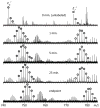
Figure 3
Evolution of isotopic distribution of c6+ and z7+ fragments produced upon ECD of the precursor ion representing the doubly protonated form of peptide (Val67-Phe84).
Figure 4
Figure 4
Backbone protection maps deduced from a ladder of z- (A) and c- (B) fragments generated upon ECD of a triply protonated peptide (Val67-Phe84), and from the entire complement of ECD fragments generated upon dissociation of the doubly protonated ion representing the same peptide (C). Diagram D shows the overall protection maps that combine the data presented in panels A-C. Bars of different colors represent varying exchange time in solution (as indicated in panel D).
Figure 5
Figure 5
A: comparison of the cumulative protection for several shorter segments within the peptic fragment (Val67-Phe84) obtained with HDX MS and deduced from the overlapping peptic fragments. B: comparison of deuterium incorporation in a shorter peptic fragment (Tyr71-Phe84) and a congruent fragment z14+ derived from a longer peptic fragment (Val67-Phe84).

References

    1. Engen JR. Analysis of protein conformation and dynamics by hydrogen/deuterium exchange MS. Anal Chem. 2009;81:7870–7875. - PMC - PubMed
    1. Konermann L, Pan J, Liu YH. Hydrogen exchange mass spectrometry for studying protein structure and dynamics. Chem Soc Rev. 2011;40:1224–1234. - PubMed
    1. Skinner JJ, Lim WK, Bedard S, Black BE, Englander SW. Protein dynamics viewed by hydrogen exchange. Protein Sci. 2012;21:996–1005. - PMC - PubMed
    1. Kaltashov IA, Bobst CE, Abzalimov RR. Mass spectrometry-based methods to study protein architecture and dynamics. Protein Sci. 2013;22:530–544. - PMC - PubMed
    1. Miranker A, Robinson CV, Radford SE, Dobson CM. Investigation of protein folding by mass spectrometry. FASEB J. 1996;10:93–101. - PubMed

Publication types

LinkOut - more resources