. 2013 Aug 26;2(8):e66.
doi: 10.1038/oncsis.2013.29.

Gremlin-1 associates with fibrillin microfibrils in vivo and regulates mesothelioma cell survival through transcription factor slug

Affiliations

Gremlin-1 associates with fibrillin microfibrils in vivo and regulates mesothelioma cell survival through transcription factor slug

J A Tamminen et al. Oncogenesis. .

Abstract

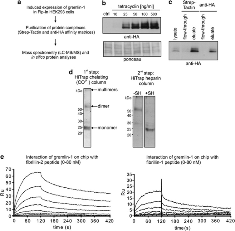
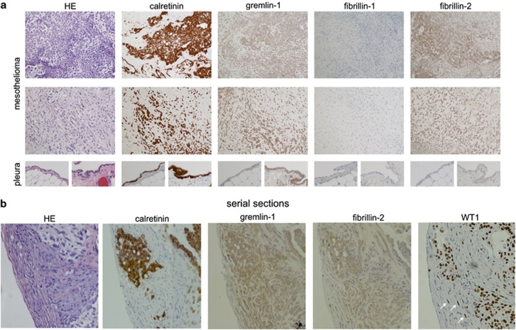
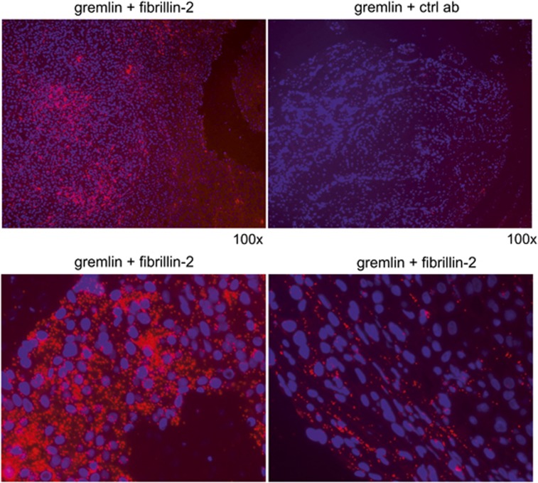
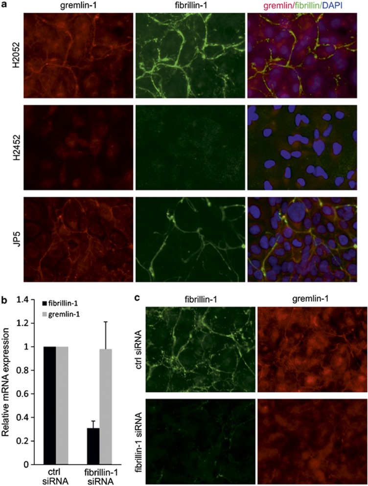
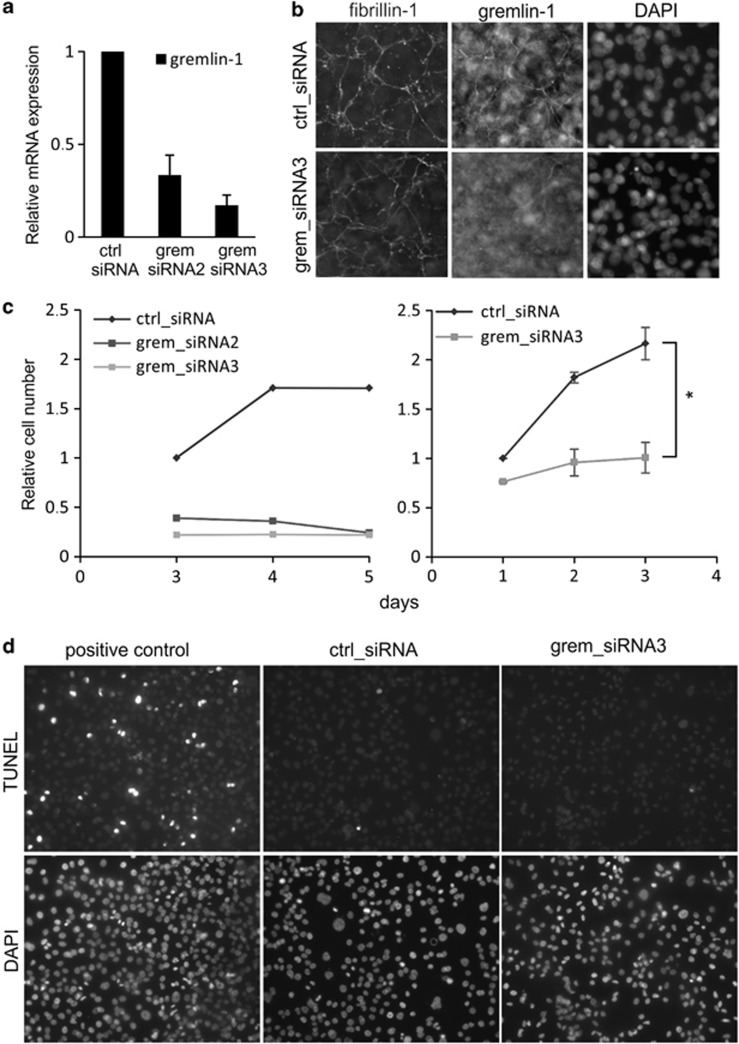
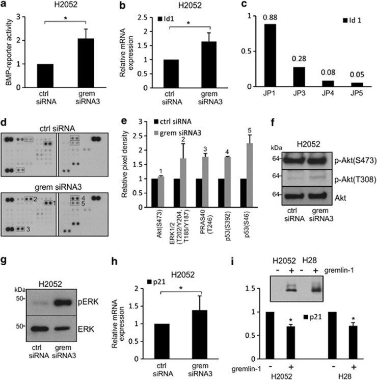
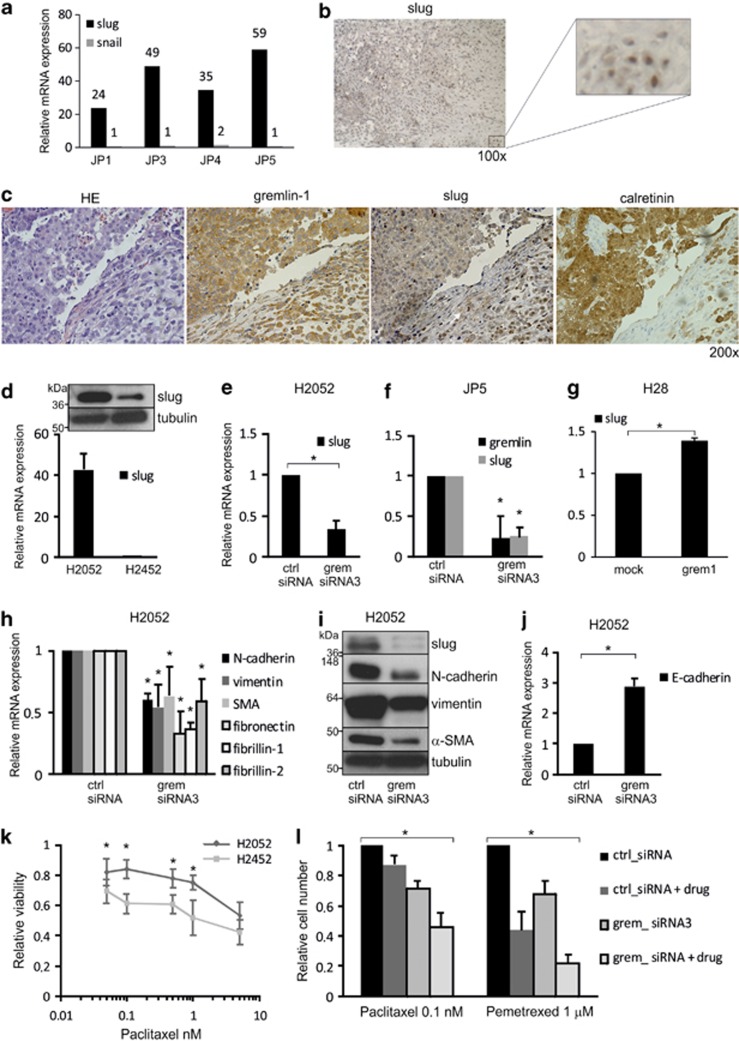
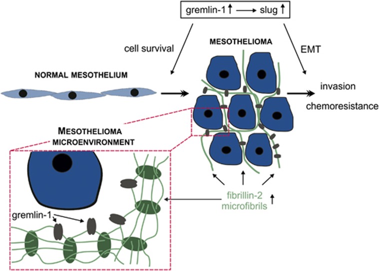
Malignant mesothelioma is a form of cancer that is highly resistant to conventional cancer therapy for which no major therapeutic advances have been introduced. Here, we identify gremlin-1, a known bone morphogenetic protein inhibitor crucial for embryonic development, as a potential therapeutic target for mesothelioma. We found high expression levels of gremlin-1 in the mesothelioma tumor tissue, as well as in primary mesothelioma cells cultured from pleural effusion samples. Downregulation of gremlin-1 expression by siRNA-mediated silencing in a mesothelioma cell line inhibited cell proliferation. This was associated with downregulation of the transcription factor slug as well as mesenchymal proteins linked to cancer epithelial-to-mesenchymal transition. Further, resistance to paclitaxel-induced cell death was associated with high gremlin-1 and slug expression. Treatment of gremlin-1-silenced mesothelioma cells with paclitaxel or pemetrexed resulted in efficient loss of cell survival. Finally, our data suggest that concomitant upregulation of fibrillin-2 in mesothelioma provides a mechanism for extracellular localization of gremlin-1 to the tumor microenvironment. This was supported by the demonstration of interactions between gremlin-1, and fibrillin-1 and -2 peptides as well as by colocalization of gremlin-1 to fibrillin microfibrils in cells and tumor tissue samples. Our data suggest that gremlin-1 is also a potential target for overcoming drug resistance in mesothelioma.


Figures

Figure 1
Figure 1
Gremlin-1 interacts with fibrillin-1 and -2. (a) Gremlin-1 protein interaction screen outline. (b) Flip-In HEK293/gremlin-1 cells were treated with increasing concentrations of tetracycline for 24 h followed by immunoblotting analyses using anti-HA antibodies. Ponceau staining of proteins indicates equal loading. (c) Immunoblotting analyses of gremlin-1, using anti-HA antibodies, after Strep-tactin and anti-HA affinity purification steps. (d) Purification of gremlin-1 from conditioned media of stably transfected HEK293 cells. After elution from a CoCl2-loaded HiTrap column using a C-terminally placed His6-affinity-tag protein, bands corresponding to monomeric, dimeric and multimeric gremlin-1 were detected via SDS–PAGE followed by Coomassie staining. A subsequent second purification step via a HiTrap heparin column yielded mostly dimeric gremlin-1, which shifted to the position of monomeric gremlin under reducing conditions (+SH). (e) Interaction studies between gremlin-1 and N-terminal peptides of fibrillin-1 and -2 using a surface plasmon resonance technology. Gremlin-1 was immobilized on a sensor chip and fibrillin-1 and -2 peptides in a concentration range from 80 to 0 nM were flown over as analytes. Affinity constants (Kd) for both interactions were about 10 nM (Table 1), indicating a high affinity interaction between gremlin-1 and the main building blocks of fibrillin microfibrils.
Figure 2
Figure 2
Overexpression of gremlin-1 and fibrillin-2 in mesothelioma. (a) Immunohistochemical staining of mesothelioma and control pleura samples using calretinin, gremlin-1, fibrillin-1 and fibrillin-2 antibodies. Hematoxylin and eosin (HE) staining is shown on the left. Abundant gremlin-1 and fibrillin-2 immunoreactivity is observed in both calretinin-negative and -positive tumor areas. Normal mesothelium (pleura, left panel) shows very low levels of staining, whereas reactive normal mesothelium (pleura, right panel) shows faint fibrillin-2 and moderate gremlin-1 staining. (b) Immunohistochemical staining of serial mesothelioma sections suggests similar staining patterns for gremlin-1 and fibrillin-2. Staining was observed also in calretinin-negative stromal-like areas, which contained isolated Wilms' tumor protein (WT1)-positive tumor cells (white arrows).
Figure 3
Figure 3
Colocalization analyses of gremlin-1 with fibrillin-2 in mesothelioma tumor tissue. Proximity ligation assay was used to detect colocalization of gremlin-1 with fibrillin-2 in mesothelioma tumor tissue. Positive colocalization signals (red dots) were abundant throughout the tumor tissue. Negative control with gremlin antibody plus mouse isotype control is shown on the upper right panel.
Figure 4
Figure 4
Primary mesothelioma cells express high levels of gremlin-1. Primary mesothelioma cells (JP cells) were isolated from pleural effusion samples. (a) Immunofluorescence staining suggests that the cells were positive for the mesothelial marker calretinin. Co-staining with cytokeratine (CK)-7 and vimentin antibodies suggests that the tumor cells co-express these marker proteins. (b) Primary mesothelioma cells were analyzed for gremlin-1, fibrillin-1 and -2 and BMP-2 mRNA expression by quantitative RT–PCR. The levels were normalized to the expression levels of TATA-binding protein and are expressed relative to the expression levels in Met5A cells (immortalized, non-tumorigenic mesothelial cells), which were set to 1. (c) Established mesothelioma cell lines were analyzed for gremlin-1, fibrillin-1 and -2 and BMP-2 mRNA expression by quantitative RT–PCR. The error bars represent s.d. (n=2).
Figure 5
Figure 5
Gremlin-1 and fibrillin-1 co-localize in mesothelioma cell ECM in vitro. (a) Mesothelioma cell lines (H2052 and H2452) and primary mesothelioma cells (JP5) were co-stained for gremlin-1 and fibrillin-1 and analyzed using immunofluorescence microscopy. A fibrillar staining pattern was observed in H2052 and JP5 cells. (b) In control (ctrl) and fibrillin-1 siRNA-transfected H2052 cells, the mRNA expression levels of gremlin-1 and fibrillin-1 were analyzed by quantitative RT-PCR. The levels were normalized to the expression levels of TATA-binding protein and are expressed relative to each control, which was set to 1. The error bars represent s.d. (n=2). (c) Double-immunofluorescence staining of siRNA-treated H2052 cells with gremlin-1 and fibrillin-1 antibodies. Note the lack of fibrillar gremlin-1 staining in fibrillin-1-silenced cells.
Figure 6
Figure 6
Gremlin-1 silencing impairs H2052 mesothelioma cell proliferation. (a) The efficiency of gremlin-1 siRNA silencing was analyzed with quantitative RT-PCR as in figure 5. The error bars represent s.d. (n=4). (b) Double immunofluorescence staining of siRNA-treated H2052 cells with gremlin-1 and fibrillin-1 antibodies. Note the lack of fibrillar gremlin-1 staining, which is suggestive of efficient silencing using gremlin siRNA3. (c) H2052 cell proliferation was analyzed by counting cells 1–5 days after siRNA transfection. Both gremlin siRNAs reduced dramatically the number of cells 3–5 days after transfection (left panel, a representative experiment is shown). Same effect was seen also at earlier time points (right panel). The error bars represent s.d. (n=3, *P=0.05). (d) Silencing gremlin-1 by siRNA3 did not induce apoptosis as measured using TUNEL staining. DNase I-treated cells (control) show positive staining. Nuclei are stained with DAPI.
Figure 7
Figure 7
Cellular signaling in gremlin-1-silenced cells. (a) BMP-dependent reporter activity was analyzed in control and gremlin-1 siRNA-treated H2052 cells. The levels are expressed relative to control, which was set to 1. The error bars represent s.d. (n=4, *P<0.01). (b) Id1 mRNA expression levels were analyzed by quantitative RT–PCR. The levels are expressed relative to control. n=3, *P<0.05. (c) Id1 mRNA expression levels in primary mesothelioma cells. The levels are expressed relative to the expression levels in Met5A cells. (d) Cell lysates from control and gremlin-1 siRNA-treated H2052 cells were analyzed using a commercial phospho-protein array. (e) Quantification of alterations in the amounts of phospho-proteins (n=2). Changes were also verified by immunoblotting analyses of Akt (f) and ERK (g) pathway proteins. Molecular weight markers are indicated on the left. Expression levels of p21 (Cip1/Waf1), a p53 target gene, were analyzed in gremlin-1 siRNA-treated (h) and gremlin-1-transfected cells (i) by quantitative RT-PCR. The levels are expressed relative to control, which was set to 1. n=3, *P<0.05. I, inset. Gremlin-1 overexpression in H2052 and H28 cells was verified by immunoblotting using an antibody against the V5 tag.
Figure 8
Figure 8
Gremlin-1 regulates slug expression and EMT phenotype. (a) Primary human mesothelioma cells (JP) were analyzed for snail and slug mRNA expression by quantitative RT-PCR. The levels were normalized to the expression levels of TATA-binding protein and are expressed relative to the expression levels in Met5A cells, which were set to 1. (b, c) Immunohistochemical staining of a mesothelioma section using slug antibodies shows mostly diffuse and granular cytoplasmic staining in the tumor tissue. Nuclear staining was also observed in certain tumor areas (b, right panel). (c) Analyses of serial mesothelioma tissue sections suggested that slug and gremlin staining colocalized in the same tumor areas. (d) Slug mRNA expression levels in H2052 and H2452 cells were analyzed by quantitative RT–PCR. The levels are expressed relative to H2452 expression levels, which were set to 1. The error bars represent s.d. (n=5). INSET: immunoblotting analyses of slug. Tubulin is used as a loading control. Molecular weight markers are indicated on the left. (eg) Slug mRNA expression levels were analyzed in control and gremlin-1 siRNA-treated H2052 and JP5 cells or H28 cells transfected with gremlin-1 (grem1) or control vector (mock). The levels are expressed relative to control, which was set to 1. The error bars represent s.d. (JP5, n=3, *P<0.05; H2052, n=4, *P<0.01; H28, n=3, *P=<0.05). (h) N-cadherin, vimentin, α-SMA, fibronectin and fibrillin mRNA expression levels were analyzed in control and gremlin siRNA-treated H2052 cells by quantitative RT-PCR. The error bars represent s.d. (n⩾3, *P<0.05). (i) Immunoblotting analyses of slug, N-cadherin, vimentin and α-SMA in control and gremlin-1 siRNA-treated H2052 cells. Tubulin is used as a loading control. (j) E-cadherin mRNA expression levels were analyzed in control and gremlin siRNA-treated H2052 cells by quantitative RT–PCR. Error bars represent s.d. (n=3, *P<0.05). (k) H2052 and H2452 cells were treated with the indicated concentrations of paclitaxel after which cell proliferation/viability was analyzed with MTT assay (n=4, *P<0.05). (l) Quantification of cell numbers after combined treatment with control or gremlin-1 siRNA and paclitaxel (0.1 nM, 24 h) or pemetrexed (1 μM, 48 h). Cell numbers were counted 3 days after transfection (n⩾4, *P=0.002).
Figure 9
Figure 9
A model for the functions of gremlin-1 and fibrillin-2 in mesothelioma. Gremlin-1 induces proliferation and epithelial-to-mesenchymal transition (EMT) in mesothelioma cells. EMT is mediated by induction of the transcription factor slug and leads to chemoresistance and acquisition of invasive properties. Gremlin-1 is targeted to the tumor microenvironment through binding to fibrillin-2 microfibrils.

References

    1. Mossman BT, Bignon J, Corn M, Seaton A, Gee JB. Asbestos: scientific developments and implications for public policy. Science. 1990;247:294–301. - PubMed
    1. Lanphear BP, Buncher CR. Latent period for malignant mesothelioma of occupational origin. J Occup Med. 1992;34:718–721. - PubMed
    1. Huuskonen MS, Rantanen J. Finnish Institute of Occupational Health (FIOH): prevention and detection of asbestos-related diseases, 1987-2005. Am J Ind Med. 2006;49:215–220. - PubMed
    1. Vorobiof DA, Mafafo K. Malignant pleural mesothelioma: medical treatment update. Clin Lung Cancer. 2009;10:112–117. - PubMed
    1. Dormoy V, Jacqmin D, Lang H, Massfelder T. From development to cancer: lessons from the kidney to uncover new therapeutic targets. Anticancer Res. 2012;32:3609–3617. - PubMed