. 2013 Oct 11;288(41):29313-22.
doi: 10.1074/jbc.M113.476929. Epub 2013 Aug 26.

More than just a cargo adapter, melanophilin prolongs and slows processive runs of myosin Va

Affiliations

More than just a cargo adapter, melanophilin prolongs and slows processive runs of myosin Va

Maria Sckolnick et al. J Biol Chem. .

Abstract

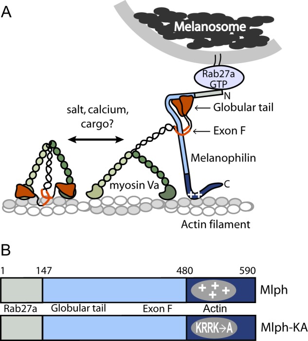
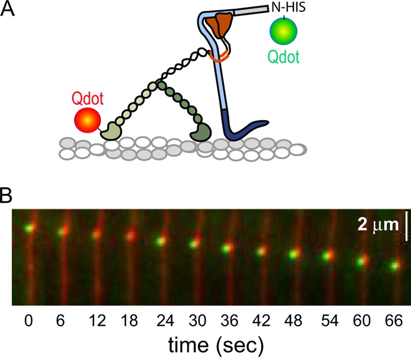
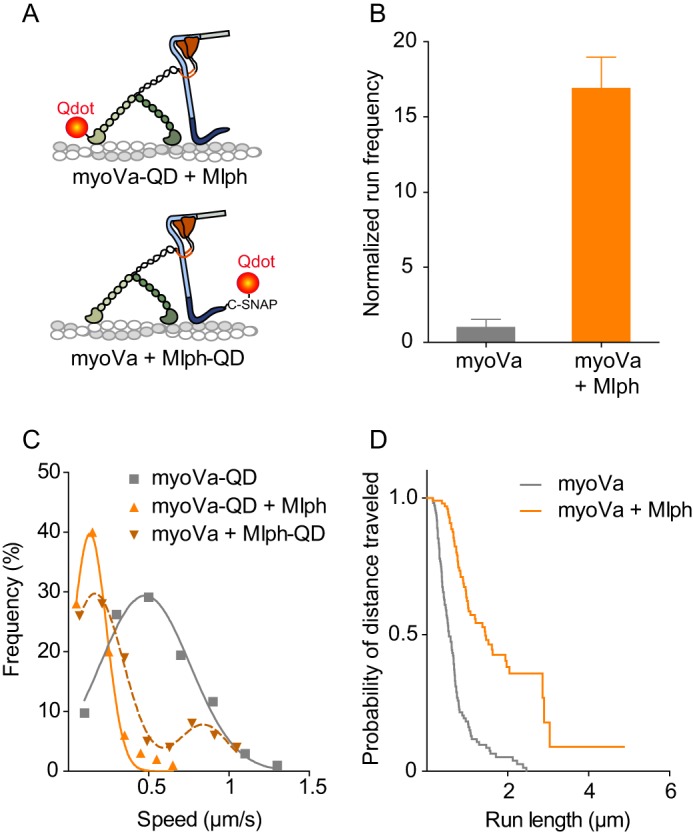
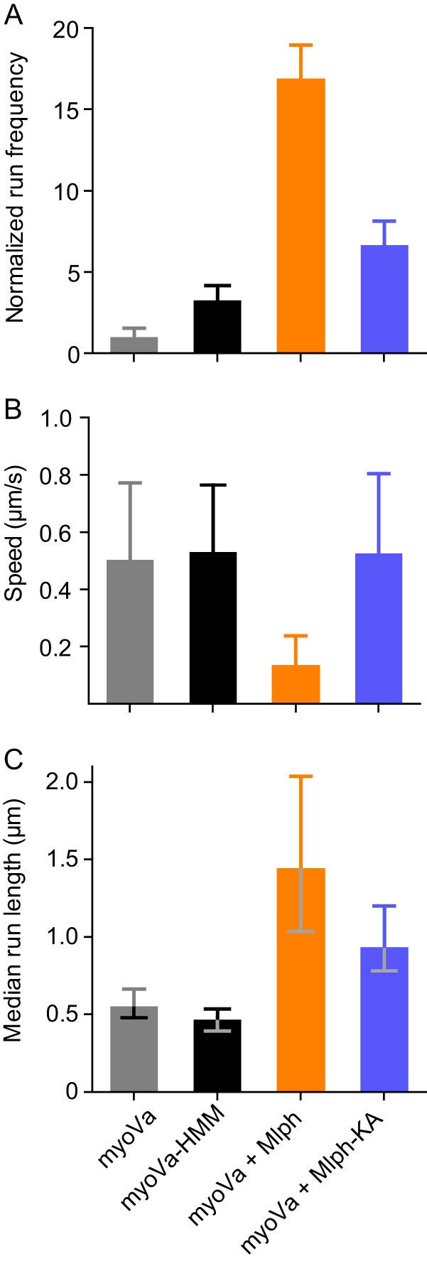
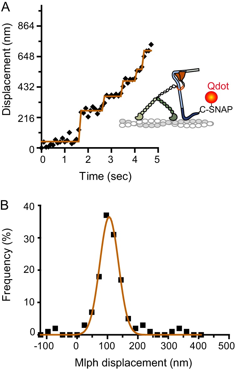
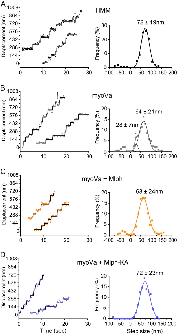
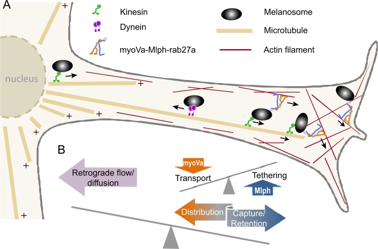
Myosin Va (myoVa) is a molecular motor that processively transports cargo along actin tracks. One well studied cargo in vivo is the melanosome, a pigment organelle that is moved first by kinesin on microtubules and then handed off to myoVa for transport in the actin-rich dendritic periphery of melanocytes. Melanophilin (Mlph) is the adapter protein that links Rab27a-melanosomes to myoVa. Using total internal reflection fluorescence microscopy and quantum dot-labeled full-length myoVa, we show at the single-molecule level that Mlph increases the number of processively moving myoVa motors by 17-fold. Surprisingly, myoVa-Mlph moves ~4-fold slower than myoVa alone and with twice the run length. These two changes greatly increase the time spent on actin, a property likely to enhance the transfer of melanosomes to the adjacent keratinocyte. In contrast to the variable stepping pattern of full-length myoVa, the myoVa-Mlph complex shows a normal gating pattern between the heads typical of a fully active motor and consistent with a cargo-dependent activation mechanism. The Mlph-dependent changes in myoVa depend on a positively charged cluster of amino acids in the actin binding domain of Mlph, suggesting that Mlph acts as a "tether" that links the motor to the track. Our results provide a molecular explanation for the uncharacteristically slow speed of melanosome movement by myoVa in vivo. More generally, these data show that proteins that link motors to cargo can modify motor properties to enhance their biological role.

Keywords: Gating; Intracellular Trafficking; Melanophilin; Myosin; Nanotechnology; Processivity; Protein Dynamics; Regulation; Slac-2a; Tethering.


Figures

FIGURE 1.
FIGURE 1.
Schematic model of the myoVa-melanosome transport complex and the domain structure of Mlph. A, Mlph linking myoVa to melanosomes via Rab27-GTP. MyoVa exists in an equilibrium between a folded, inhibited state and an extended, active conformation, depending on the ionic strength and calcium concentration in vitro. Binding of cargo has been hypothesized to be the physiologic activator of myoVa. Binding of Mlph to myoVa requires both the globular tail and exon F. Mlph also binds via electrostatic interactions to actin (indicated by plus symbols on Mlph). B, domain structure of Mlph. The N-terminal domain binds to Rab 27a, the intrinsically disordered central portion to myoVa, and the C-terminal region to actin. A mutant Mlph (Mlph-KA) contains four point mutations (K493A/R495A/R496A/K497A) that ablate the positive charge necessary to interact strongly with actin.
FIGURE 2.
FIGURE 2.
SDS-gels of purified proteins and ability of Mlph to bind actin. A, purified full-length myoVa with an N-terminal biotin tag showing the myoVa heavy chain (HC) and calmodulin (CaM); 4–12% gradient SDS-gel. B, left lane, marker proteins with indicated molecular masses. Middle lane, WT-Mlph. Right lane, mutant Mlph-KA, 12% SDS-gel. C, co-sedimentation of actin with WT-Mlph or Mlph-KA showing that Mlph-KA has a decreased ability to interact with actin. S, supernatant; P, pellet; 12% SDS-gel.
FIGURE 3.
FIGURE 3.
Salt-dependent conformational transition of myoVa. Analytical ultracentrifugation was used to follow the ionic strength dependence of the folded, inhibited conformation (∼14 S) to the active, extended (∼10 S) form. Intermediate S values represent an equilibrium between bent and extended molecules. Individual data points represent separate experiments. Horizontal bar indicates the average value.
FIGURE 4.
FIGURE 4.
Mlph binds to myoVa in single-molecule assays. A, myoVa with a biotin tag on the motor domain was labeled with a 565-nm streptavidin-Qdot, and Mlph with an N-terminal His tag with a 655-nm anti-HIS Qdot. B, simultaneous movement of both red and green Qdots along rhodamine-phalloidin-labeled actin filaments was observed by TIRF microscopy. Speed, ∼30 nm/s. Conditions: 150 mm KCl, pH 7.4, 2 mm MgATP.
FIGURE 5.
FIGURE 5.
Comparison of myoVa single-molecule kinetics in the presence or absence of Mlph. A, illustration of labeling approaches for the myoVa-Mlph complex. Upper panel, myoVa with an N-terminal biotin tag was labeled with a 655-nm streptavidin-Qdot. Mlph was unlabeled. Lower panel, Mlph with a C-terminal SNAP-biotin tag was labeled with a 655-nm streptavidin-Qdot. MyoVa was unlabeled. B, normalized frequency of motors starting to move within a fixed time frame and area. Values are normalized to actin length and the number of runs with myoVa under the same conditions. Error bars are S.D. (n = 11 for myoVa, and n = 5 for myoVa-Mlph) (t test, p < 0.001). C, histogram and Gaussian fit of speeds for myoVa alone (v = 502 ± 269 nm/s, n = 103; gray squares and line) or in the presence of a 10-fold molar excess of unlabeled Mlph (v = 135 ± 103 nm/s, n = 100; orange triangles and solid line) (t test, p < 0.001). Labeling strategy is depicted in A, upper. Alternatively, an unlabeled motor was used, and the Qdot was attached to the C terminus of Mlph and myoVa was unlabeled (brown dashed line and inverted triangles). This labeling strategy is depicted in A, lower. These data best fit two Gaussians (v1 = 155 ± 188 nm/s; v2 = 860 ± 149 nm/s, n = 100). The small fraction of runs with a higher velocity implied that the tethering capacity of some labeled Mlph was compromised by the labeling. D, Kaplan-Meier plot of run lengths showing the percent of motors that have traveled at least a certain distance. MyoVa alone (median run length 550 nm, n = 116; gray line) or in the presence of a 10-fold molar excess of Mlph (median run length 1440 nm, n = 100; orange) (log-rank test, p < 0.001). Conditions: 150 mm KCl, pH 7.4, 2 mm MgATP.
FIGURE 6.
FIGURE 6.
Comparison of single-molecule properties with different motors and different Mlph constructs. A, normalized frequency of motors starting to move within a fixed time frame and area. Values are normalized to actin length and number of runs with myoVa under the same conditions. Error bars are S.D. For myoVa-HMM versus myoVa-Mlph-KA, p < 0.01; all other pairs, p < 0.001. B, mean speed as determined by the fit of a Gaussian distribution to the histogram. Error bars are S.D. Differences between myoVa-Mlph and all other constructs are statistically significant (t test, p < 0.001). C, median run length as determined by Kaplan-Meyer survival statistics. Error bars are 95% confidence intervals. Difference between myoVa-HMM and myoVa is not significant. All other differences are statistically significant (log-rank test). For myoVa-Mlph versus myoVa-Mlph-KA, p < 0.01; all other pairs are p < 0.001.
FIGURE 7.
FIGURE 7.
Step-like movement of a Qdot bound to the C terminus of Mlph, near the actin binding site, in complex with unlabeled myoVa. A, Mlph-C-SNAP-biotin was bound to a streptavidin-Qdot. MyoVa did not contain a biotin tag. A representative displacement versus time trace is shown. B, the histogram of Mlph displacement was fit with a single Gaussian (d = 106 ± 31 nm, n = 131).
FIGURE 8.
FIGURE 8.
Stepping pattern of myoVa in the presence or absence of Mlph. Representative displacement versus time traces and histograms of step sizes fit with Gaussian distributions are shown. A, myoVa-HMM has a mean step size of 71.9 ± 19.4 nm (n = 161) and shows occasional back steps (arrow). B, myoVa shows two step populations with average sizes of 27.8 ± 6.5 nm and 64.0 ± 20.5 nm (n = 245). Dashed line shows the individual Gaussian fits to the slower shoulder and main peak. Arrow in the left panel points to a back step. C, in the presence of Mlph the distribution becomes uniform with an average step size of 63.0 ± 24.0 nm (n = 246). D, in the presence of Mlph-KA step sizes are uniform and show the same size as myoVa-HMM (71.7 ± 22.4 nm, n = 137). Conditions: 150 mm KCl, pH 7.4, 2–4 μm MgATP.
FIGURE 9.
FIGURE 9.
Model of strategies for correct melanosome localization in the cell. A, schematic diagram of a melanosome dendritic protrusion. Mature melanosomes can undergo bidirectional transport along microtubules by using either kinesin for anterograde movement or dynein for retrograde transport. At the interface with the cortical actin network at the cell periphery melanosomes are transferred and tethered to actin by the action of myoVa and Mlph. Transport through the cortical actin mesh to the plasma membrane requires both the tethering action of Mlph as well as the motor properties of myoVa. B, tethering to actin has to both outweigh the propensity of the microtubule-based transporters to carry melanosomes back to the cell body and prevent dissociation and diffusion away from the cell cortex. MyoVa-directed transport has to counterbalance tethering forces to allow for adequate transport and distribution of melanosomes within the cell cortex.

References

    1. Arnold D. B. (2009) Actin and microtubule-based cytoskeletal cues direct polarized targeting of proteins in neurons. Sci. Signal. 2, pe49. - PMC - PubMed
    1. Hume A. N., Seabra M. C. (2011) Melanosomes on the move: a model to understand organelle dynamics. Biochem. Soc. Trans. 39, 1191–1196 - PubMed
    1. Wu X., Bowers B., Rao K., Wei Q., Hammer J. A. (1998) Visualization of melanosome dynamics within wild-type and dilute melanocytes suggests a paradigm for myosin V function in vivo. J. Cell Biol. 143, 1899–1918 - PMC - PubMed
    1. Wu X. S., Tsan G. L., Hammer J. A. (2005) Melanophilin and myosin Va track the microtubule plus end on EB1. J. Cell Biol. 171, 201–207 - PMC - PubMed
    1. Marks M. S., Seabra M. C. (2001) The melanosome: membrane dynamics in black and white. Nat. Rev. Mol. Cell Biol. 2, 738–748 - PubMed

Publication types

MeSH terms