. 2013 Sep;123(9):4023-35.
doi: 10.1172/JCI45361. Epub 2013 Aug 27.

Chronic epithelial kidney injury molecule-1 expression causes murine kidney fibrosis

Affiliations

Chronic epithelial kidney injury molecule-1 expression causes murine kidney fibrosis

Benjamin D Humphreys et al. J Clin Invest. 2013 Sep.

Abstract

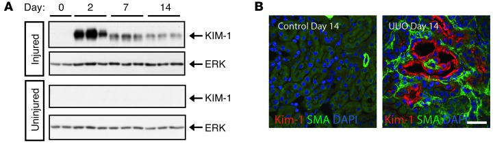
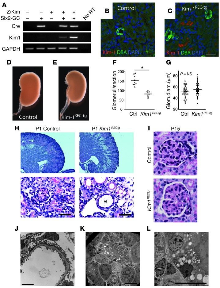
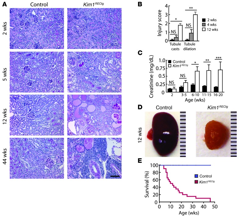
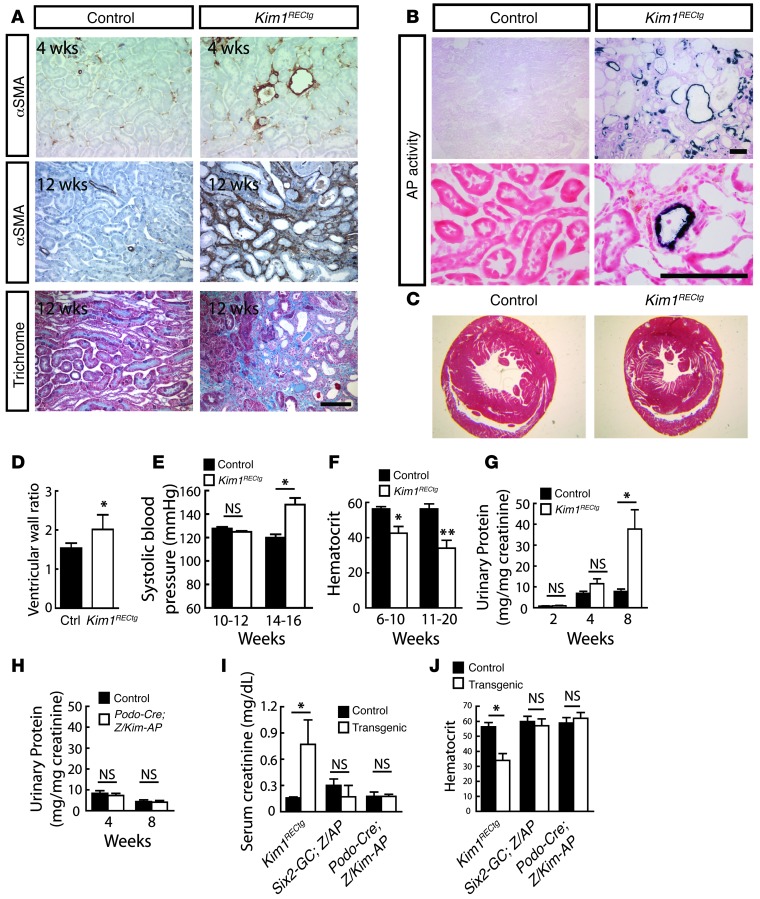
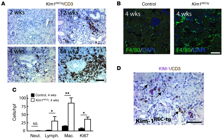
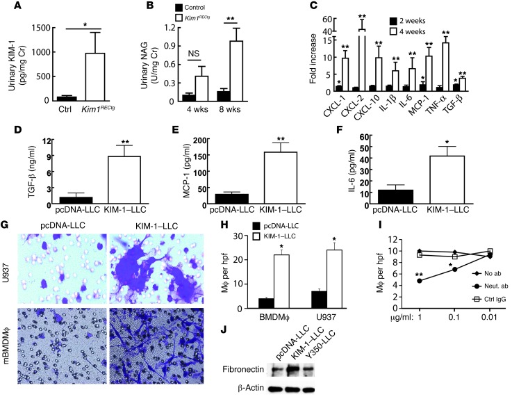
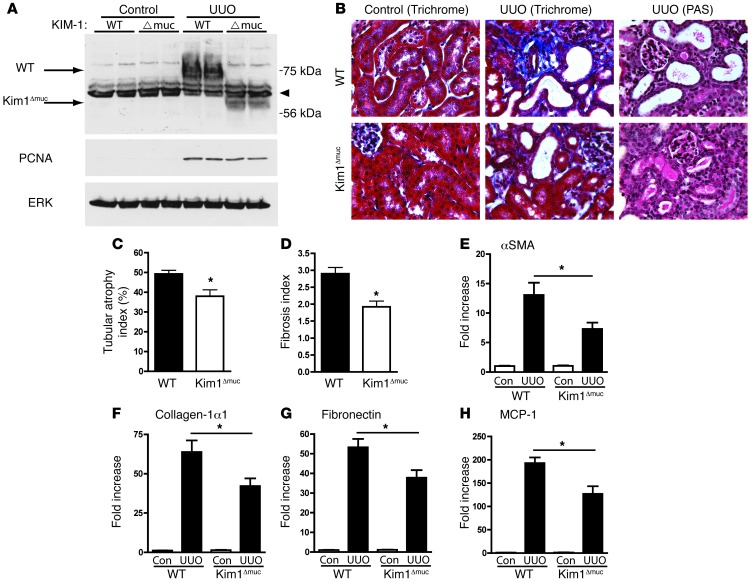
Acute kidney injury predisposes patients to the development of both chronic kidney disease and end-stage renal failure, but the molecular details underlying this important clinical association remain obscure. We report that kidney injury molecule-1 (KIM-1), an epithelial phosphatidylserine receptor expressed transiently after acute injury and chronically in fibrotic renal disease, promotes kidney fibrosis. Conditional expression of KIM-1 in renal epithelial cells (Kim1(RECtg)) in the absence of an injury stimulus resulted in focal epithelial vacuolization at birth, but otherwise normal tubule histology and kidney function. By 4 weeks of age, Kim1(RECtg) mice developed spontaneous and progressive interstitial kidney inflammation with fibrosis, leading to renal failure with anemia, proteinuria, hyperphosphatemia, hypertension, cardiac hypertrophy, and death, analogous to progressive kidney disease in humans. Kim1(RECtg) kidneys had elevated expression of proinflammatory monocyte chemotactic protein-1 (MCP-1) at early time points. Heterologous expression of KIM-1 in an immortalized proximal tubule cell line triggered MCP-1 secretion and increased MCP-1-dependent macrophage chemotaxis. In mice expressing a mutant, truncated KIM-1 polypeptide, experimental kidney fibrosis was ameliorated with reduced levels of MCP-1, consistent with a profibrotic role for native KIM-1. Thus, sustained KIM-1 expression promotes kidney fibrosis and provides a link between acute and recurrent injury with progressive chronic kidney disease.


Figures

Figure 1
Figure 1. KIM-1 is highly induced in fibrotic kidney injury adjacent to interstitial myofibroblasts.
(A) KIM-1 protein is highly induced by 2 days after ureteral ligation, with persistent expression at days 7 and 14 as assessed by Western blot. (B) Tubular KIM-1 expression in the UUO renal fibrosis model. After fibrotic injury, KIM-1–positive epithelia are adjacent to SMA-positive interstitial myofibroblasts. Scale bar: 10 μm.
Figure 2
Figure 2. Generation and initial characterization of Kim1RECtg mice.
(A) Schematic of the Z/Kim1-AP transgene used for generation of transgenic mice. (B) Transient transfection of Cos7 cells with the Z/Kim1-AP (Z/Kim1) plasmid in the absence or presence of a plasmid directing expression of a GFPCre fusion protein verifies that LacZ is expressed before Cre-dependent recombination, and AP after. Original magnification, ×400. (C) KIM-1 protein is detected only in lysates of Cos7 cells cotransfected with Z/Kim1-AP and GFPCre plasmids. (D) Wholemount X-gal stain of a Z/Kim1-AP–positive mouse and littermate control (age 4 weeks) shows mosaic LacZ activity throughout cortex. (E) WT mice express neither LacZ nor AP, whereas Z/Kim1-AP mice (age 4 weeks) exhibit mosaic LacZ expression but no AP expression. Kidney sections from bigenic Kim1RECtg mice show reduced LacZ expression and activation of AP expression in the cortex. Scale bar: 250 μm.
Figure 3
Figure 3. Tubular KIM-1 expression and phenotype of Kim1RECtg kidneys.
(A) Kim1 mRNA is present in P1 kidneys only in bigenic Kim1RECtg mice. (B and C) At P14, kidneys from control mice do not express KIM-1 protein, whereas kidneys from Kim1RECtg mice exhibit appropriate apical expression of KIM-1 in proximal tubules. KIM-1, red; Dolichos biflorus lectin (DBA), green. Scale bar: 20 μm. (D and E) At birth, Kim1RECtg kidneys are smaller in size and 23% smaller in weight compared with those of littermate controls. (F) Kim1RECtg mice (n = 8) had 46% fewer nephrons than controls at P14 (n = 9); *P = 0.0001. (G) Glomerular diameter was not different between controls and Kim1RECtg mice. (H) Low- and high-power views of P1 kidneys from control or Kim1RECtg kidneys reveal thinned cortex and occasional cystic glomerular changes (*) in Kim1RECtg. Scale bar: 25 μm. (I) At P15, glomeruli of control and Kim1RECtg kidneys were similar, without cystic dilation. Original magnification, ×400. (J) Electron microscopy of P14 Kim1RECtg kidneys revealed normal glomerular architecture. Scale bar: 2 μm. In tubules, occasional focal coarse epithelial vacuoles were visible (K and L), suggestive of epithelial injury. Scale bars: 10 μm.
Figure 4
Figure 4. Spontaneous inflammation, tubule injury, renal failure, and death in Kim1RECtg mice.
(A) Periodic acid-schiff stain of control or Kim1RECtg kidneys between 2 and 44 weeks of age reveals no difference in histology at 2 weeks, but progressive interstitial mononuclear cell infiltration, tubule dilation, epithelial simplification and interstitial expansion in Kim1RECtg kidneys only at later time points. Scale bar, 50 μm. (B) Tubule cast and injury scores rise with age in Kim1RECtg kidneys (n = 3) compared with littermate controls (n = 3). *P = 0.001, **P = 0.0009, NS (not significant). (C) Serum creatinine begins to rise between 3 and 5 weeks and rises progressively in Kim1RECtg kidneys (n = 4–8 per time point) compared with controls (n = 5–11 per time point), *P = 0.002, **P = 0.006, ***P = 0.004. (D) A Kim1RECtg kidney is markedly reduced in size and fibrotic at 10 weeks compared with littermate control kidney. (E) Kim1RECtg mice die spontaneously at a median age of 11 weeks (n = 20, both groups), P < 0.0001.
Figure 5
Figure 5. CKD phenotype in Kim1RECtg.
(A) Focal fibrotic changes at 4 weeks in Kim1RECtg that become progressively more severe with time. Scale bar: 50 μm. (B) AP expression identifies cells that have undergone Cre-mediated recombination. No AP-positive cells were found in fibrotic interstitium. Scale bar: 50 μm. (C) Concentric left ventricular hypertrophy in aged Kim1RECtg, trichrome stain. (D) Ventricular wall ratio (outer to inner diameter) was increased in aged (range, 12–45 weeks) Kim1RECtg (n = 5 for each group). *P = 0.03. (E) Younger Kim1RECtg (n = 3) do not have hypertension, but older Kim1RECtg (n = 5) do develop hypertension. *P = 0.0002. (F) Hematocrit in control (n = 10) or Kim1RECtg (n = 5) mice between 6 and 10 weeks or control (n = 13) and Kim1RECtg (n = 7) mice measured between 10 and 20 weeks of age. *P = 0.001; **P = 0.0001. (G) Total urinary protein is elevated in 8-week-old Kim1RECtg (n = 3–5) but not at 2 or 4 weeks compared with littermate controls (n = 4–6). *P = 0.01. (H) Urinary protein is not elevated in mice with expression of KIM-1 in podocytes alone at either 4 or 8 weeks. (I) Serum creatinine 13- to 20-week-old mice comparing control (n = 12) and Kim1RECtg (n = 8), control (n = 6) and Six2-GC;Z/AP (n = 7), or control (n = 4) and Podocin-Cre;Z/Kim1-AP (n = 4). *P = 0.006, NS. (J) Anemia was seen in Kim1RECtg but not control mice or mice in which KIM-1 was expressed in podocytes. Control refers to mice with neither transgene or 1 transgene for all groups. *P < 0.0001, NS.
Figure 6
Figure 6. Leukocyte infiltration in Kim1RECtg.
(A) CD3+ lymphocyte infiltration begins at 4 weeks in Kim1RECtg and is observed in tubulointerstitium, especially surrounding vessels in older animals. Scale bar: 50 μm. (B) F4/80-positive peritubular macrophages and dendritic cells are increased at 4 weeks in Kim1RECtg. Scale bar: 50 μm. (C) Quantitation of infiltrating leukocytes and Ki67+ proliferating cells at 4 weeks in Kim1RECtg compared with control kidneys (n = 3 for each). *P < 0.0001; **P = 0.0003. Neut., neutrophil; Lymph., lymphocyte; Mac., macrophage. (D) CD3-positive lymphocytes (brown) are frequently identified adjacent to KIM-1–positive (purple, *) tubules. Scale bar: 25 μm.
Figure 7
Figure 7. Persistent KIM-1 expression induces epithelial damage and proinflammatory cytokines in kidney epithelia in vivo and in vitro.
(A) Shed KIM-1 is detectable in urine of Kim1RECtg at 4 weeks of age compared with controls. *P = 0.03. (B) Evidence of tubular injury in Kim1RECtg is also reflected by increasing urinary NAG in 4- and 8-week-old Kim1RECtg. *P < 0.01. (C) qPCR of kidney cortex cytokines, presented as fold increase in Kim1RECtg (n = 3) compared with controls (n = 3) at 2 weeks (black bars) or 4 weeks of age (white bars). At 2 weeks, there is mild induction of CXCL-1, MCP-1, and TGF-β, with much higher levels of mRNA expression for all soluble cytokines by 4 weeks of age. *P < 0.05; **P < 0.006. (DF) Porcine proximal tubule epithelial cells stably transfected with either pcDNA3 control plasmid (pcDNA-LLC) or KIM-1 plasmid (KIM-1–LLC) spontaneously express TGF-β, MCP-1, and IL-6 (n = 3 for each). *P < 0.05; **P < 0.01. (G and H) Boyden chamber assay with pcDNA-LLC or KIM-1–LLC seeded on the bottom well and either U937 or mBMDM applied to top filter. Migration was measured after 3 hours. *P < 0.05. Original magnification, ×500. (I) Neutralizing antibody against MCP-1 abrogated KIM-1–LLC–dependent migration but not control IgG. *P < 0.05; **P < 0.01. (J) Cellular lysates from pcDNA-LLC, KIM-1–LLC, or LLC-PK1 cells transfected with KIM-1–Y350F (Y350-LLC) probed for fibronectin by Western blot demonstrate strong induction of fibronectin in KIM-1, but not Y350F–KIM-1–transfected cells.
Figure 8
Figure 8. The functional mutant Kim1Δmuc is protected from kidney fibrosis.
(A) Control and Kim1Δmuc mice were subjected to UUO and sacrificed at day 2. Kidney lysates reveal KIM-1 protein at 75 kDa in the control, but at 55 kDa in the Kim1Δmuc, corresponding to the deletion of exon 3 (arrows). The arrowhead identifies a nonspecific Ig band. Proliferating cell nuclear antigen (PCNA) staining reflects increased cell proliferation after UUO. (B) Control or Kim1Δmuc kidney sections before or after UUO. There is reduced interstitial collagen in Kim1Δmuc reflected by Masson’s trichrome stain, and reduced tubular injury (PAS). (C and D) Quantification of tubular atrophy and fibrosis index, respectively (n = 5 kidneys each condition). (EG) qPCR of kidney cortex fibrosis, presented as fold increase of Kim1RECtg (n = 5) compared with control (n = 5) at 10 days after UUO. *P < 0.05. (H) Reduced MCP-1 mRNA by qPCR in Kim1Δmuc compared with control (n = 5). *P < 0.05.

Comment in

References

    1. Clarkson MR, Friedewald JJ, Eustace H, Rabb H. Brenner and Rector’s The Kidney. 2008. Acute kidney injury. In: Brenner BM, ed. pp. 943–986. 8th ed. Philadelphia, Pennsylvania, USA: Saunders Elsevier;
    1. Ishani A, et al. Acute kidney injury increases risk of ESRD among elderly. J Am Soc Nephrol. 2009;20(1):223–228. doi: 10.1681/ASN.2007080837. - DOI - PMC - PubMed
    1. Wald R, et al. Chronic dialysis and death among survivors of acute kidney injury requiring dialysis. JAMA. 2009;302(11):1179–1185. doi: 10.1001/jama.2009.1322. - DOI - PubMed
    1. Lafrance JP, Miller DR. Acute kidney injury associates with increased long-term mortality. J Am Soc Nephrol. 2010;21(2):345–352. doi: 10.1681/ASN.2009060636. - DOI - PMC - PubMed
    1. Hsu CY, McCulloch CE, Fan D, Ordoñez JD, Chertow GM, Go AS. Community-based incidence of acute renal failure. Kidney Int. 2007;72(2):208–212. doi: 10.1038/sj.ki.5002297. - DOI - PMC - PubMed

Publication types

MeSH terms