. 2014 Aug;66(4):687-97.
doi: 10.1007/s10616-013-9622-y. Epub 2013 Aug 27.

Establishment of a pancreatic β cell proliferation model in vitro and a platform for diabetes drug screening

Affiliations

Establishment of a pancreatic β cell proliferation model in vitro and a platform for diabetes drug screening

Jing Jia et al. Cytotechnology. 2014 Aug.

Abstract

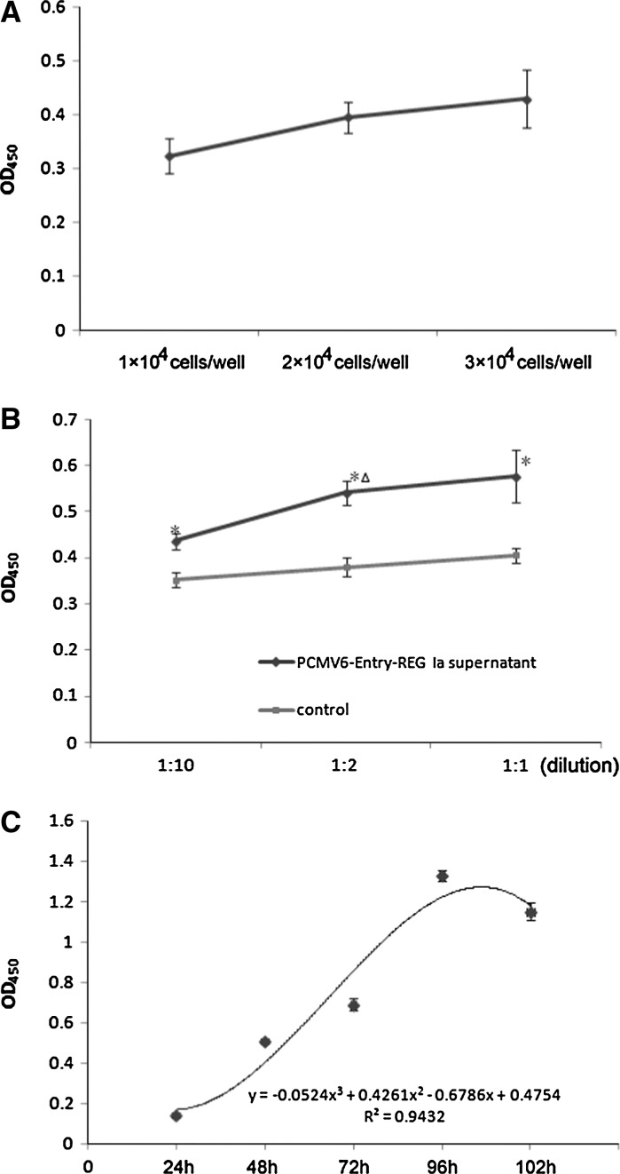
Diabetes, a disease resulting from loss of functional β cells, is globally an increasingly important condition. Based on the islet-differentiation ability of ductal epithelial cells and stimulating β cell proliferation ability of the Reg Iα gene, we aimed to establish an in vitro pancreatic β cell proliferation model for screening therapeutic drugs of diabetes in the future. Pancreatic ductal epithelial cells were isolated from male Wistar rats, and induced to differentiate into pancreatic β cells. Immunofluorescence staining assay, western blot, RT-PCR analysis, and dithizone staining were used to characterize the cells. Rat Reg Iα protein was transiently expressed in vitro by transfection of HEK 293 cells with the PCMV6-entry-REG Ia plasmid, and expression was verified by RT-PCR analysis, proliferation assay, and apoptosis assay. The pancreatic β cell proliferation model was further validated by a proliferation assay using differentiated pancreatic β cells treated with transfection supernatant. Finally, we have successfully established an in vitro pancreatic β cells proliferation model using transiently expressed rat Reg Iα protein and differentiated pancreatic β cells from pancreatic ductal epithelial cells. This model could be used as a platform to screen new drugs for islet neogenesis to cure diabetes, especially Chinese herbal drugs in the future.


Figures

Fig. 1
Fig. 1
Pancreatic ductal epithelial cells were cultured until 80–90 % confluence (A, bar = 50 μm, magnification 100 x). For validation, CK19 was stained green and detected by immunofluorescence staining assay (B, bar = 50 μm, magnification 200 x), and the protein levels of CK19 and pan-cytokeratin were evaluated by Western blot assay (C). mRNA levels of PDX-1 and beta actin were detected in these cells by RT-PCR analysis, while mRNAs of Reg Iα and insulin were not detected (D)
Fig. 2
Fig. 2
The in vitro cultured pancreatic ductal epithelial cells were induced to differentiate into pancreatic β cells. The cells gradually became round and detached from the culture surface (A, bar = 20 μm, magnification 100x). After 6 weeks, the differentiated β cells were stained red by dithizone (DTZ) staining (B, bar = 20 μm, magnification 400x), and the insulin protein was detected by immunofluorescence staining assay (C, bar = 20 μm, magnification 100x) and Western blot assay (D) in these differentiated pancreatic β cells
Fig. 3
Fig. 3
The transient expression of Reg Ia mRNA was detected by RT-PCR assay (A). The biological activity of Reg Ia protein was assessed by cell proliferation assay using CFSE (B) and apoptosis assay (C) by double staining of annexin V and PI, with PCMV6-Entry transfection supernatant as control. After 96 h, the cell proliferation indexes (14.01 vs 11.83), division peaks (7 vs 5), and the percentages of cells reached generation 6th (23.87 vs 12.39 %) were measured and compared. The apoptosis ratios (bottom right: 10.45 vs 13.60 %), necrosis ratios (top right 12.17 vs 23.30 %), and viable cells ratios (bottom left 76.14 vs 58.13 %) were also measured and compared
Fig. 4
Fig. 4
Establishment of pancreatic β cell proliferation model. A Cell proliferation assay by the WST-1 method was used to select seeded cell density from 1 x 10⁴ cells/well, 2 x 10⁴ cells/well, 3 x 10⁴ cells/well. 48 h later, OD450 values of the three groups were tested (0.324 ± 0.033, 0.40 ± 0.029, 0.43 ± 0.053, respectively). B Cell proliferation assay by the WST-1 method was used to select the dilution of the transfection supernatants (1:10, 1:2, and 1:1). 48 h later, OD450 values were measured and compared between the groups (0.44 ± 0.018 (1:10), 0.54 ± 0.026 (1:2), 0.58 ± 0.056 (1:1) versus controls: 0.35 ± 0.016 (1:10), 0.38 ± 0.021 (1:2), 0.41 ± 0.017 (1:1)). *P < 0.05 versus control. Δ P < 0.05 versus 1:10 group. C Cell proliferation assay by the WST-1 method was used to establish the growth curve of pancreatic β cells under the effect of Reg Iα. Differentiated pancreatic β cells (1 x 10⁴ cells/well) were incubated with transfection supernatants (1:2 diluted) in 96 well plates for 102 h. The number of viable cells was represented by OD450 values measureded every 24 h (0.14 ± 0.015 (24 h), 0.51 ± 0.013 (48 h), 0.69 ± 0.031 (72 h), 1.33 ± 0.026 (96 h), 1.15 ± 0.042 (102 h))

Similar articles

Cited by

References

    1. Agrawal R, Sethiya NK, Mishra SH. Antidiabetic activity of alkaloids of Aerva lanata roots on streptozotocin-nicotinamide induced type-II diabetes in rats. Pharm Biol. 2013;51:635–642. doi: 10.3109/13880209.2012.761244. - DOI - PubMed
    1. Akiyama T, Takasawa S, Nata K, Kobayashi S, Abe M, Shervani NJ, Ikeda T, Nakagawa K, Unno M, Matsuno S, Okamoto H. Activation of Reg gene, a gene for insulin-producing beta-cell regeneration: poly(ADP-ribose) polymerase binds Reg promoter and regulates the transcription by autopoly(ADP-ribosyl)ation. Proc Natl Acad Sci USA. 2001;98:48–53. - PMC - PubMed
    1. Bonner-Weir S, Taneja M, Weir GC, Tatarkiewicz K, Song KH, Sharma A, O’Neil JJ. In vitro cultivation of human islets from expanded ductal tissue. Proc Natl Acad Sci USA. 2000;97:7999–8004. doi: 10.1073/pnas.97.14.7999. - DOI - PMC - PubMed
    1. Bonner-Weir S, Toschi E, Inada A, Reitz P, Fonseca SY, Aye T, Sharma A. The pancreatic ductal epithelium serves as a potential pool of progenitor cells. Pediatr Diabetes. 2004;5:16–22. doi: 10.1111/j.1399-543X.2004.00075.x. - DOI - PubMed
    1. Bretzel RG, Hering BJ, Federlin KF. Islet cell transplantation in diabetes mellitus—from bench to bedside. Exp Clin Endocrinol Diabetes. 1995;103:143–159. doi: 10.1055/s-0029-1211413. - DOI - PubMed

LinkOut - more resources