. 2013 Nov;33(21):4140-51.
doi: 10.1128/MCB.00410-13. Epub 2013 Aug 26.

Multipoint binding of the SLP-76 SH2 domain to ADAP is critical for oligomerization of SLP-76 signaling complexes in stimulated T cells

Affiliations

Multipoint binding of the SLP-76 SH2 domain to ADAP is critical for oligomerization of SLP-76 signaling complexes in stimulated T cells

Nathan P Coussens et al. Mol Cell Biol. 2013 Nov.

Abstract

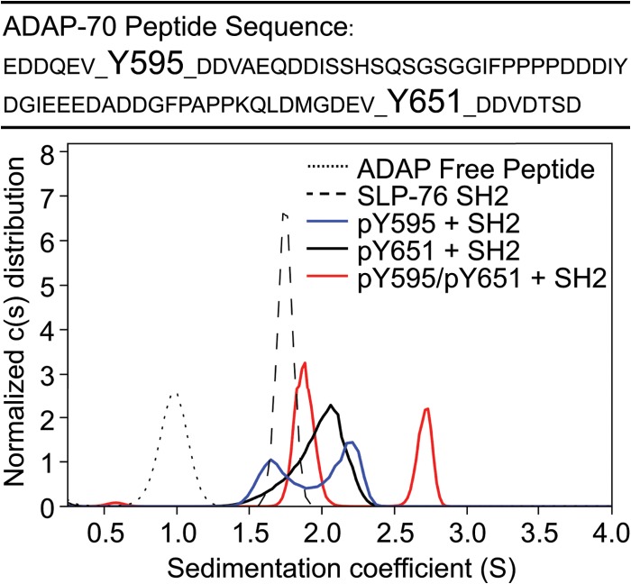
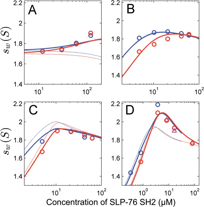
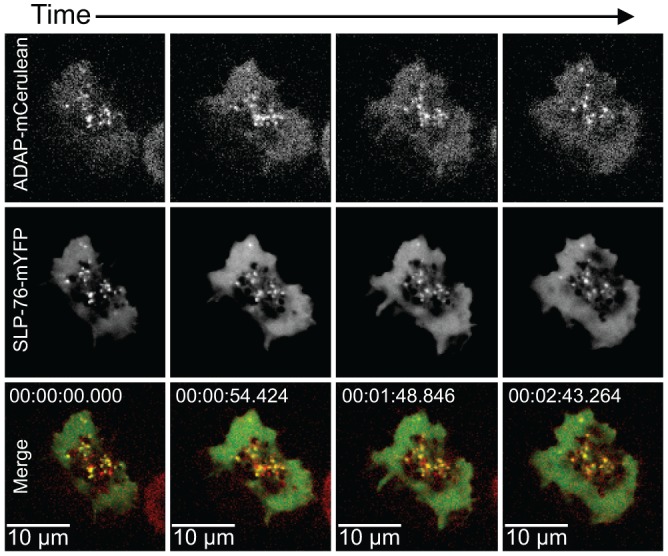
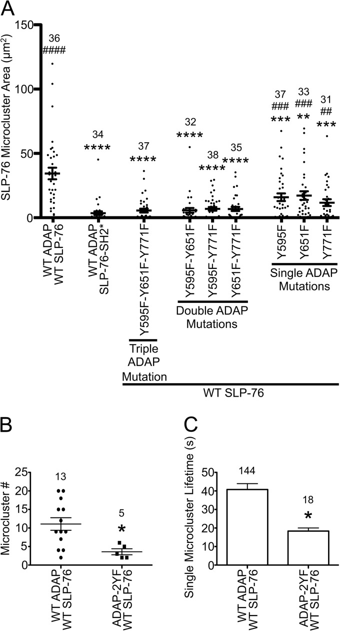
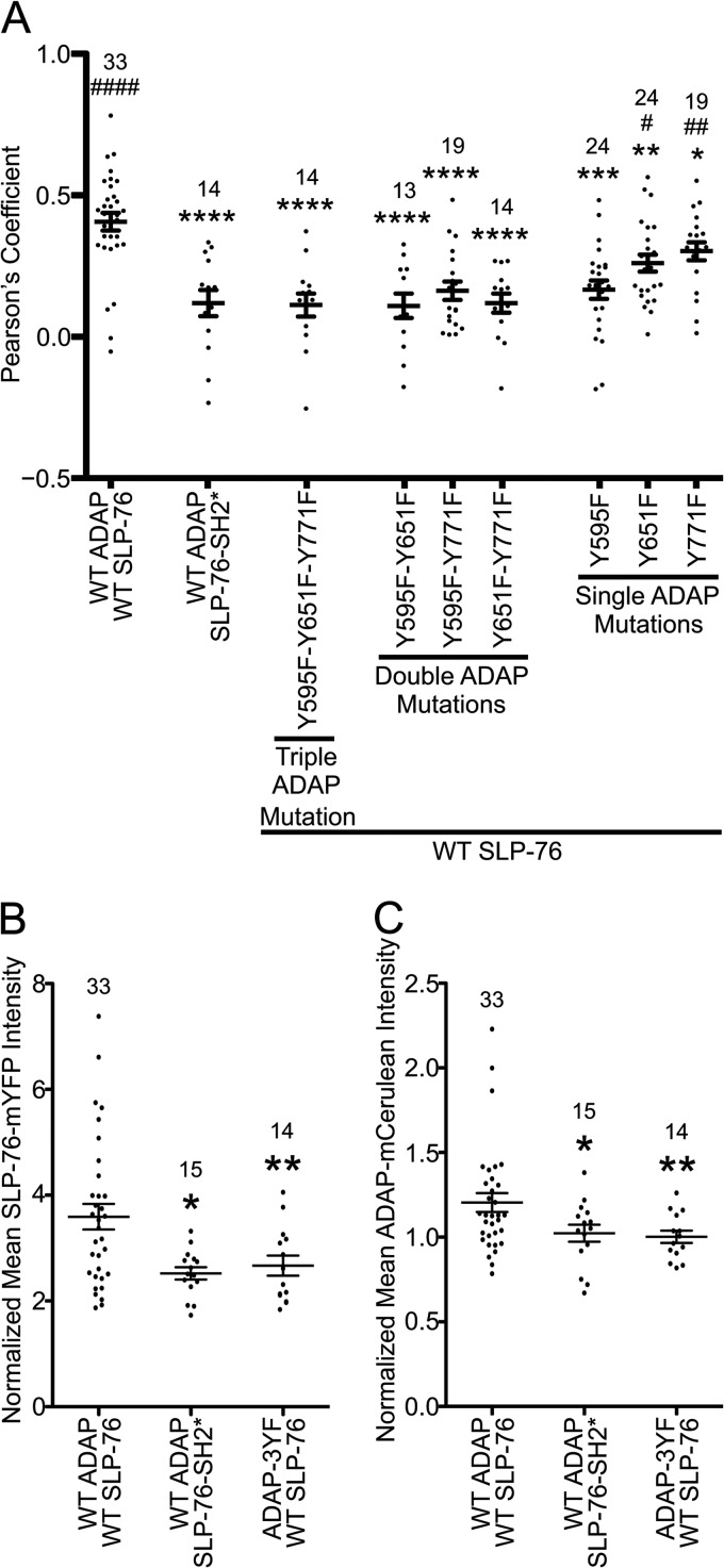
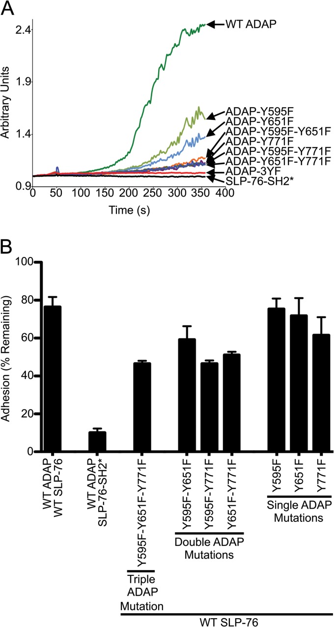
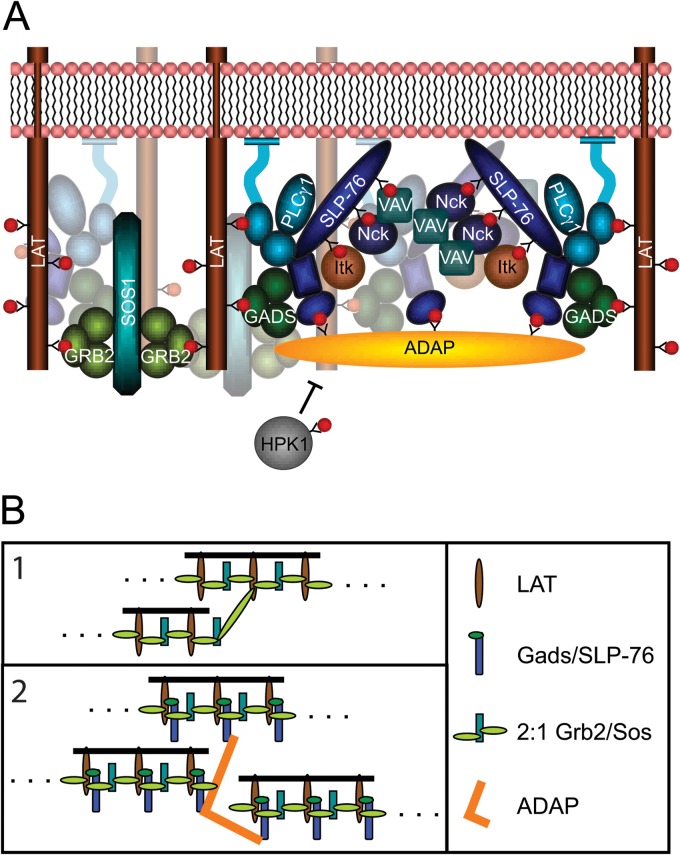
The adapter molecules SLP-76 and LAT play central roles in T cell activation by recruiting enzymes and other adapters into multiprotein complexes that coordinate highly regulated signal transduction pathways. While many of the associated proteins have been characterized, less is known concerning the mechanisms of assembly for these dynamic and potentially heterogeneous signaling complexes. Following T cell receptor (TCR) stimulation, SLP-76 is found in structures called microclusters, which contain many signaling complexes. Previous studies showed that a mutation to the SLP-76 C-terminal SH2 domain nearly abolished SLP-76 microclusters, suggesting that the SH2 domain facilitates incorporation of signaling complexes into microclusters. S. C. Bunnell, A. L. Singer, D. I. Hong, B. H. Jacque, M. S. Jordan, M. C. Seminario, V. A. Barr, G. A. Koretzky, and L. E. Samelson, Mol. Cell. Biol., 26:7155-7166, 2006). Using biophysical methods, we demonstrate that the adapter, ADAP, contains three binding sites for SLP-76, and that multipoint binding to ADAP fragments oligomerizes the SLP-76 SH2 domain in vitro. These results were complemented with confocal imaging and functional studies of cells expressing ADAP with various mutations. Our results demonstrate that all three binding sites are critical for SLP-76 microcluster assembly, but any combination of two sites will partially induce microclusters. These data support a model whereby multipoint binding of SLP-76 to ADAP facilitates the assembly of SLP-76 microclusters. This model has implications for the regulation of SLP-76 and LAT microclusters and, as a result, T cell signaling.


Figures

Fig 1
Fig 1
Sedimentation coefficient distributions of 30 μM equimolar mixtures of SLP-76 SH2 with phosphorylated and nonphosphorylated 70-amino-acid ADAP fragments (solid lines) or individual components (broken lines). The primary sequence of the 70-amino-acid peptides is shown at the top, with the positions of Y595 and Y651 indicated.
Fig 2
Fig 2
Global analysis of weighted-average sedimentation coefficients (sw) as a function of SLP-76 SH2 concentration in mixtures with ADAP peptides. Symbols indicate experimentally determined sw values, with red circles for interference optical detection and triangles and blue circles for absorbance detection. Lines represent different best-fit models for the experimental data as described below. (A) Mixtures of nonphosphorylated ADAP-70 peptide (at concentrations ranging from 2.5 to 25 μM) and a 5-fold molar excess of SLP-76 SH2. Thick lines represent a model allowing for binding to both nonphosphorylated sites, while the thin lines represent a model lacking binding to the nonphosphorylated sites (leading to a 1.6-fold increase in the RMSD). (B to D) A global analysis of the sw data for all singly and doubly phosphorylated peptides. This model assumes that the affinity of SLP-76 SH2 to each site is equivalent among different peptides and only dependent on the phosphorylation state. The affinity of SLP-76 SH2 to pY651, as determined by ITC, was used as a constraint in this model. Thick lines are the global best fit to a model accounting for weak binding to nonphosphorylated sites and allowing for simultaneous binding to both phosphorylated sites in ADAP-70-pY595-pY651. The thin lines are the global best fit to a model lacking the potential for simultaneous occupancy of both sites (leading to a 2.1-fold increase in RMSD of the fit). (B) ADAP-70-pY595 at 9.1 μM (circles). The thick and thin lines superimpose exactly, so the thin lines are not visible (indicating a similar fit to both models). (C) ADAP-70-pY651 at 10 μM (circles). (D) ADAP-70-pY595-pY651 at 2.3 μM (circles and triangles).
Fig 3
Fig 3
SLP-76 and ADAP translocate together in punctae. SLP-76-deficient Jurkat T cells (J14) expressing wild-type SLP-76-mYFP and wild-type ADAP-mCerulean proteins were plated onto coverslips coated with stimulatory antibodies and imaged with a spinning-disk confocal system. Live-cell imaging began shortly after the cell contacted the coverslip, and the first image was defined as t = 0. The images are maximum-intensity projections of z-stacks (four sections 0.5 μm apart) of selected time points. For each time point, the ADAP-mCerulean fluorescence signal is shown at the top and the SLP-76-mYFP fluorescence signal is shown in the center. In the merged images shown at the bottom, comigration (shown in yellow) of SLP-76 (shown in green) and ADAP (shown in red) is apparent.
Fig 4
Fig 4
Assembly and persistence of SLP-76 microclusters requires multiple ADAP binding sites. (A) Area of TCR-induced SLP-76 microclusters in stable cell lines. The indicated cell lines were plated on stimulatory coverslips, fixed after incubation at 37°C for 3 min, and imaged with a spinning-disk confocal system. Single z-slices with the highest intensity punctae at the cell surface were chosen, and the total area of SLP-76 microclusters in each cell was calculated using Imaris software and plotted. Means are presented along with error bars representing ±SEM. Significant deviations from the values of cells expressing wild-type proteins or SLP-76-SH2* are indicated, respectively, as follows: * or #, P = 0.01 to 0.05; ** or ##, P = 0.001 to 0.01; *** or ###, P = 0.0001 to 0.001; **** or ####, P < 0.0001. The number of cells analyzed for each sample is shown above the symbols. (B and C) Analysis of SLP-76 microclusters from live-cell imaging. SLP-76-deficient Jurkat T cells (J14) expressing wild-type SLP-76-mYFP and either wild-type ADAP-mCerulean or ADAP-Y595F-Y651F-mCerulean proteins were imaged with a spinning-disk confocal system. Live-cell imaging began shortly after the cell contacted the coverslip. Movies were prepared with Slidebook software from z-stacks by making a maximum-intensity projection of each time point and assembling a sequence of all projections. Microcluster lifetimes were determined using particle tracking from Slidebook software. Mutations to Y595 and Y651 reduce the number (B) and lifetime (C) of SLP-76 microclusters in live cells.
Fig 5
Fig 5
Altered molecular composition of SLP-76 microclusters with a SLP-76 SH2 mutation or ADAP Y-F mutations. The single z-slices selected to determine the area of SLP-76 microclusters shown in Fig. 4A were further analyzed. (A) Colocalization between SLP-76 and ADAP was determined by Imaris software as described in Materials and Methods. Wild-type SLP-76 and ADAP proteins colocalize in the SLP-76 punctae, but mutations to either SLP-76 or ADAP reduce the Pearson's coefficient between the two proteins. Means are presented along with error bars representing ±SEM. Significant deviations from the values of cells expressing wild-type proteins or SLP-76-SH2* are indicated, respectively, as follows: * or #, P = 0.01 to 0.05; ** or ##, P = 0.001 to 0.01; *** or ###, P = 0.0001 to 0.001; **** or ####, P < 0.0001. The number of cells analyzed for each sample is shown above the symbols. (B and C) Recruitment of SLP-76-mYFP and ADAP-mCerulean into SLP-76 punctae. The average fluorescence of wild-type or mutated SLP-76-mYFP and ADAP-mCerulean in punctae was calculated and normalized to the average fluorescence in a cytoplasmic region of the same z-slice. Mutations to either SLP-76 or ADAP reduce the amount of SLP-76 (B) and ADAP (C) recruited into punctae.
Fig 6
Fig 6
Mutations to the SLP-76 SH2 domain or the three ADAP binding sites cause defects in calcium signaling and adhesion. (A) Calcium signaling is reduced in cell lines expressing SLP-76 or ADAP mutations. SLP-76-deficient Jurkat T cell lines (J14) stably expressing wild-type or mutated versions of SLP-76 and ADAP (as indicated) were stimulated with anti-CD3 (OKT3), and the Ca2+ influx was measured as described in Materials and Methods. (B) The percentage of cells retained in the wells of 96-well plates coated with anti-CD3 (OKT3) and recombinant human VCAM-1 is shown. The percentage of cells remaining was determined after high shear stress was applied. Averages from 8 replicates were calculated from each experiment, and the means ± SEM from two independent experiments are shown.
Fig 7
Fig 7
(A) Multipoint binding of SLP-76 to ADAP induces oligomerization of SLP-76 upon TCR stimulation. The binding of ADAP favors SLP-76 clustering and interferes with the binding of HPK1. (B) Separate models for oligomerization of LAT and SLP-76/LAT complexes. (1) Interactions between bivalent LAT and Grb2/Sos complexes can induce oligomerization of LAT into chains. However, if LAT binds 3 Grb2/Sos complexes, the oligomerization potential increases. (Adapted from reference with permission.) (2) Association of the Gads/SLP-76 complex with LAT reduces the number of Grb2 binding sites to two, allowing only the assembly of chains. Multipoint binding of SLP-76 to ADAP oligomerizes SLP-76 and increases the oligomerization potential of LAT complexes.

Similar articles

Cited by

References

    1. Balagopalan L, Coussens NP, Sherman E, Samelson LE, Sommers CL. 2010. The LAT story: a tale of cooperativity, coordination, and choreography. Cold Spring Harb. Perspect. Biol. 2:a005512.10.1101/cshperspect.a005512 - DOI - PMC - PubMed
    1. Bartelt RR, Houtman JC. 2013. The adaptor protein LAT serves as an integration node for signaling pathways that drive T cell activation. Wiley Interdiscip. Rev. Syst. Biol. Med. 5:101–110 - PMC - PubMed
    1. Barda-Saad M, Shirasu N, Pauker MH, Hassan N, Perl O, Balbo A, Yamaguchi H, Houtman JC, Appella E, Schuck P, Samelson LE. 2010. Cooperative interactions at the SLP-76 complex are critical for actin polymerization. EMBO J. 29:2315–2328 - PMC - PubMed
    1. Sangani D, Venien-Bryan C, Harder T. 2009. Phosphotyrosine-dependent in vitro reconstitution of recombinant LAT-nucleated multiprotein signalling complexes on liposomes. Mol. Membr. Biol. 26:159–170 - PubMed
    1. Houtman JC, Higashimoto Y, Dimasi N, Cho S, Yamaguchi H, Bowden B, Regan C, Malchiodi EL, Mariuzza R, Schuck P, Appella E, Samelson LE. 2004. Binding specificity of multiprotein signaling complexes is determined by both cooperative interactions and affinity preferences. Biochemistry 43:4170–4178 - PubMed

Publication types

MeSH terms