

Circulating acylcarnitines as biomarkers of mitochondrial dysfunction after acetaminophen overdose in mice and humans
- PMID: 23979652
- PMCID: PMC3946727
- DOI: 10.1007/s00204-013-1118-1
Circulating acylcarnitines as biomarkers of mitochondrial dysfunction after acetaminophen overdose in mice and humans
Abstract
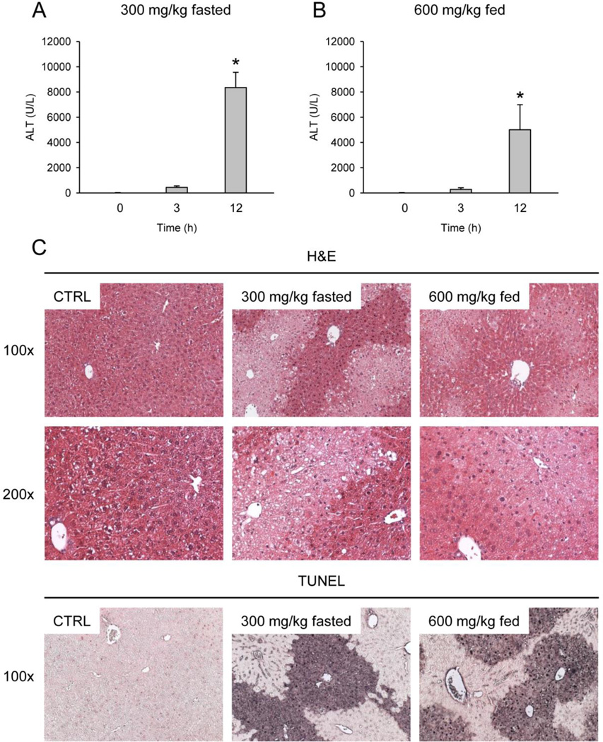
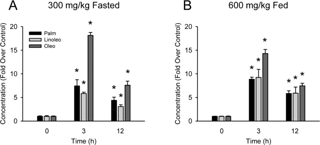
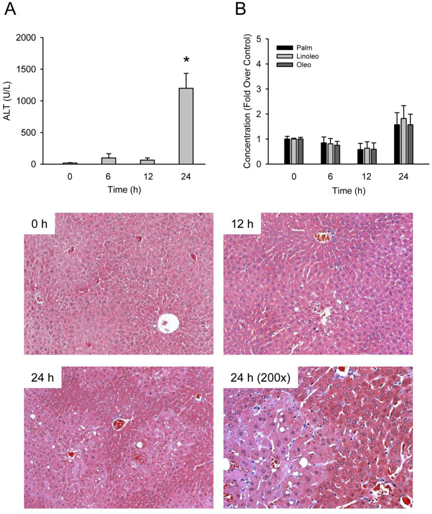
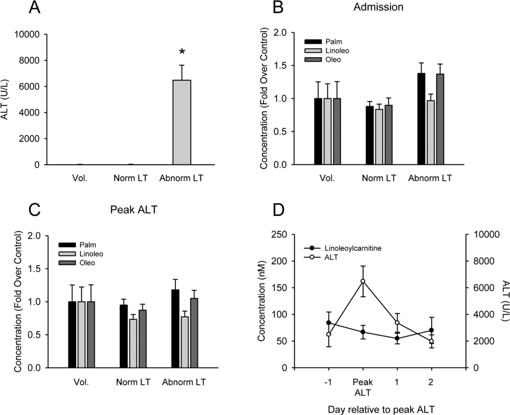
Acetaminophen (APAP) is a widely used analgesic. However, APAP overdose is hepatotoxic and is the primary cause of acute liver failure in the developed world. The mechanism of APAP-induced liver injury begins with protein binding and involves mitochondrial dysfunction and oxidative stress. Recent efforts to discover blood biomarkers of mitochondrial damage have identified increased plasma glutamate dehydrogenase activity and mitochondrial DNA concentration in APAP overdose patients. However, a problem with these markers is that they are too large to be released from cells without cell death or loss of membrane integrity. Metabolomic studies are more likely to reveal biomarkers that are useful at early time points, before injury begins. Similar to earlier work, our metabolomic studies revealed that acylcarnitines are elevated in serum from mice after treatment with toxic doses of APAP. Importantly, a comparison with furosemide demonstrated that increased serum acylcarnitines are specific for mitochondrial dysfunction. However, when we measured these compounds in plasma from humans with liver injury after APAP overdose, we could not detect any significant differences from control groups. Further experiments with mice showed that N-acetylcysteine, the antidote for APAP overdose in humans, can reduce acylcarnitine levels in serum. Altogether, our data do not support the clinical measurement of acylcarnitines in blood after APAP overdose due to the standard N-acetylcysteine treatment in patients, but strongly suggest that acylcarnitines would be useful mechanistic biomarkers in other forms of liver injury involving mitochondrial dysfunction.
Conflict of interest statement
CONFLICT OF INTEREST DISCLOSURE
The authors declare no competing financial interest.
Figures


References
-
- Antoine DJ, Dear JW, Starkey-Lewis P, Platt V, Coyle J, Masson M, Thanacoody RH, Gray AJ, Webb DJ, Moggs JG, Bateman DN, Goldring CE, Park BK. Mechanistic biomarkers provide early and sensitive detection of acetaminophen-induced acute liver injury at first presentation to hospital. Hepatology. 2013;58:777–787. - PMC - PubMed
-
- Bajt ML, Cover C, Lemasters JJ, Jaeschke H. Nuclear translocation of endonuclease G and apoptotis-inducing factor during acetaminophen-induced liver cell injury. Toxicol Sci. 2006;94:217–225. - PubMed
-
- Bajt ML, Farhood A, Lemasters JJ, Jaeschke H. Mitochondrial bax translocation accelerates DNA fragmentation and cell necrosis in a murine model of acetaminophen hepatotoxicity. J Pharmacol Exp Ther. 2008;324:8–14. - PubMed
-
- Bajt ML, Knight TR, Farhood A, Jaeschke H. Scavenging peroxynitrite with glutathione promotes regeneration and enhances survival during acetaminophen-induced liver injury in mice. J Pharmacol Exp Ther. 2003;307:67–73. - PubMed
-
- Bajt ML, Knight TR, Lemasters JJ, Jaeschke H. Acetaminophen-induced oxidant stress and cell injury in cultured mouse hepatotocytes: protection by N-acetylcysteine. Toxicol Sci. 2004;80:343–349. - PubMed
Publication types
MeSH terms
Substances
Grants and funding
LinkOut - more resources
Full Text Sources
Other Literature Sources
Medical
