

doi: 10.1534/g3.113.007310.
Characterization of two ENU-induced mutations affecting mouse skeletal morphology
Affiliations
- PMID: 23979929
- PMCID: PMC3789799
- DOI: 10.1534/g3.113.007310
Item in Clipboard
Characterization of two ENU-induced mutations affecting mouse skeletal morphology
G3 (Bethesda).
.
Abstract
Using the N-ethyl-N-nitrosourea (ENU) mutagenesis screen, we have identified two skeletal morphology mutants, Skm1 and Skm2. Positional cloning and candidate gene sequencing localized the causative point mutations within the genes coding for natriuretic peptide receptor C (NPR-C) and filamin b (FLNB), respectively. Mice that carry a mutation in Npr3 exhibit a skeletal overgrowth phenotype, resulting in an elongated body and kyphosis. Skm2 mice, carrying a mutation in Flnb, present with scoliosis and lordosis. These mutant mice will serve as useful models for the study of vertebral malformations.
Figures
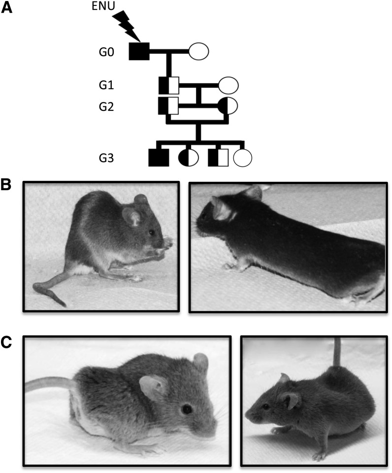
N-ethyl-N-nitrosourea (ENU) breeding scheme and visible morphology of skeletal morphology mutants (Skm). (A) Schematic representation of the breeding scheme used during the identification of deviant pedigrees. 129S1 male G0 mutagenized mice were crossed to 129X1 females and the resulting G1 males were outcrossed to DBA/2J females to generate G2 progeny. Visible skeletal malformations in Skm1 (B) mice and Skm2 (C) mice. (B) Skm1 mice show kyphotic spines and kinked tails (left) and elongated bodies (right). (C) Skm2 mice exhibit severe scoliosis and lordosis.
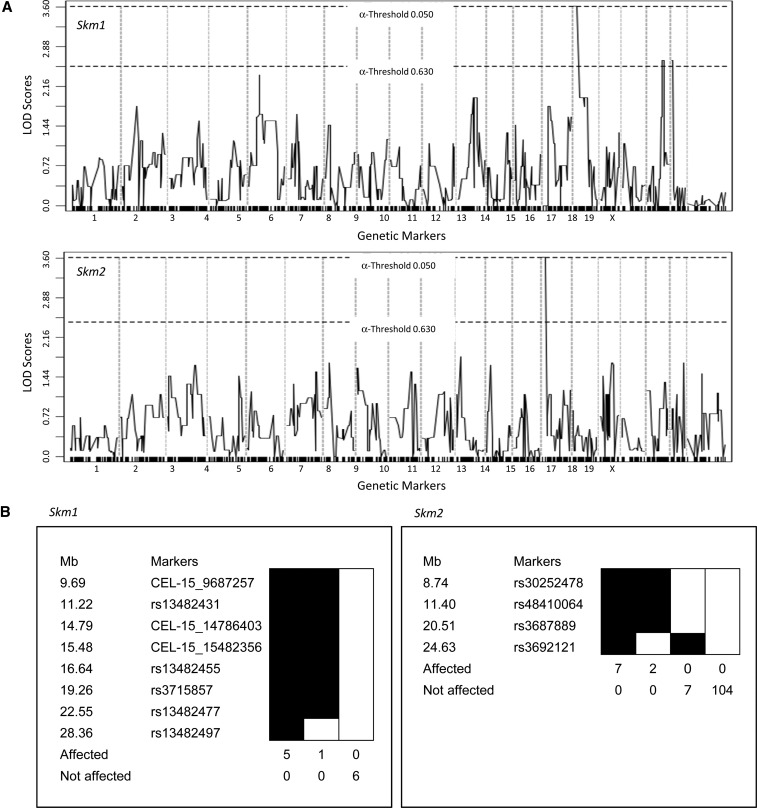
Mapping of the mutant pedigrees. (A) Genome-wide linkage analysis of the visible mutants (Skm1, left panel; Skm2, right panel) was conducted with 12 animals (6 mutant and 6 normal) using polymorphic markers informative for the 129S1 and DBA/2J parents. LOD scores above the threshold line were considered significant. (B) Haplotype analysis of the proximal region of chromosome 15 for Skm1 (left panel) and of chromosome 14 for Skm2 (right panel). Positions of the markers (Mb) are relative to the centromere.
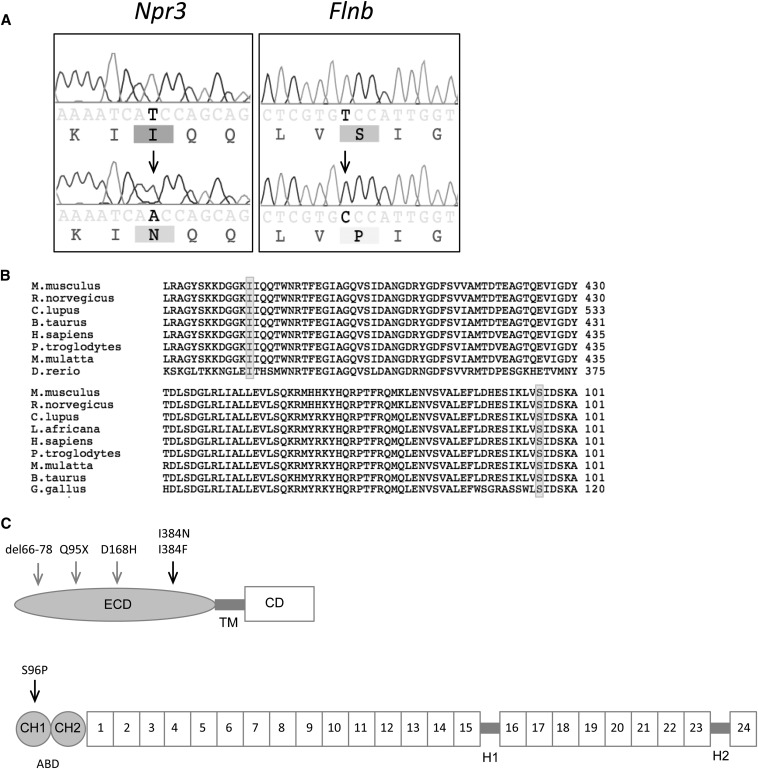
Identification of mutations underlying skeletal morphology mutant 1 (Skm1) and skeletal morphology mutant 2 (Skm2). (A) Genomic DNA sequence chromatograms from normal (upper) and mutant (lower) Skm1 and Skm2 mice (left panel and right panel, respectively). The location of the mutation is indicated by the arrowhead. (B) Alignment of the amino acid sequence for NPR3 (top) and FLNB (bottom) orthologs. The location of the mutation is indicated by shading. (C) Schematic representation of NPR3 (top) and FLNB (bottom).The arrows indicate location of Skm mutations (black) and other reported mutations (gray). NPR3 contains an extracellular domain (ECD), transmembrane (TM) domain, and cytoplasmic domain (CD). FLNB comprises an actin-binding domain (ABD) containing two calponin homology (CH) domains at the amino terminus, followed by 24 filamin repeat domains that are separated by two hinge (1H-2) regions.
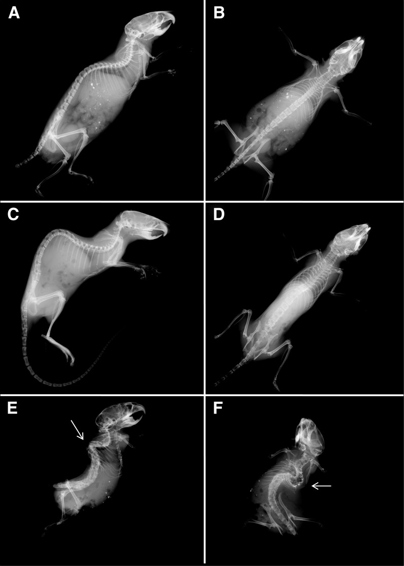
Skeletal abnormalities in skeletal morphology mutant (Skm) mice. Radiographs of normal (A, B), Skm1 (C, D), and Skm2 (E, F) mice in lateral and supine positions. Skm1 mice in the lateral position showing kyphosis (C) and in the supine position showing elongated body (D). Skm2 mice in the supine (E) and lateral (F) positions. Scoliotic and lordotic curvatures of the vertebral columns are indicated by arrowhead relative to wild-type littermates.
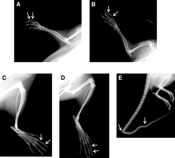
Arachnodactyly and kinked tail in skeletal morphology mutant 1 (Skm1). Radiographs of normal (A–C) and Skm1 (B–D) manus (A, B) and pes (C, D). Phalanges are indicated by arrowheads. (E) Radiograph of the tail of Skm1. The hemivertebrae and fused vertebrae are indicated by arrowheads.
References
-
- Hensinger R. N., 2009. Congenital scoliosis: etiology and associations. Spine 34: 1745–1750 - PubMed
-
- Kesling K.L., Reinker K.A. 1997. Scoliosis in twins. A meta-analysis of the literature and report of six cases. Spine 22: 2009–2015. - PubMed
-
- Krakow D., Robertson S. P., King L. M., Morgan T., Sebald E. T., et al. , 2004. Mutations in the gene encoding filamin B disrupt vertebral segmentation, joint formation and skeletogenesis. Nat. Genet. 36: 405–410 - PubMed
Publication types
MeSH terms
Substances
Grants and funding
LinkOut - more resources
Full Text Sources
Other Literature Sources
Molecular Biology Databases
Miscellaneous