. 2013 Sep 10;110(37):14843-8.
doi: 10.1073/pnas.1309482110. Epub 2013 Aug 26.

Cellular capsules as a tool for multicellular spheroid production and for investigating the mechanics of tumor progression in vitro

Affiliations

Cellular capsules as a tool for multicellular spheroid production and for investigating the mechanics of tumor progression in vitro

Kévin Alessandri et al. Proc Natl Acad Sci U S A. .

Abstract

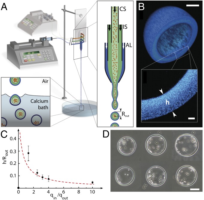
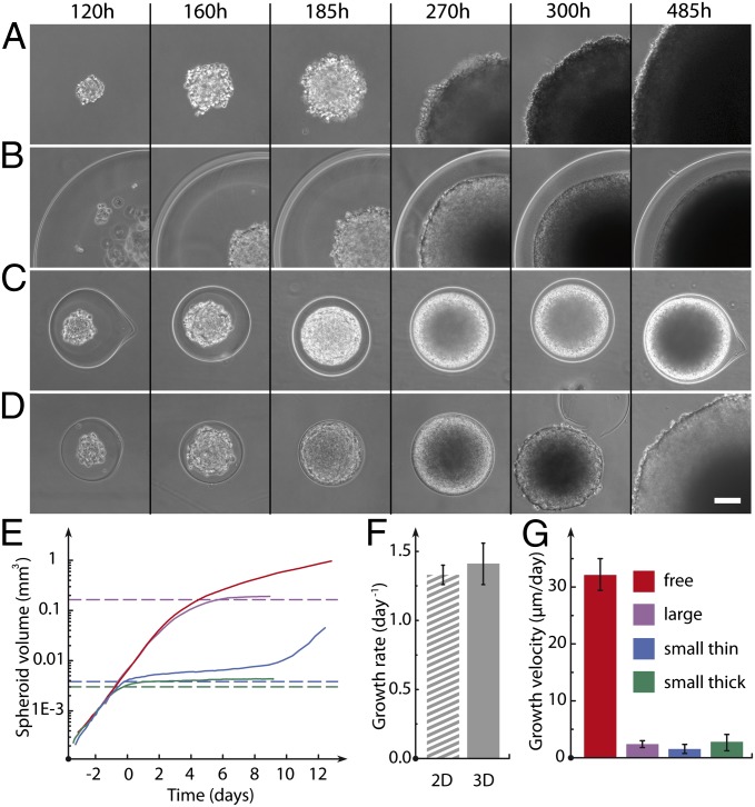
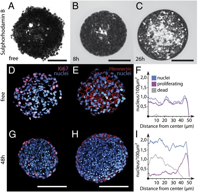
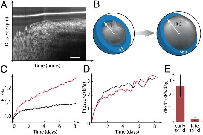
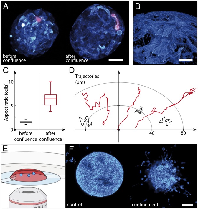
Deciphering the multifactorial determinants of tumor progression requires standardized high-throughput preparation of 3D in vitro cellular assays. We present a simple microfluidic method based on the encapsulation and growth of cells inside permeable, elastic, hollow microspheres. We show that this approach enables mass production of size-controlled multicellular spheroids. Due to their geometry and elasticity, these microcapsules can uniquely serve as quantitative mechanical sensors to measure the pressure exerted by the expanding spheroid. By monitoring the growth of individual encapsulated spheroids after confluence, we dissect the dynamics of pressure buildup toward a steady-state value, consistent with the concept of homeostatic pressure. In turn, these confining conditions are observed to increase the cellular density and affect the cellular organization of the spheroid. Postconfluent spheroids exhibit a necrotic core cemented by a blend of extracellular material and surrounded by a rim of proliferating hypermotile cells. By performing invasion assays in a collagen matrix, we report that peripheral cells readily escape preconfined spheroids and cell-cell cohesivity is maintained for freely growing spheroids, suggesting that mechanical cues from the surrounding microenvironment may trigger cell invasion from a growing tumor. Overall, our technology offers a unique avenue to produce in vitro cell-based assays useful for developing new anticancer therapies and to investigate the interplay between mechanics and growth in tumor evolution.

Keywords: mechanotransduction; microfluidics; tissue mechanics; tumor growth.


Conflict of interest statement

The authors declare no conflict of interest.

Figures

Fig. 1.
Fig. 1.
Operating principle of the microfluidic device and characterization of the alginate microcapsules. (A) Schematic of the microfluidic platform, which is composed of an external fluidic injection system, coextrusion microdevice, and off-chip gelation bath. An enlarged view of the chip (Right) shows the three-way configuration, with cell suspension (CS), intermediate solution (IS), and alginate solution (AL), respectively, flowing into the coaligned capillaries. The inlets of the chip are connected to three syringes controlled by two syringe pumps. The compound liquid microdroplets fall into a 100-mM calcium bath. Calcium-mediated gelation of the alginate shell freezes the structure of the capsule, and cells remain encapsulated. (B) Confocal image of an alginate capsule stained with high-molecular-weight fluorescent dextran. (Inset) Magnified view of the capsule wall. (C) Plot of the capsule aspect ratio h/Rout as a function of the ratio between the inner flow rate qin = qCS + qIS and outer flow rate qout = qAL. Black dots represent experimental data. The red dashed line is the theoretical curve derived from volume conservation (SI Materials and Methods). Notations are indicated in A and B. (D) Representative phase-contrast micrographs of individual capsules encapsulating cells. (Scale bars: B, 50 μm; D, 100 μm.)
Fig. 2.
Fig. 2.
Confined and free growth of spheroids. Sequences of phase-contrast micrographs show free growth of a spheroid (A) and confined growth of a spheroid encapsulated in large and thick (B; h = 100 μm, R = 407 μm), small and thick (C; h = 38 μm, R = 151 μm), and small and thin (D; h = 7 μm, R = 129 μm) alginate shells. The thin capsule ultimately bursts. Time is recorded from encapsulation. (Scale bars: 50 μm.) (E) Representative log-linear time plots of spheroid volume in conditions of free growth (red) and confined growth in a large capsule (purple), in a small and thick capsule (blue), and in a small and thin capsule (green). (F) Growth rate for CT26 cells cultured in a Petri dish (2D) and as spheroids (3D) before confluence (early stages). The 3D growth rate is derived from MCS volume increase normalized by the actual volume. (G) Growth velocity measured in the late stages of CT26 spheroid growth.
Fig. 3.
Fig. 3.
Imaging of the internal cellular organization of growing spheroids under elastic confinement. Snapshots taken by two-photon imaging of free (A) and encapsulated (B and C) spheroids stained with the polar dye SRB are shown. Time t = 0 corresponds to confluence. Confocal images of free (D and E) and confined (G and H) spheroids after cryosection and immunolabeling for DAPI (blue), KI67 (magenta), and fibronectin (red). Quantification of cell nucleus (blue), proliferating cell (purple), and dead cell (gray) radial densities for free (F) and confined (I) CT26 spheroids. (Scale bars: AC, 50 μm; D, E, G, and H, 100 μm.)
Fig. 4.
Fig. 4.
Quantitative mechanical analysis of postconfluent spheroid growth. (A) Phase-contrast intensity plot as a function of time and radial distance from the MCS center. The bright line is the outer wall of the capsule. (Scale bars: 20 h and 50 μm.) (B) Drawing of encapsulated spheroid and capsule deformation suggesting the use of the shell as a mechanical sensor. R0 and h0 are the initial capsule radius and shell thickness, respectively. (C) Representative time plots show the influence of capsule stiffness (via shell thickness) on MCS growth. The radius of the spheroid Rin is normalized to the undeformed inner radius R0 vs. time (h = 8 μm, red; h = 28 μm, black). (D) Pressure exerted by the spheroid on the capsule wall vs. time (h = 8 μm, red; h = 28 μm, black). (E) Statistical analysis (n = 40, including 23 thin capsules and 17 thick capsules). The rate of pressure increase within 1 d following confluence and at later stages is shown.
Fig. 5.
Fig. 5.
Impact of elastic confinement on cell motility at the periphery of growing spheroids. (A) Confocal live imaging of an encapsulated spheroid grown from cells stably transfected with LifeAct-mCherry. Maximum intensity projections of the confocal stacks [a hot look-up table acquired using Fiji is shown (cyan)]. One representative cell is colored magenta before and after confluence to highlight the features quantified in C. (B) Enlarged view of the surface of a fixed spheroid imaged by confocal microscopy after staining with phalloidin-Alexa 488 (Hot LUT, cyan). (C) Box plot shows the aspect ratio of cells before and after confluence (n = 94, five spheroids). (D) Representative projected trajectories of the center of mass of cells moving at the periphery of spheroids. The time interval is 15 min. (E) Cartoon describing cell migration assay. Spheroids (blue) are embedded in collagen matrix (red) in a Petri dish. (F) Confocal image taken 48 h after implantation in collagen for spheroids grown freely (control) and in a capsule past confluence after shell dissolution (confined). (Scale bars: A, 50 μm; B, 10 μm; F, 100 μm.)

References

    1. Cukierman E, Pankov R, Stevens DR, Yamada KM. Taking cell-matrix adhesions to the third dimension. Science. 2001;294(5547):1708–1712. - PubMed
    1. Sutherland RM. Cell and environment interactions in tumor microregions: The multicell spheroid model. Science. 1988;240(4849):177–184. - PubMed
    1. Lee GY, Kenny PA, Lee EH, Bissell MJ. Three-dimensional culture models of normal and malignant breast epithelial cells. Nat Methods. 2007;4(4):359–365. - PMC - PubMed
    1. Kim JB, Stein R, O’Hare MJ. Three-dimensional in vitro tissue culture models of breast cancer—A review. Breast Cancer Res Treat. 2004;85(3):281–291. - PubMed
    1. Lee M-Y, et al. Three-dimensional cellular microarray for high-throughput toxicology assays. Proc Natl Acad Sci USA. 2008;105(1):59–63. - PMC - PubMed

Publication types

MeSH terms