. 2013 Sep 9;14(9):3010-6.
doi: 10.1021/bm400447z. Epub 2013 Aug 27.

Cell patterning with mucin biopolymers

Affiliations

Cell patterning with mucin biopolymers

T Crouzier et al. Biomacromolecules. .

Abstract

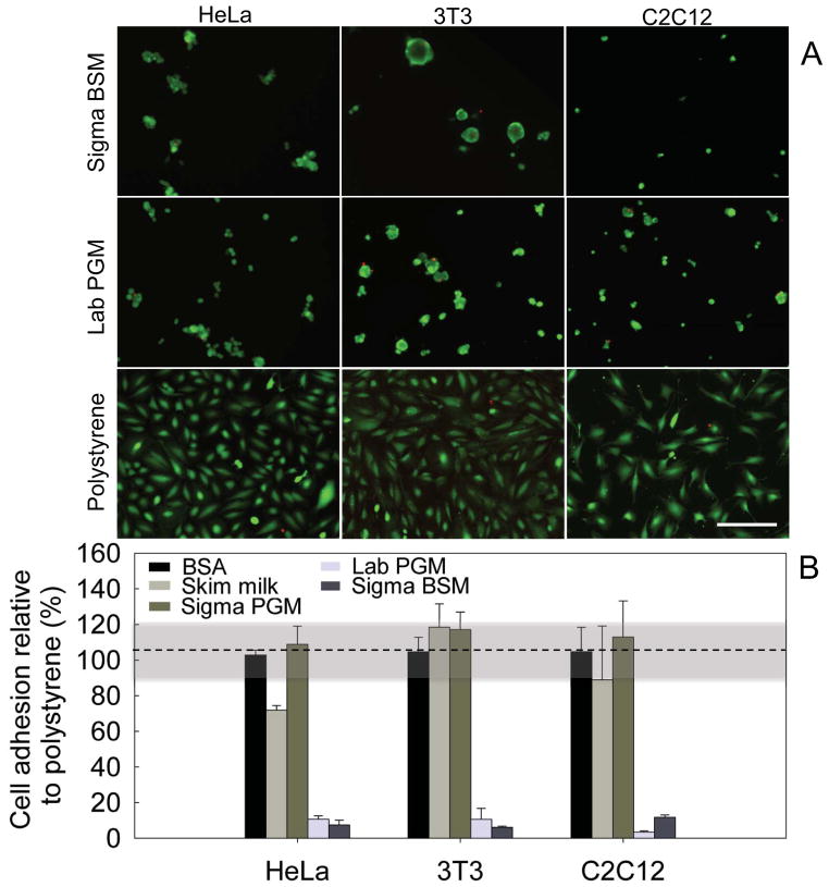
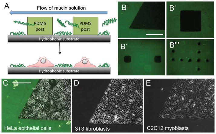
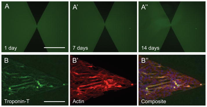
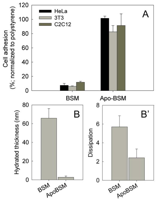
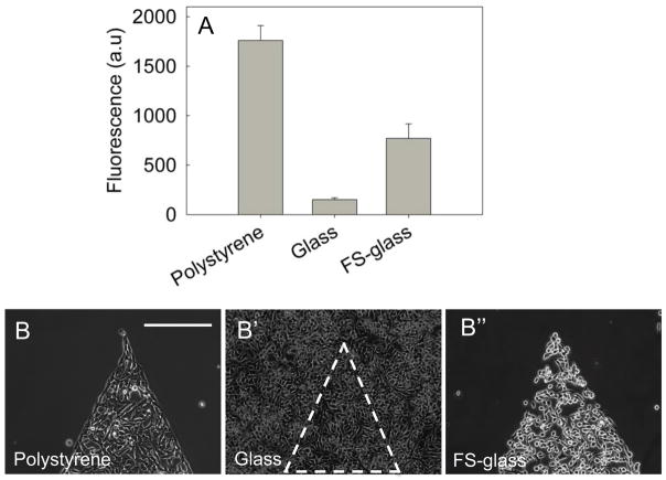
The precise spatial control of cell adhesion to surfaces is an endeavor that has enabled discoveries in cell biology and new possibilities in tissue engineering. The generation of cell-repellent surfaces currently requires advanced chemistry techniques and could be simplified. Here we show that mucins, glycoproteins of high structural and chemical complexity, spontaneously adsorb on hydrophobic substrates to form coatings that prevent the surface adhesion of mammalian epithelial cells, fibroblasts, and myoblasts. These mucin coatings can be patterned with micrometer precision using a microfluidic device, and are stable enough to support myoblast differentiation over seven days. Moreover, our data indicate that the cell-repellent effect is dependent on mucin-associated glycans because their removal results in a loss of effective cell-repulsion. Last, we show that a critical surface density of mucins, which is required to achieve cell-repulsion, is efficiently obtained on hydrophobic surfaces, but not on hydrophilic glass surfaces. However, this limitation can be overcome by coating glass with hydrophobic fluorosilane. We conclude that mucin biopolymers are attractive candidates to control cell adhesion on surfaces.


Figures

Figure 1
Figure 1. Mucin coatings are cytophobic
(A) HeLa epithelial cells, 3T3 fibroblast cells and C2C12 myoblasts were seeded on a polystyrene surface, or on coatings generated from BSM (Sigma) or PGM (in-lab purified) and labeled with a live (green)/dead (red) stain. Scale bars: 200μm.” (B) Quantification of cell adhesion on mucin coatings generated with commercial BSM and PGM (Sigma BSM/PGM), in-lab purified PGM (Lab PGM), Skim milk, and BSA. The highlighted region between 90–120% is the standard deviation of the reference adhesion to polystyrene.
Figure 2
Figure 2. Mucin coatings for cell patterning
(A) Mucin coatings were patterned using a microfluidic device with posts masking defined areas of the surface. (B) Patterns of mucin-free areas (in black) can be generated in different sizes and shapes. When seeded on the patterned surfaces, the epithelial cells (C), fibroblasts (D) and myoblasts (E) accumulated in the uncoated regions, avoiding the mucin coatings. Scale bars: 250μm.”
Figure 3
Figure 3. Mucin coatings and their cytophobic effect are robust
(A) Mucin patterns immersed in cell culture media for 14 days showed no alteration in their shape or fluorescence intensity. C2C12 myoblasts that were cultured on the coatings for 7-days expressed troponin-T, which is a marker for early myogenic differentiation. (B′) Actin staining reveals that cells are confined within the pattern. A Troponin-T/Actin overlay is shown in (B″). Scale bars: 250μm.”
Figure 4
Figure 4. Mucin glycans are essential for the cytophobic effect
(A) Coatings generated from de-glycosylated mucins (Apo-BSM) failed to repel all three cell types tested. Glycan removal from mucins resulted in altered properties of the coatings as probed by QCM-D measurements. (B) Glycosylated mucins yielded thicker coatings and more (B′) dissipation, which is diagnostic for soft materials.
Figure 5
Figure 5. Mucin coatings form preferentially on hydrophobic surfaces
(A) The relative amount of mucins adsorbed to various surfaces was estimated by measuring the fluorescence intensity of coatings generated with fluorescently labeled mucins. Mucins failed to adsorb on glass efficiently, however, treating glass with fluorosilane (FS-glass) reversed this effect and promoted the formation of mucin coatings. Accordingly, mucin-mediated cell-repulsion was observed on polystyrene (B) and on fluorosilane-treated glass (B″), but not on untreated microscope glass slides (B′).

References

    1. Anderson JM, Rodriguez A, Chang DT. Semin Immunol. 2008;20:86–100. - PMC - PubMed
    1. Wan LQ, Ronaldson K, Park M, Taylor G, Zhang Y, Gimble JM, Vunjak-Novakovic G. Proc Natl Acad Sci. 2011;108:12295–12300. - PMC - PubMed
    1. Popescu DC, Lems R, Rossi NAA, Yeoh CT, Loos J, Holder SJ, Bouten CVC, Sommerdijk NAJM. Adv Mater. 2005;17:2324–2329.
    1. Tseng Q, Duchemin-Pelletier E, Deshiere A, Balland M, Guillou H, Filhol O, Théry M. Proc Natl Acad Sci. 2012;109:1506–1511. - PMC - PubMed
    1. Fukuda J, Khademhosseini A, Yeh J, Eng G, Cheng J, Farokhzad OC, Langer R. Biomaterials. 2006;27:1479–1486. - PubMed

Publication types

LinkOut - more resources