. 2013 Nov;223(5):489-94.
doi: 10.1111/joa.12094. Epub 2013 Aug 27.

What is the origin of the arterial vascularization of the corpora cavernosa? A computer-assisted anatomic dissection study

Affiliations

What is the origin of the arterial vascularization of the corpora cavernosa? A computer-assisted anatomic dissection study

Djibril Diallo et al. J Anat. 2013 Nov.

Abstract

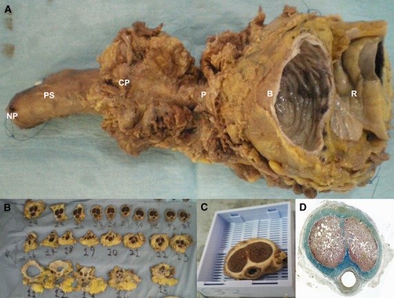
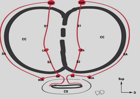
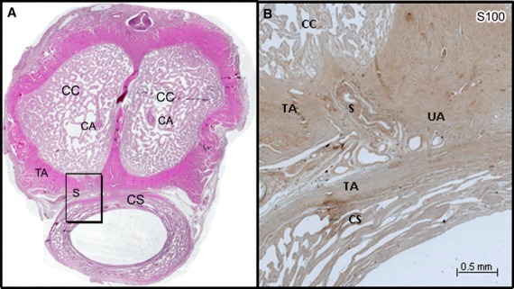
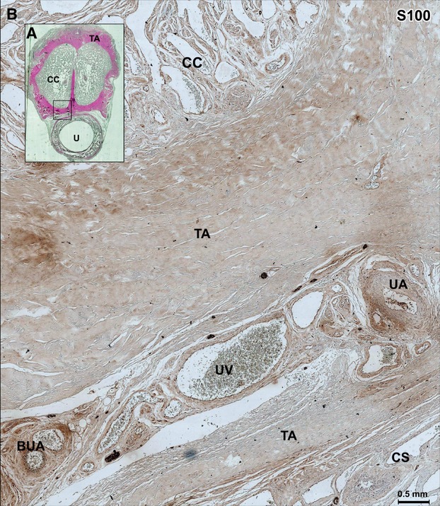
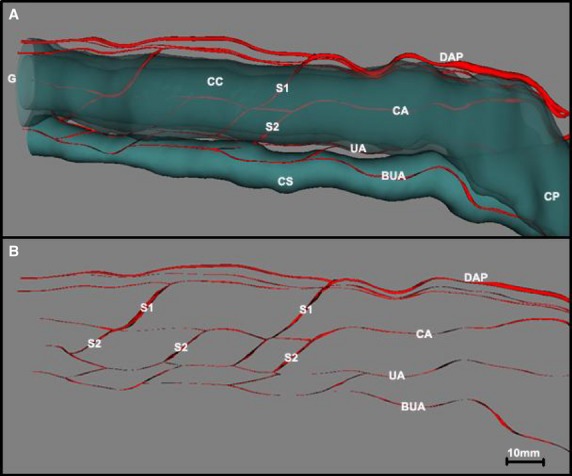
The purpose of this study was to identify the microscopic arterial vascularization of the corpora cavernosa (CC) of the penis using computer-assisted anatomic dissection (CAAD), determine the contribution of the different penile arteries towards this vascularization, detail the nature of cavernospongiosum shunts, and locate the anastomoses between these different arteries. Tissue specimens were taken from five donors who donated their bodies to science. The specimens were fixed in 10% formalin and sliced into a series of five 5-μm sections at intervals of 200 μm. The first section was stained with hematoxylin-eosin or Masson's trichrome and the second with anti-protein S100. The cavernous artery of the penis is not the only source of arterial vascularization of the CC. In four of the five cases studied, we found two to four perforating branches arising from the dorsal arteries of the penis that join up with the cavernous artery of the penis or that are solely responsible for the vascularization of the distal third of the penis. The bulbo-urethral and urethral arteries are situated outside of the tunica albuginea of the corpus spongiosum on their lateral and dorsal sides. The anastomoses do not occur between the cavernous artery of the penis and the corpus spongiosum but between the cavernous artery of the penis and the urethral artery on the surface of the tunica albuginea. All of these arteries are accompanied by nerve branches. The CC were found to be vascularized by both cavernous and dorsal arteries of the penis. Intrapenile vascularization is organized around four arterial axes, which are anastomosed by multiple neurovascular shunts.

Keywords: cavernous artery; cavernous urethral shunt; corpora cavernosa; erection; penis.


Figures

Fig. 1
Fig. 1
(A) Pelvic block taken from 70-year-old adult male from the bladder (B) to the neck of the penis (NP). (B) The specimen was cut into 1-cm macroscopic blocks. (C) These blocks were put in mega-cassettes (code: 38VSP59040) before being embedded in paraffin. (D) Histological section of the previous block stained by TreM and scanned at high resolution (3200 dots inch−1, dpi). R, rectum; P, prostate; CP, crura of penis; PS, penile shaft.
Fig. 2
Fig. 2
Schematic representation of the arteries of the penis and their anastomoses. The dorsal arteries of the penis (DAP) have superficial anastomoses (SA) (outside of the tunica albuginea TA) with the bulbo-urethral arteries (BUA). They give off deep branches (S1) which run through the tunica albuginea to join up with the cavernous arteries (CA) inside the corpora cavernosa (CC). The urethral arteries (UA) and the bulbo-urethral arteries are situated outside of the TA of the corpus spongiosum (CS). The anastomoses (S2) between the CAs and the UAs occurred outside of the CS, hence the term ‘cavernous urethral shunts’ used to designate these anastomoses.
Fig. 3
Fig. 3
(A) Histological section of the penis of a 68-year-old man stained by HE and scanned at high resolution (3200 dpi). (B) Microscopic aspect (×10) of the shunt (S) between the CA and the UA (frame in A) immunostained with anti-protein S100. The shunt came from the CA, passed on the ventral surface of the tunica albuginea (TA) of the corpora cavernosa (CC) to join UA located on the dorsal surface of the corpus spongiosum (CS) between the two tunicae albugineae (TA) and not in the CS, hence our use of the term ‘cavernosal urethral shunt’.
Fig. 4
Fig. 4
(A) Histological section through the body of the penis in 68-year-old man, stained by HE. (B) Microscopic aspect (×10) of the next section immunolabeled with S100 showing the space between the urethral artery (UA) (after its anastomosis with the shunt arising from the cavernous artery, as shown above) and the bulbo-urethral artery (BUA) (frame in A). The UA is situated on the dorsal side and outside of the corpus spongiosum (CS) between the two tunicae albugineae (TA), which receive the arterial shunt arising from the cavernous artery. The term ‘cavernous urethral shunts’ is thus a more appropriate designation for this arterial anastomosis. CC, corpora cavernosa; TA, tunica albuginea; UV, urethral vein.
Fig. 5
Fig. 5
(A) Three-dimensional reconstruction of the penis of a 72-year-old human cadaver showing the trajectory of the arteries of the penis with regard to the corpora cavernosa (CC) and the corpus spongiosum (CS). (B) The same image after the CC and CS have been made transparent, showing the different anastomoses between the arteries of the penis. The dorsal arteries of the penis (DAP) played a role in the vascularization of the CC via the perforating branches (S1), which ran through the tunica albuginea (TA) to anastomose with the cavernous arteries (CA) or to ensure solely the vascularization of the one-third distal part of the CC. The bulbo-urethral arteries (BUA) and the urethral arteries (UA), highly anastomosed with each other, were situated outside of the TA of the CS. These were the UAs that received the shunt (S2) stemming from the CA, reinforcing the vascularization of the CS and the urethra. CP, crura of penis; G, glans.

References

    1. Alsaid B, Bessede T, Karam I, et al. Coexistence of adrenergic and cholinergic nerves in the inferior hypogastric plexus: anatomical and immunohistochemical study with 3D reconstruction in human male fetus. J Anat. 2009;214:645–654. - PMC - PubMed
    1. Benoit G, Droupy S, Quillard J, et al. Supra and infralevator neurovascular pathways to the penile CC. J Anat. 1999;195(Pt 4):605–615. - PMC - PubMed
    1. Bookstein JJ, Lang EV. Penile magnification pharmacoarteriography: details of intrapenile arterial anatomy. Am J Roentgenol. 1987;148:883–888. - PubMed
    1. Conti G. The erection of the human penis and its morphologico-vascular basis. Acta Anat (Basel) 1952;14:217–262. - PubMed
    1. Costabile RA, Spevak M, Fishman IJ, et al. Efficacy and safety of transurethral alprostadil in patients with erectile dysfunction following radical prostatectomy. J Urol. 1998;160:1325–1328. - PubMed

LinkOut - more resources