. 2013 Oct;2(10):758-65.
doi: 10.5966/sctm.2012-0156. Epub 2013 Aug 27.

Long-term maintenance of limbal epithelial progenitor cells using rho kinase inhibitor and keratinocyte growth factor

Affiliations

Long-term maintenance of limbal epithelial progenitor cells using rho kinase inhibitor and keratinocyte growth factor

Hideyuki Miyashita et al. Stem Cells Transl Med. 2013 Oct.

Abstract

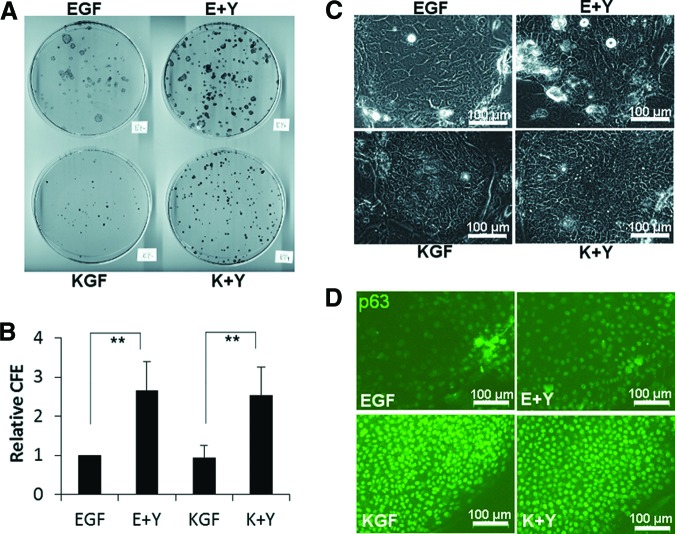
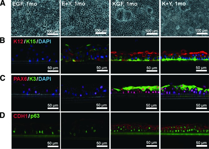
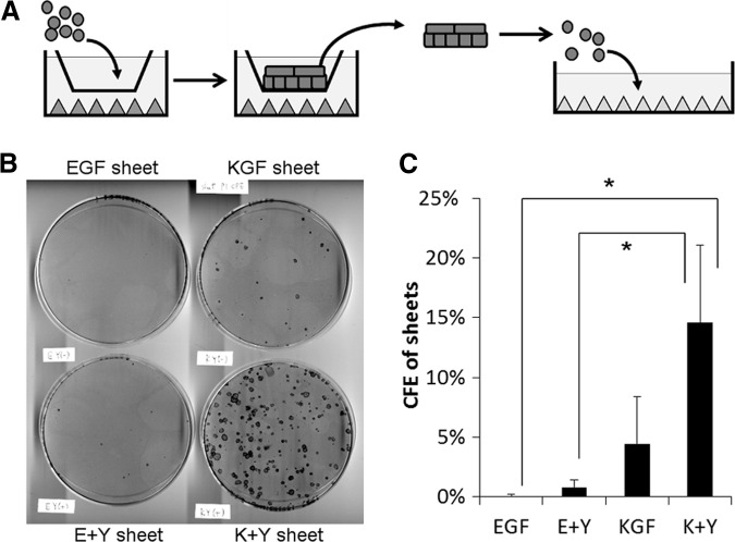
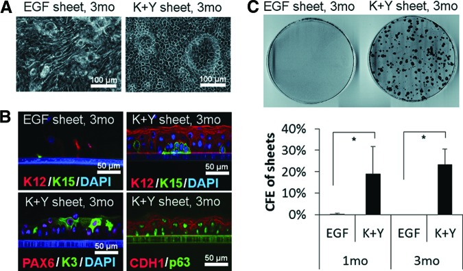
Corneal epithelial stem cells are located in the limbus, the junction between the cornea and the conjunctiva. A limbal epithelium model in vitro would be useful for the study of epithelial stem cells, as well as improving the quality of cultivated epithelial sheets for the treatment of limbal stem cell deficiency. In this study, we succeeded in constructing a limbal epithelium-like structure that could be maintained for at least 5 months in vitro. We modified conventional medium by replacing epidermal growth factor with keratinocyte growth factor (KGF) and adding Y-27632, a rho kinase inhibitor. Using this medium, epithelial cells freshly isolated from human limbus were cocultured with human mesenchymal stem cell-derived feeder cells. Cells formed a stratified layer without air exposure, and both basal and suprabasal layers maintained their unique morphologies for up to 5 months. Basal layers expressed the progenitor marker p63 uniformly and K15 heterogeneously. Expressions of PAX6, K3, and K12 indicated that cell sheets underwent normal differentiation in the corneal epithelium lineage. Although medium was changed daily after day 7, cell debris was observed every day, suggesting that cell sheets underwent turnover. Furthermore, secondary colonies were observed from cells dissociated from 1-month and 3-month cultured sheets. In conclusion, human limbal epithelial cell sheet cultures with KGF and Y-27632 maintained stratification, high expression of both stem/progenitor markers and differentiation markers, and colony-forming cells long-term. This protocol may be useful as an in vitro limbal epithelial model for basic studies.

Keywords: Adult stem cells; Cell culture; Colony formation; Differentiation; Experimental models; Long-term repopulation; Stem cell culture.


Figures

Figure 1.
Figure 1.
The effects of EGF, KGF, and Y-27632 on the colony formation of human limbal epithelial cells. (A): Rhodamine B-stained 100-mm dish. (B): Relative CFE; n = 7. **, p < .01. CFE was normalized as CFE of EGF = 1. (C): Phase contrast micrograph of colonies at day 7. (D): Immunostaining of colonies at day 10 using anti-p63 antibody (green). Scale bars = 100 μm (C, D). Abbreviations: CFE, colony forming efficiency; E+Y, epidermal growth factor and Y-27632; EGF, epidermal growth factor; K+Y, keratinocyte growth factor and Y-27632; KGF, keratinocyte growth factor.
Figure 2.
Figure 2.
The effects of EGF, KGF, and Y-27632 on the morphology of human limbal epithelial cell sheets. (A): Phase contrast micrograph of cells cultured for 1 month in the indicated medium. (B–D): Cryosections stained with antibodies specific for K15 (green, [B]), K12 (red, [B]), K3 (green, [C]), PAX6 (red, [C]), p63 (green, [D]), and CDH1 (red, [D]). Phase contrast images were merged with immunofluorescence images. Where indicated, cell nuclei were stained with DAPI. Culture conditions were the same as for the top panel. Scale bars = 100 μm (A) and 50 μm (B–D). Abbreviations: DAPI, 4′,6-diamidino-2-phenylindole; E+Y, epidermal growth factor and Y-27632; EGF, epidermal growth factor; K+Y, keratinocyte growth factor and Y-27632; KGF, keratinocyte growth factor; mo, month.
Figure 3.
Figure 3.
The effects of EGF, KGF, and Y-27632 on the colony forming ability of human limbal epithelial cell sheets. (A): Scheme of colony formation assay. Cell sheets cultured with EGF, E+Y, KGF, or K+Y medium were dissociated by enzyme treatment, followed by subculture on the indicator dishes at clonal density. All dishes were fed with same medium. (B): Rhodamine B-stained indicator dishes. (C): CFE of sheets; n = 3. *, p < .05. Abbreviations: CFE, colony forming efficiency; E+Y, epidermal growth factor and Y-27632; EGF, epidermal growth factor; K+Y, keratinocyte growth factor and Y-27632; KGF, keratinocyte growth factor.
Figure 4.
Figure 4.
Morphology and colony forming ability of human limbal epithelial cell sheets cultured for 3 months. (A): Phase contrast micrograph of cell sheets cultured with EGF medium or K+Y medium for 3 months. (B): Immunohistochemistry of 3-month cultured EGF sheets and K+Y sheets using the specific antibodies indicated. Phase contrast images were merged with immunofluorescence images. (C): Colony formation assays of cells dissociated from 3-month cultured EGF sheets and K+Y sheets. Top: Rhodamine B-stained indicator dishes. Bottom: CFE of 1-month cultured sheets and 3-month cultured sheets. Scale bars = 100 μm (A) and 50 μm (B). Abbreviations: CFE, colony forming efficiency; EGF, epidermal growth factor; K+Y, keratinocyte growth factor and Y-27632; mo, months.

References

    1. Ordonez P, Di Girolamo N. Limbal epithelial stem cells: Role of the niche microenvironment. Stem Cells. 2012;30:100–107. - PubMed
    1. Yoshida S, Shimmura S, Kawakita T, et al. Cytokeratin 15 can be used to identify the limbal phenotype in normal and diseased ocular surfaces. Invest Ophthalmol Vis Sci. 2006;47:4780–4786. - PubMed
    1. Pellegrini G, Dellambra E, Golisano O, et al. p63 identifies keratinocyte stem cells. Proc Natl Acad Sci USA. 2001;98:3156–3161. - PMC - PubMed
    1. Chen Z, de Paiva CS, Luo L, et al. Characterization of putative stem cell phenotype in human limbal epithelia. Stem Cells. 2004;22:355–366. - PMC - PubMed
    1. Watanabe K, Nishida K, Yamato M, et al. Human limbal epithelium contains side population cells expressing the ATP-binding cassette transporter ABCG2. FEBS Lett. 2004;565:6–10. - PubMed

Publication types

MeSH terms

Substances