. 2013 Dec;27(12):5010-21.
doi: 10.1096/fj.13-236380. Epub 2013 Aug 27.

Extracellular histones are essential effectors of C5aR- and C5L2-mediated tissue damage and inflammation in acute lung injury

Affiliations

Extracellular histones are essential effectors of C5aR- and C5L2-mediated tissue damage and inflammation in acute lung injury

Markus Bosmann et al. FASEB J. 2013 Dec.

Abstract

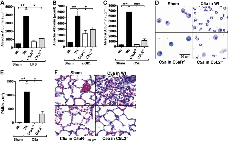
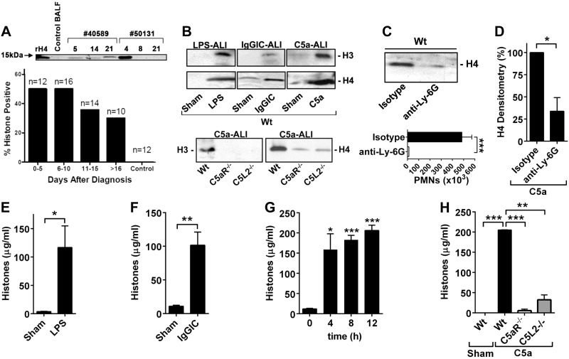
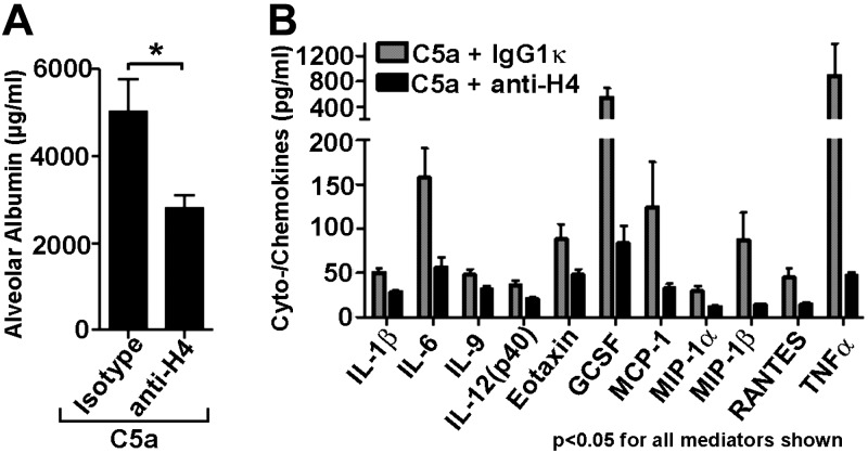
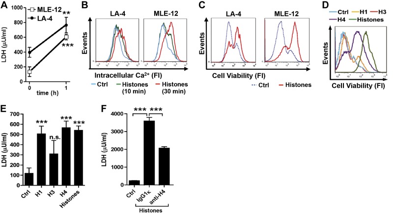
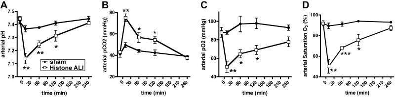
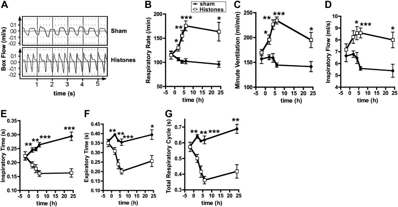
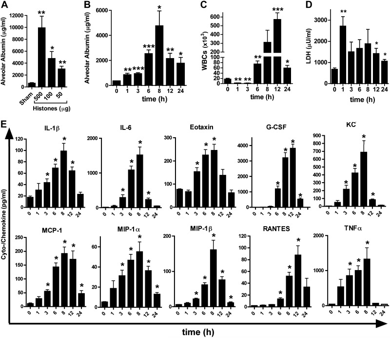
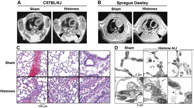
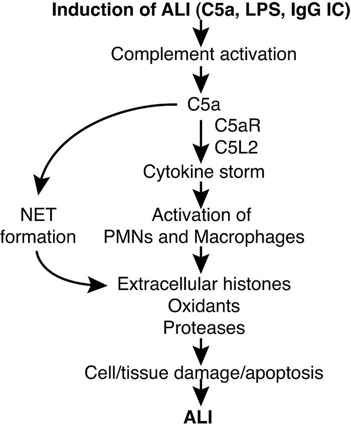
We investigated how complement activation promotes tissue injury and organ dysfunction during acute inflammation. Three models of acute lung injury (ALI) induced by LPS, IgG immune complexes, or C5a were used in C57BL/6 mice, all models requiring availability of both C5a receptors (C5aR and C5L2) for full development of ALI. Ligation of C5aR and C5L2 with C5a triggered the appearance of histones (H3 and H4) in bronchoalveolar lavage fluid (BALF). BALF from humans with ALI contained H4 histone. Histones were absent in control BALF from healthy volunteers. In mice with ALI, in vivo neutralization of H4 with IgG antibody reduced the intensity of ALI. Neutrophil depletion in mice with ALI markedly reduced H4 presence in BALF and was highly protective. The direct lung damaging effects of extracellular histones were demonstrated by airway administration of histones into mice and rats (Sprague-Dawley), which resulted in ALI that was C5a receptor-independent, and associated with intense inflammation, PMN accumulation, damage/destruction of alveolar epithelial cells, together with release into lung of cytokines/chemokines. High-resolution magnetic resonance imaging demonstrated lung damage, edema and consolidation in histone-injured lungs. These studies confirm the destructive C5a-dependent effects in lung linked to appearance of extracellular histones.

Keywords: complement; cytotoxic factors.


Figures

Figure 1.
Figure 1.
Essential roles of both C5aR and C5L2 during the 3 models of ALI. A) Comparison of alveolar permeability after LPS-ALI in C57BL/6J (Wt), C5aR−/−, or C5L2−/− mice. Sham-treated mice received PBS i.t., ELISA for BALF mouse albumin. B) Alveolar permeability after IgGIC-ALI in C57BL/6J (Wt), C5aR−/−, or C5L2−/− mice (ELISA). C) Alveolar permeability after C5a-ALI in C57BL/6J (Wt), C5aR−/−, or C5L2−/− mice (ELISA). D) Cytospins of BALF cells after C5a-ALI in C57BL/6J (Wt), C5aR−/−, or C5L2−/− mice, Wright-Giemsa stains. E) PMN counts in BALF after C5a-ALI in C57BL/6J (Wt), C5aR−/−, or C5L2−/− mice. F) Histopathology after C5a-ALI in C57BL/6J (Wt), C5aR−/−, or C5L2−/− mice (H&E stains). Duration of all ALIs shown was 8 h, and 500 ng i.t. rmC5a was used for C5a-ALI. Data from each experiment shown are representative of n ≥ 7 mice/group. Error bars = sem. *P < 0.05, **P < 0.01, ***P < 0.001; Student's t test.
Figure 2.
Figure 2.
Detection of extracellular histones during ALI in humans or mice. A) Top panel: Western blotting of histone H4 in BALF from 2 humans with ALI/ARDS (40589; 50131) at sequential time points (4–21 d), a healthy volunteer (Ctrl BALF, negative control), or recombinant H4 (rH4, positive control). Bottom panel: presence of histone H4 in BALF from humans with ALI/ARDS and healthy volunteers (Western blotting). Percentage values of positive samples are indicated as bars for different time points after diagnosis of ALI/ARDS, with total numbers of analyzed samples at top of each bar. B) Histones H3 and H4 in BALF after ALI models in Wt, C5aR−/−, or C5L2−/− mice, 8 h; Western blotting. C) Top panel: histone H4 detection by Western blotting in BALF of PMN-depleted Wt mice 8 h after C5a-ALI. Depletion of PMNs was achieved by monoclonal anti-Ly-6G antibody (200 μg i.p., −24 h) using isotype IgG as control. Bottom panel: absence of BALF PMNs after anti-Ly-6G treatment. D) Densitometry of three independent Western blots for histone H4 in BALF of C5a-ALI (8 h) in Wt mice, when PMNs were depleted as described above. E) ELISA for extracellular histones in BALF of sham-treated Wt mice or in Wt mice after LPS-ALI, 8 h. F) Extracellular histones in BALF after IgGIC-ALI in Wt mice, 8 h; ELISA. G) Time course of histone release in BALF following C5a-ALI, ELISA. H) Detection of extracellular histones in BALF of Wt, C5aR−/− and C5L2−/− mice after C5a-ALI, 8 h; ELISA. Data are representative of the indicated numbers of human samples (A) or n = 5 mice/group (B–H). Error bars = sem. *P < 0.05; Student's t test.
Figure 3.
Figure 3.
Protective effects of neutralization of extracellular histones during ALI. A) Reduced alveolar permeability disturbances after C5a-ALI with treatment of neutralizing anti-H4 antibody (BWA3, 250 μg i.v. + 50 μg i.t.) or isotype control IgG1κ, 8 h; ELISA. B) Multiplex bead-based assay of mediators in BALF after C5a-ALI with treatment of anti-H4 antibody or control IgG1κ, 8 h; ELISA. Only mediators with significant differences (P<0.05, 11 of 23 mediators) between groups are shown. C5a was used as 500 ng/mouse i.t. Data were acquired using n ≥ 15 mice/group. Error bars = sem. *P < 0.05; Student's t test.
Figure 4.
Figure 4.
Cytotoxic effects of extracellular histones on alveolar epithelial cells. A) LDH release from LA-4 and MLE-12 lung alveolar epithelial cells exposed to purified histones (50 μg/ml, 1 h), chromogenic assay. B) [Ca2+]i staining in untreated (Ctrl) LA-4 and MLE-12 cells or after histones (50 μg/ml); flow cytometry. C) Cell viability in LA-4 and MLE-12 cells exposed to histones (50 μg/ml, 1 h); cells were stained with a fixable cell viability dye, eFluor450; flow cytometry. D) Cell viability of MLE-12 cells after incubation with histone H1, histone H3, histone H4, purified histones (all 50 μg/ml), or untreated controls, 1 h; flow cytometry. E) LDH concentrations in supernatant fluids from MLE-12 cells after 1 h incubation with histone H1, histone H3, histone H4, or purified histones from calf thymus (all 50 μg/ml). F) LDH assay in MLE-12 cell culture supernatant fluids. MLE-12 cells were left as untreated controls or incubated for 60 min with purified histones (12.5 μg/ml) premixed (30 min, 37°C) with either IgG1κ isotype control antibody (250 μg/ml) or anti-histone H4 antibody (BWA3; 250 μg/ml). Data are representative of 3 independent experiments. Error bars = sem. n.s., not significant. **P < 0.01, ***P < 0.001; Student's t test.
Figure 5.
Figure 5.
Administration of extracellular histones into airways induces severe disturbances in alveolar-capillary gas exchange. Purified histones (50 μg/g body weight) were administered i.t. to Sprague-Dawley rats with implanted carotid artery catheters. Sham-treated animals only received PBS i.t. Serial blood gas analyses were performed at baseline and at intervals following the intervention. A) Arterial pH. B) Arterial pCO2. C) Arterial pO2. D) Arterial oxygen saturation, SatO2. Data are representative of n ≥ 4 rats/group. Error bars = sem. *P < 0.05, **P < 0.01, ***P < 0.001; Student's t test.
Figure 6.
Figure 6.
Whole-body plethysmography following airway administration of histones. Sprague-Dawley rats received purified histones (50 μg/g body weight i.t.) or PBS i.t (sham treatment). Respiration was monitored before the intervention and at intervals during the following 24 h: A) Typical box flows (raw data) during whole-body plethysmography of rats after airway exposure to purified histones compared to sham-treated rats. B) Respiratory rates. C) Minute ventilation. D) Inspiratory flow times. E) Inspiratory times. F) Expiratory flow times. G) Total respiratory cycle. Data are representative of n ≥ 4 rats/group. Error bars = sem. *P < 0.05, **P < 0.01, ***P < 0.001; Student's t test.
Figure 7.
Figure 7.
Induction of acute inflammation in lungs by extracellular histones. A) Disruption of alveolar permeability in C57BL/6J mice after intratracheal deposition of purified histones at different doses, 8 h; BALF albumin ELISA. B) Time course for alveolar permeability after exposure to purified histones (100 μg i.t.), C57BL/6J mice; ELISA. C) White blood cell (WBC) content (mainly PMNs) in BALF from C57BL/6J mice after purified histones from the experiment described in B. D) LDH in BALF from the experiment in B; chromogenic assay. E) Mediators in C57BL/6J mice after treatment with purified histones (100 μg i.t.), with BALF harvested at different time points; multiplex bead-based assay. Data are representative of n ≥ 5 mice/group. Error bars = sem. *P < 0.05, **P < 0.01, ***P < 0.001; Student's t test.
Figure 8.
Figure 8.
Extracellular histones mediate lung consolidation and acute histopathology in lungs. A) High-resolution MRI of lungs from C57BL/6J mice after PBS i.t. (sham treatment) or purified histones (20 μg/g body weight), 6 h. Arrowheads indicate signal-intense intrapulmonary areas consistent with consolidation of lung tissue; axial proton density/T1-weighted images; slice thickness: 1 mm. B) High-resolution MRI of lungs from Sprague-Dawley rats 6 h after PBS i.t. (sham) or purified histones (20 μg/g body weight). Arrowheads indicate signal-intense intrapulmonary areas of lung infiltrations; slice thickness: 2 mm. C) Histopathology of lungs from C57BL/6J mice after treatment with purified histones (20 μg/g body weight i.t.) compared to sham-treated lungs, 6 h, H&E staining. D) Transmission electron microscopy of normal lungs (sham treatment) from C57BL/6J mice compared to lungs after exposure to purified histones (100 μg i.t.), 8 h; ×2000. Arrow indicates blebbing of type II pneumocyte; arrowheads indicate fibrin deposits. Data are representative of n ≥ 5 animals/group.
Figure 9.
Figure 9.
Schematic overview of the proposed pathophysiologic events in ALI. LPS and IgG immune complexes trigger complement activation, including release of C5a. Ligation of C5a with C5aR and C5L2 activates PMNs and macrophages for mediating the formation of NETs in the case of PMNs. Extracellular histones derived from NETs or dying nonleukocytic cells perpetuate detrimental cell/tissue injury, resulting in greater severity of ALI.

References

    1. Goss C. H., Brower R. G., Hudson L. D., Rubenfeld G. D. (2003) Incidence of acute lung injury in the United States. Crit. Care Med. 31, 1607–1611 - PubMed
    1. Rubenfeld G. D., Caldwell E., Peabody E., Weaver J., Martin D. P., Neff M., Stern E. J., Hudson L. D. (2005) Incidence and outcomes of acute lung injury. N Engl. J. Med. 353, 1685–1693 - PubMed
    1. Erickson S. E., Martin G. S., Davis J. L., Matthay M. A., Eisner M. D. (2009) Recent trends in acute lung injury mortality: 1996–2005. Crit. Care Med. 37, 1574–1579 - PMC - PubMed
    1. Matthay M. A., Ware L. B., Zimmerman G. A. (2012) The acute respiratory distress syndrome. J. Clin. Invest. 122, 2731–2740 - PMC - PubMed
    1. Matthay M. A., Zemans R. L. (2011) The acute respiratory distress syndrome: pathogenesis and treatment. Ann. Rev. Pathol. 6, 147–163 - PMC - PubMed

Publication types

MeSH terms