. 2014 Mar 1;134(5):1091-101.
doi: 10.1002/ijc.28450. Epub 2013 Sep 18.

Lymphokine-activated killer and dendritic cell carriage enhances oncolytic reovirus therapy for ovarian cancer by overcoming antibody neutralization in ascites

Affiliations
Free PMC article

Lymphokine-activated killer and dendritic cell carriage enhances oncolytic reovirus therapy for ovarian cancer by overcoming antibody neutralization in ascites

V A Jennings et al. Int J Cancer. .
Free PMC article

Abstract

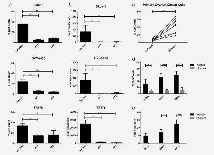
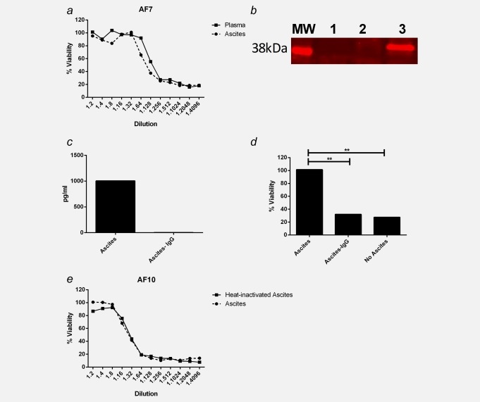
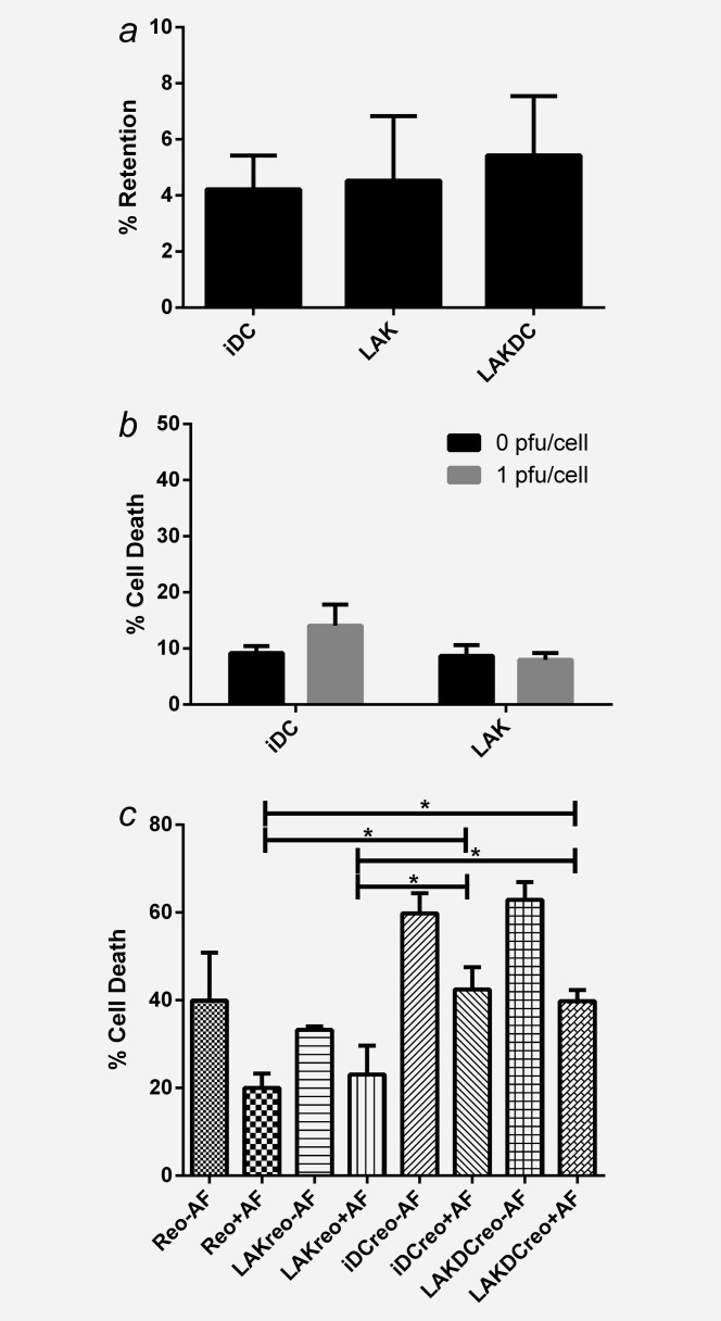
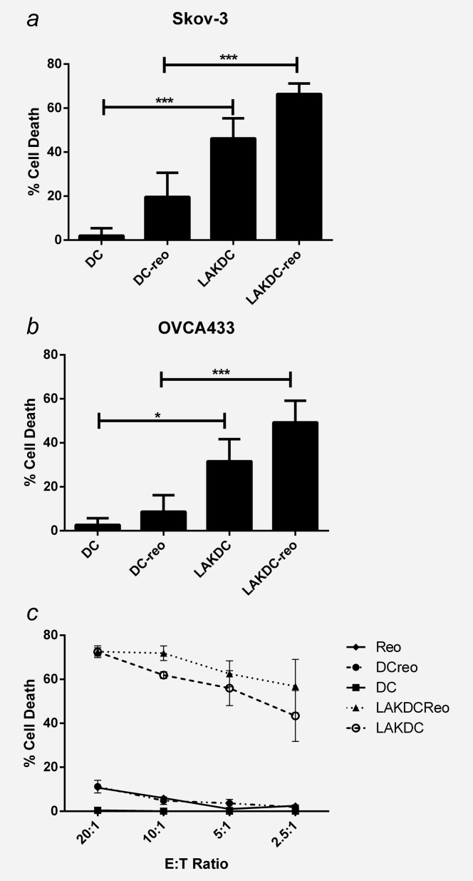
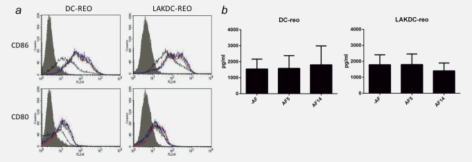
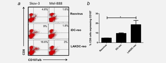
Reovirus is an oncolytic virus (OV), which acts by both direct tumor cell killing and priming of antitumor immunity. A major obstacle for effective oncolytic virotherapy is effective delivery of OV to tumor cells. Ovarian cancer is often confined to the peritoneal cavity and therefore i.p. delivery of reovirus may provide the ideal locoregional delivery, avoiding systemic dissemination. However, ovarian cancer is associated with an accumulation of ascitic fluid, which may interfere with oncolytic viral therapy. Here, we investigated the effect of ascites on reovirus-induced oncolysis against primary ovarian cancer cells and ovarian cancer cell lines. In the absence of ascites, reovirus was cytotoxic against ovarian cancer cells; however, cytotoxicity was abrogated in the presence of ascitic fluid. Neutralizing antibodies (NAb) were identified as the cause of this inhibition. Loading OV onto cell carriers may facilitate virus delivery in the presence of NAb and immune cells which have their own antitumor effector activity are particularly appealing. Immature dendritic cells (iDC), Lymphokine-activated killer (LAK) cells and LAKDC cocultures were tested as potential carriers for reovirus for tumor cell killing and immune cell priming. Reovirus-loaded LAKDC, and to a lesser degree iDC, were able to: (i) protect from NAb and hand-off reovirus for tumor cell killing; (ii) induce a proinflammatory cytokine milieu (IFNɣ, IL-12, IFNα and TNFα) and (iii) generate an innate and specific antitumor adaptive immune response. Hence, LAKDC pulsed with reovirus represent a novel, clinically practical treatment for ovarian cancer to maximise both direct and innate/adaptive immune-mediated tumor cell killing.

Keywords: antitumor immunity; malignant ascites; neutralizing antibodies; reovirus.


Figures

Figure 1
Figure 1
Malignant ascites inhibits reovirus-induced oncolysis. Ovarian cancer cell lines, Skov-3, OVCA433 and TR175 were infected with reovirus (1 pfu/cell) ± 2.5% ascitic fluid (2 separate samples; AF1 and 2). (a) Cell viability was determined using the Live/dead® viability assay and flow cytometry 48 hr postinfection (p.i.). Graphs show the mean percentage cell death + SEM from three independent experiments. (b) Reovirus replication was determined by plaque assay using L929 cells and fold increase was calculated from initial input reovirus. Graphs show mean fold increase of reovirus titres + SEM at 72 hr p.i. from three independent experiments. (c) Primary ascites-derived ovarian cancer cells from 10 patients were infected ± reovirus (10 pfu/cell) and cell viability was determined 48 hr p.i. using the Live/dead® assay. (d) Four primary samples were infected with reovirus (10 pfu/cell) ± 2.5% autologous ascitic fluid (samples AF3, 4, 5 and 6) for 24, 48 and 72 hr and cell viability determined as above. Graph shows the mean percentage cell death + SEM from four patient samples. (e) Reovirus replication was determined in primary ascites-derived ovarian cancer cells. Cells were infected with reovirus (1 pfu/cell) for 24, 48 and 72 hr ± 2.5% autologous ascitic fluid (samples AF3, 4 and 7) and reovirus replication determined as in (b). Graph shows the mean + SEM from three patient samples (*p < 0.05, **p < 0.01, ***p < 0.001 and ****p < 0.0001).
Figure 2
Figure 2
Reovirus inhibition is IgG-mediated. (a) L929 cells were infected with reovirus (0.05 pfu/cell) for 48 hr in the presence of serial dilutions of matched patient plasma and ascites (AF7). L929 cell viability was determined by MTT assay and normalized to uninfected control. Graph is representative of five patient samples. (b) IgG was immunoprecipitated from ascites, which had been incubated ± reovirus (5 × 10⁶ pfu) using protein A resin, then probed for reovirus σ3 protein (41 kDa MW) by western blotting. MW: molecular weight marker; Lane1: ascites (AF3) + protein A resin (no reovirus); Lane 2: protein A resin + reovirus [no ascites (IgG)]; Lane 3: ascites (AF3) + reovirus + protein A resin. Blot is representative of six ascitic samples. IgG-depleted ascites samples were collected by centrifugation following protein A resin incubation. (c) IgG concentration in ascites before and after IgG depletion was determined by ELISA. (d) L929 cells were infected ± reovirus (0.05 pfu) either in the presence of ascites, IgG-depleted ascites or in the absence of ascites, for 48 hr. Cell viability was determined by MTT assay and normalized to uninfected control. Graphs shows representative ascitic sample, AF9, of three samples (AF1, 2 and 9). (e) L929 cells were infected with reovirus (0.05 pfu/cell) for 48 hr in the presence of serial dilutions of matched ascites ± heat inactivation (AF10). L929 cell viability was determined by MTT assay and normalized to uninfected control. Graph is representative of three patient samples (**p < 0.01).
Figure 3
Figure 3
Reovirus-loaded iDC and LAKDC can deliver reovirus for tumor cell killing in the presence of ascites. Reovirus was loaded onto iDC, LAK cells and LAKDC cocultures (1pfu/cell) and either quantified for: (a) Percentage of reovirus retention by plaque assay or (b) Cytotoxicity by Live/dead® viability assay at 48 hr postloading. Graphs show the mean + SEM of three healthy donors. (c) TR175 were treated with neat reovirus or reovirus-loaded onto iDC, LAK cells and LAKDC (iDC-reo, LAK-reo and LAKDC-reo) for 4 hr ± 2.5% ascites. Carrier cells were then removed and replaced with growth media ± ascites; TR175 cell viability was then determined 72 hr p.i. by Live/dead® viability assay. Graph shows mean percentage of cell death + SEM from five healthy donors using two ascitic fluids (AF1 and 7; *p < 0.05).
Figure 4
Figure 4
Reovirus-loaded LAKDC are more cytotoxic towards ovarian cancer cells than reovirus-loaded iDC. Cytotoxicity of reovirus-loaded iDC and LAKDC was determined by chromium release assay. (a) Skov-3 and (b) OVCA433 targets were labeled with chromium and cultured with iDC-reo or LAKDC-reo in the presence of 2.5% ascites (AF11, 12 and 13) at indicated E:T ratios for 48 hr. Graphs show the mean percentage cell death + SEM from three healthy donors. (c) Daudi cells were labeled with chromium and incubated with neat reovirus (1 pfu/cell), reovirus-loaded or -unloaded iDC and LAKDC at indicated E:T ratios for 48 hr. Graph shows the mean percentage cell death ± SEM from three healthy donors (*p < 0.05, ***p < 0.001).
Figure 5
Figure 5
LAKDC and reovirus phenotypically mature iDC and produce proinflammatory cytokines in the presence of ascites. (a) Reovirus-loaded iDC or LAKDC were cultured in the absence (red line) or presence of 2.5% AF5 (green line) or AF14 (blue line) for 48 hr. DC (CD11c+ cells) were analyzed by flow cytometry for expression of maturation/activation markers, CD80 and CD86. Histogram plots are representative of two healthy donors. Shaded gray = isotype control, black line = unloaded DC controls [either iDC (left panel) or DC within LAKDC coculture (right panel)]. (b) Reovirus-loaded iDC and LAKDC were cultured for 48 hr ± ascites (AF5 and 14) before supernatants were collected and concentrations of IFNα were determined by ELISA. Graphs show the mean + SEM of four healthy donors.
Figure 6
Figure 6
Reovirus-loaded LAKDC prime antitumor immunity in the presence of ascites. Skov-3 cells were infected with either reovirus, iDC-reo or LAKDC-reo in the presence of 2.5% ascites (AF4) for 24 hr. Nonadherent carrier cells/dead tumor cells were removed and cultured with autologous PBMC for a week. CTL were restimulated weekly and tumor specific CTL activity measured in a CD107 assay against specific Skov-3 targets or irrelevant Mel-888 targets. (a) FACS plots are representative of three donors and the percentage of CD8 cells degranulating against targets is shown. (b) Graph shows the mean percentage + SEM of CD8 cells degranulating against Skov-3 targets from three healthy donors (*p < 0.05).

References

    1. Steele L, Errington F, Prestwich R, et al. Pro-inflammatory cytokine/chemokine production by reovirus treated melanoma cells is PKR/NF-kappaB mediated and supports innate and adaptive anti-tumour immune priming. Mol Cancer. 2011;10:20. - PMC - PubMed
    1. Gujar SA, Pan D, Marcato P, et al. Oncolytic virus-initiated protective immunity against prostate cancer. Mol Ther. 2011;19:797–804. - PMC - PubMed
    1. Endo Y, Sakai R, Ouchi M, et al. Virus-mediated oncolysis induces danger signal and stimulates cytotoxic T-lymphocyte activity via proteasome activator upregulation. Oncogene. 2008;27:2375–81. - PubMed
    1. Errington F, White CL, Twigger KR, et al. Inflammatory tumour cell killing by oncolytic reovirus for the treatment of melanoma. Gene Ther. 2008;15:1257–70. - PMC - PubMed
    1. Norman KL, Coffey MC, Hirasawa K, et al. Reovirus oncolysis of human breast cancer. Hum Gene Ther. 2002;13:641–52. - PubMed

Publication types