. 2014 Apr;3(4):609-16.
doi: 10.1002/adhm.201300151. Epub 2013 Aug 27.

Rapid prototyping of concave microwells for the formation of 3D multicellular cancer aggregates for drug screening

Affiliations

Rapid prototyping of concave microwells for the formation of 3D multicellular cancer aggregates for drug screening

Ting-Yuan Tu et al. Adv Healthc Mater. 2014 Apr.

Abstract

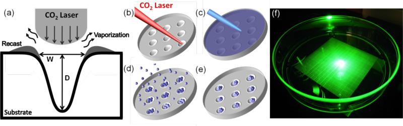
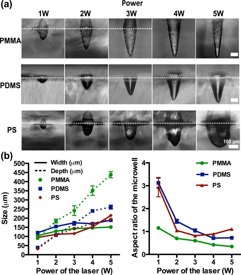
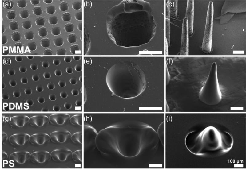
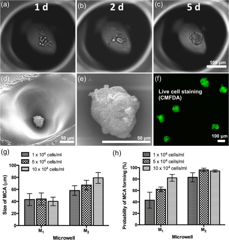
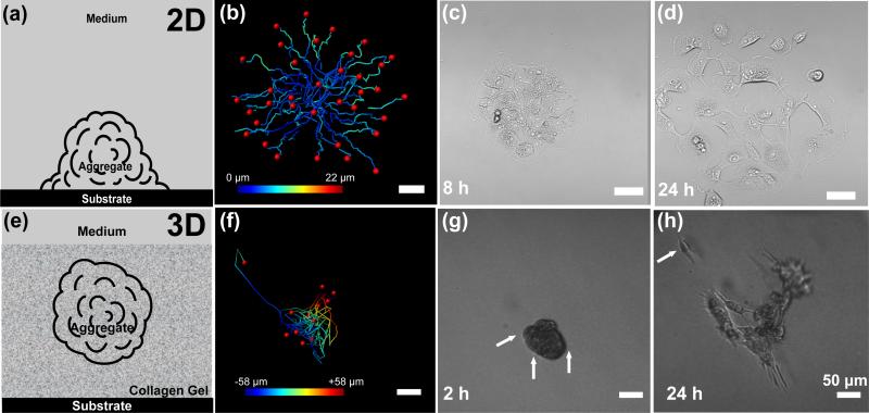
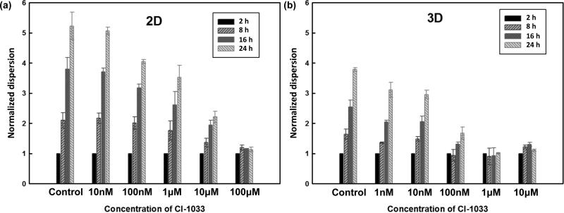
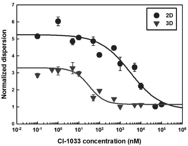
Microwell technology has revolutionized many aspects of in vitro cellular studies from 2D traditional cultures to 3D in vivo-like functional assays. However, existing lithography-based approaches are often costly and time-consuming. This study presents a rapid, low-cost prototyping method of CO2 laser ablation of a conventional untreated culture dish to create concave microwells used for generating multicellular aggregates, which can be readily available for general laboratories. Polymethylmethacrylate (PMMA), polydimethylsiloxane (PDMS), and polystyrene (PS) microwells are investigated, and each produces distinctive microwell features. Among these three materials, PS cell culture dishes produce the optimal surface smoothness and roundness. A549 lung cancer cells are grown to form cancer aggregates of controllable size from ≈40 to ≈80 μm in PS microwells. Functional assays of spheroids are performed to study migration on 2D substrates and in 3D hydrogel conditions as a step towards recapitulating the dissemination of cancer cells. Preclinical anti-cancer drug screening is investigated and reveals considerable differences between 2D and 3D conditions, indicating the importance of assay type as well as the utility of the present approach.

Keywords: drug screening; microwells; multicellular cancer aggregates; multicellular spheroids.


Figures

Figure 1
Figure 1
Schematic diagram of the microwell fabrication process for generation of 3D aggregates (a) Illustration of laser ablation process. Substrate is melted by CO2 laser energy distributed in a Gaussian profile. The ablation leaves a concave well with a recast zone at its edge. (b) Laser drilling via an XY mobile focus carriage travelling to the location of interest. (c) A thin layer of pluronic was deposited to prevent cell attachment on the substrate. (d) Cell seeding. (e) Formation of aggregates after 4-5 days. (f) A snapshot of microwell array pattern in a cell culture dish.
Figure 2
Figure 2
Characterization of microwells on PMMA, PDMS, and PS. (a) Representative microwell features with respect to a range of laser power from 1W-5W. For clarity, the reflection plane is indicated in white dotted line. (b) Evaluation of diameter, depth, and aspect ratio of microwell in different materials.
Figure 3
Figure 3
Isometric-view SEM images of microwells from three materials. A detailed surface profile of a single microwell is presented (middle), along with a negative formed by PDMS replica molding (right). (a)-(c) PMMA microwells exhibit a rough surface at the periphery and have micro- pores distributed over much of the internal surface. (d)-(f) PDMS microwells exhibit a smooth surface and a cone-shaped bottom. (g)-(i) PS microwells have smooth surface characteristics with substantial recast material around the edge and gentle curvature at the bottom.
Figure 4
Figure 4
Generation and characterization of MCAs. (a)-(c) Cell cultures are shown 1, 2, and 5 days after seeding. MCAs formed within 5 d. (d) SEM image illustrating the location of an MCA in a PS microwell. (e) A close-up SEM image of an MCA reveals the surface properties. The majority of cells clumped into a compact aggregate without distinct cell-cell interface indicating maximized intercellular adhesion. (f) CMFDA live cell staining indicated good cell viability and illustrates the shape of the MCAs. (g) Size correlation between MCAs and different cell seeding concentrations in M1 and M2 microwells. (h) Probability of MCA formation across different cell seeding densities in M1 and M2 microwells.
Figure 5
Figure 5
Schematic diagram and cell migration path over 24 h in 2D and 3D conditions. (a) Diagram of MCAs in direct contact with the substrate, where dispersion occurs on the 2D surface. (b) Single cells tracking over 24 h in 2D conditions. (c)-(d) Phase-contrast image of an MCA spreading at 8 and 24 hour in 2D conditions. (e) Diagram of MCAS in 3D suspension in native type I collagen. (f) Cell tracking over 24 h under 3D conditions. (g) Phase-contrast image of an MCA embedded in 3D collagen matrix showing early formation of pseudopodia at 2 h, indicated by arrows. (h) The same MCA broke up after 24 h, and a single escaped cell was observed (indicated by the arrow). Color bars represent the position along the Z direction.
Figure 6
Figure 6
Influence of CI-1033 on dispersion of A549 MCA in 2D and 3D conditions. Representative samples show normalized dispersion over different concentrations at 2, 8, 16 and 24 h. (a) 2D conditions. (b) 3D conditions.
Figure 7
Figure 7
Dose-response assays on dispersion of A549 MCA after 24 h CI-1033 drug treatment in 2D and 3D conditions.

References

    1. Wendt D, Riboldi SA, Cioffi M, Martin I. Adv. Mater. 2009;21:3352. - PubMed
    2. Wolf K, Wu YI, Liu Y, Geiger J, Tam E, Overall C, Stack MS, Friedl P. Nat. Cell Biol. 2007;9:893. - PubMed
    1. Hwang Y, Chung BG, Ortmann D, Hattori N, Moeller H, Khademhosseini A. Proc. Natl. Acad. Sci. U. S. A. 2009;106:16978. - PMC - PubMed
    1. Friedl P, Gilmour D. Nat. Rev. Mol. Cell Biol. 2009;10:445. - PubMed
    1. Hakanson M, Textor M, Charnley M. Integr. Biol. 2011;3:31. - PubMed
    2. Aref AR, Huang RY, Yu W, Chua KN, Sun W, Tu TY, Bai J, Sim WJ, Zervantonakis IK, Thiery JP, Kamm RD. Integr. Biol. 2013;5:381. - PMC - PubMed
    1. Chambers AF, Groom AC, MacDonald IC. Nat. Rev. Cancer. 2002;2:563. - PubMed

Publication types

MeSH terms

Substances

LinkOut - more resources