. 2013 Oct 8;110(41):E3945-54.
doi: 10.1073/pnas.1309991110. Epub 2013 Aug 27.

Polo-like kinase 2 regulates selective autophagic α-synuclein clearance and suppresses its toxicity in vivo

Affiliations

Polo-like kinase 2 regulates selective autophagic α-synuclein clearance and suppresses its toxicity in vivo

Abid Oueslati et al. Proc Natl Acad Sci U S A. .

Abstract

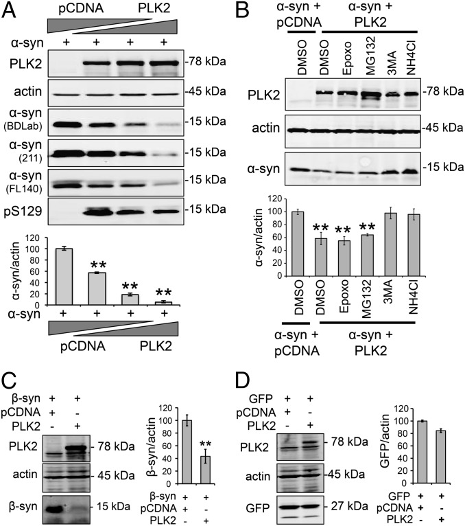
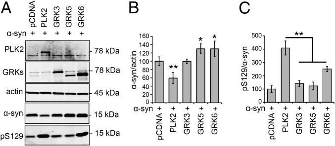
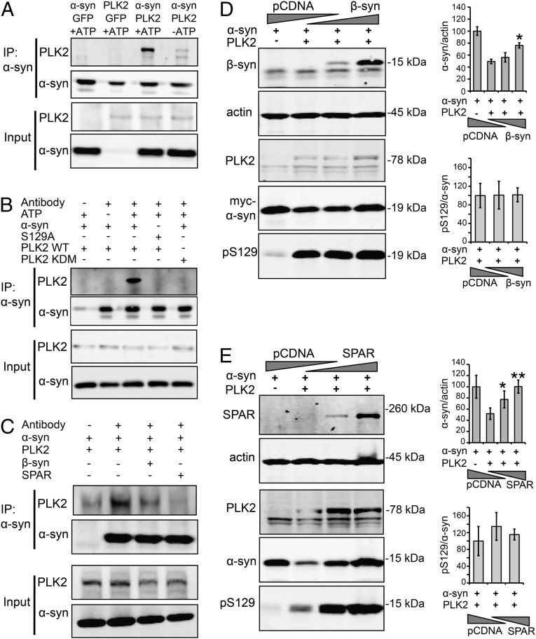
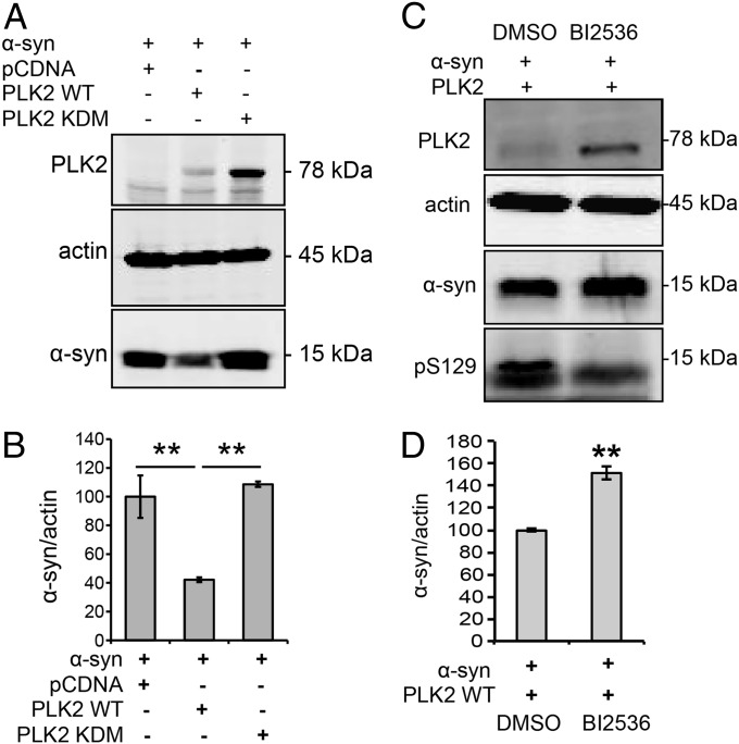
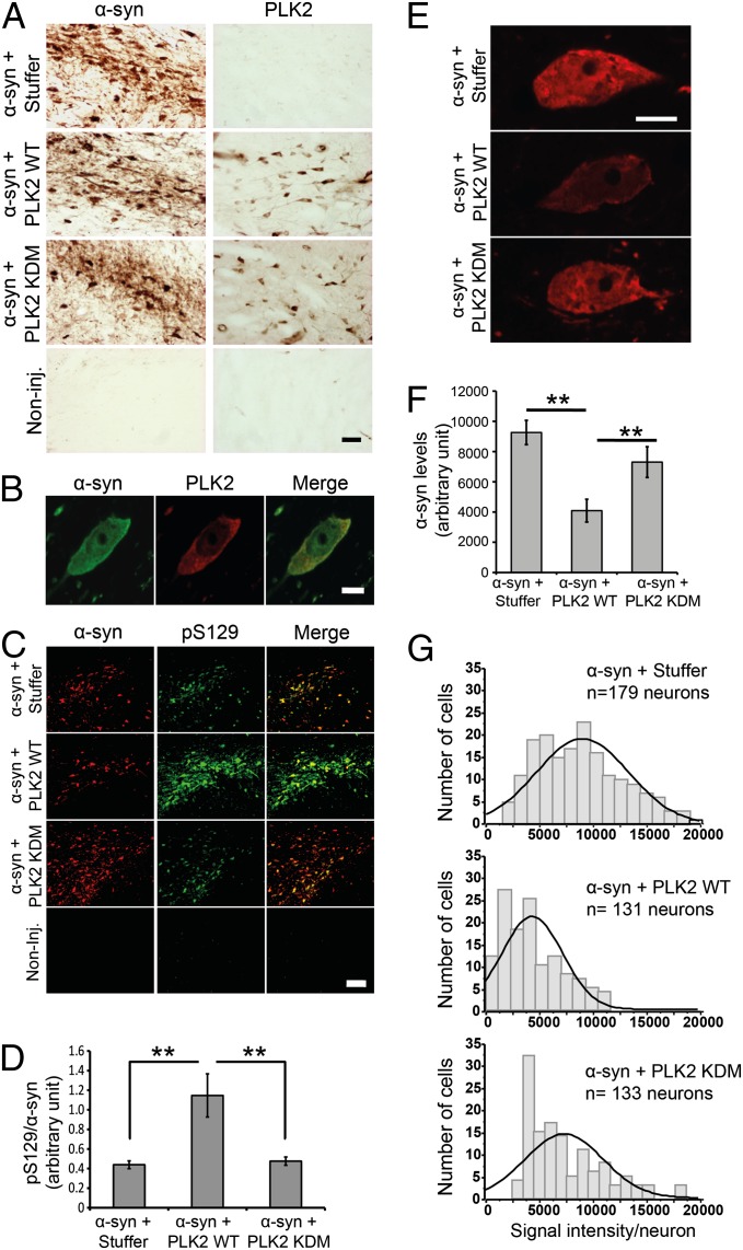
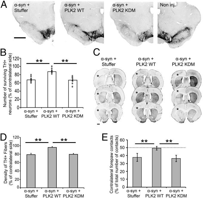
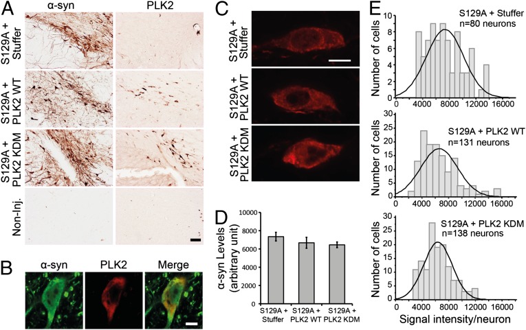
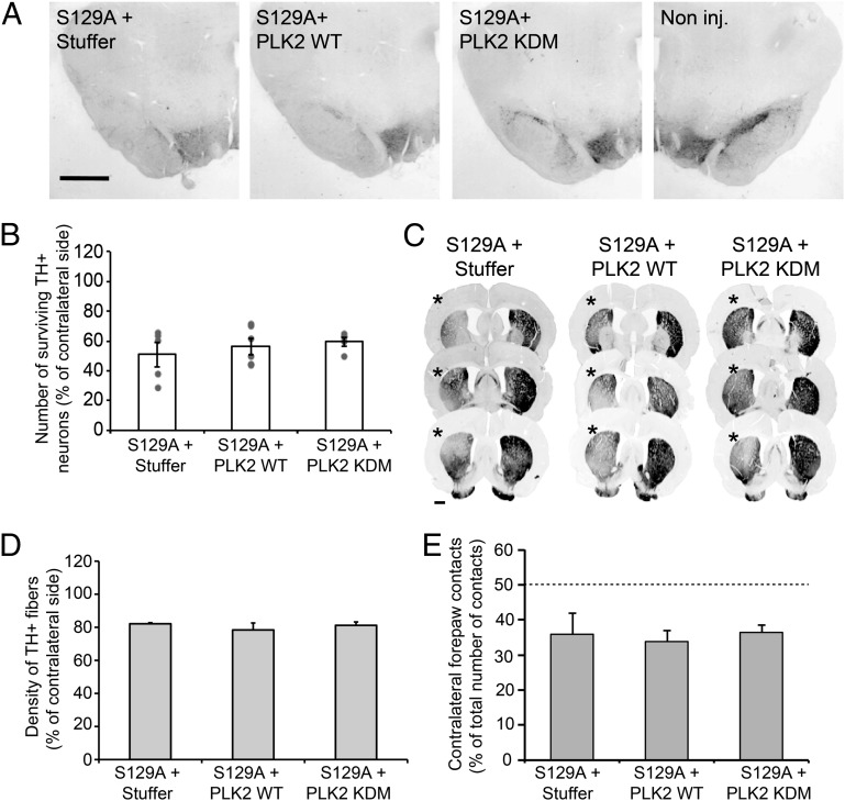
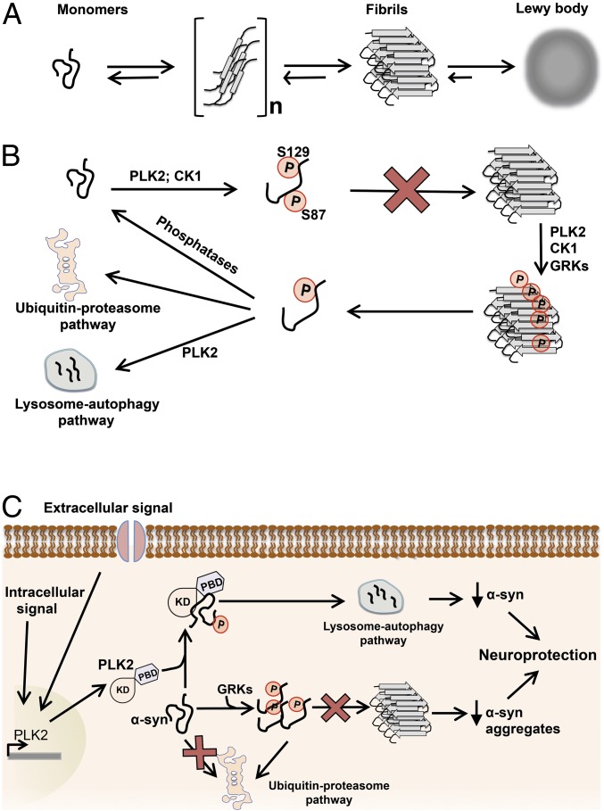
An increase in α-synuclein levels due to gene duplications/triplications or impaired degradation is sufficient to trigger its aggregation and cause familial Parkinson disease (PD). Therefore, lowering α-synuclein levels represents a viable therapeutic strategy for the treatment of PD and related synucleinopathies. Here, we report that Polo-like kinase 2 (PLK2), an enzyme up-regulated in synucleinopathy-diseased brains, interacts with, phosphorylates and enhances α-synuclein autophagic degradation in a kinase activity-dependent manner. PLK2-mediated degradation of α-synuclein requires both phosphorylation at S129 and PLK2/α-synuclein complex formation. In a rat genetic model of PD, PLK2 overexpression reduces intraneuronal human α-synuclein accumulation, suppresses dopaminergic neurodegeneration, and reverses hemiparkinsonian motor impairments induced by α-synuclein overexpression. This PLK2-mediated neuroprotective effect is also dependent on PLK2 activity and α-synuclein phosphorylation. Collectively, our findings demonstrate that PLK2 is a previously undescribed regulator of α-synuclein turnover and that modulating its kinase activity could be a viable target for the treatment of synucleinopathies.

Keywords: adeno-associated virus; animal model; serum inducible kinase.


Conflict of interest statement

Conflict of interest statement: This work has been partly supported by Merck Serono S.A., a for-profit corporation.

Figures

Fig. 1.
Fig. 1.
PLK2 overexpression enhances α-syn autophagy-mediated degradation in a cell-based assay. (A) Western blot and quantification of whole-cell lysate from HEK cells transfected with human α-syn (1 µg) and increasing amounts of PLK2 WT (0.5, 1, and 2 µg). For DNA titrations, total DNA per transfection was equalized by addition of pcDNA empty vector. The overexpression of PLK2 induced a concentration-dependent decrease of α-syn protein levels. The detection of α-syn using antibodies targeting different epitopes on α-syn ruled out the possibility of loss of epitope recognition (**P < 0.01). (B) Whole-cell lysate form HEK cells transfected with α-syn (1 µg) and PLK2 WT (0.5 µg) and treated with proteasome inhibitors (50 nM Epoxomicin and 10 µM MG132) and autophagy–lysosome inhibitors (10 mM 3MA and 25 mM NH4Cl). Western blot and optical density quantification showed that only autophagy–lysosome inhibitors reverse PLK2-induced reduction of α-syn (**P < 0.01). (C) Whole-cell lysate analysis and optical-density quantification showing that β-synuclein (β-syn) is degraded after PLK2 overexpression (**P < 0.01). (D) Whole-cell lysate analysis and optical density quantification showing that GFP levels are not affected by PLK2 overexpression.
Fig. 2.
Fig. 2.
PLK2 overexpression, but not the GRKs, mediated α-syn degradation in HEK239T cells. (A) Whole-cell analysis and Western blot illustrating the levels of total α-syn and pS129 in HEK cells transfected with α-syn and PLK2 or GRKs (GRK3, GRK5, and GRK6). (B) Optical-density quantification of α-syn signal showing that only the overexpression of PLK2 induced a decrease of α-syn levels. Interestingly, GRK5 and GRK6 induced an increase of α-syn signal (*P < 0.05; **P < 0.01). (C) Optical-density quantification of pS129 levels, relative to total α-syn signal, showing the superiority of PLK2 in phosphorylating α-syn in cell culture (**P < 0.01).
Fig. 3.
Fig. 3.
PLK2/α-syn complex formation is required for PLK2-mediated α-syn turnover. (A) Coimmunoprecipitation of α-syn and PLK2 in the presence or absence of ATP, showing that the presence of ATP is required for the stabilization of PLK2 and α-syn protein–protein interaction. (B) Coimmunoprecipitation of α-syn (WT and S129A) with PLK2 (WT and KDM), showing that the integrity of α-syn phosphorylation site S129 and PLK2 kinase domain are important for PLK2/α-syn interaction. (C) Coimmunoprecipitation of α-syn and PLK2 in the presence of β-syn or SPAR, showing that the presence of one of these two PLK2 substrates competes with α-syn for the interaction with PLK2 and reduces PLK2/α-syn coimmunoprecipitation. (D) Whole-cell lysate form HEK cells transfected with human α-syn (1 µg), PLK2 WT (0.5 µg), and increasing amounts of β-syn (1.5 and 3 µg). For DNA titrations, total DNA per transfection was equalized by addition of pcDNA empty vector. The Western blot revealed that the overexpression of β-syn reverses α-syn turnover without affecting pS129/α-syn levels (*P < 0.05). (E) Whole-cell lysate form HEK cells transfected with human α-syn (1 µg), PLK2 WT (0.5 µg), and increasing amounts of SPAR (1.5 and 3 µg). For DNA titrations, total DNA per transfection was equalized by addition of empty pcDNA empty vector. The Western blot revealed that the overexpression of SPAR reverses α-syn turnover without affecting pS129/α-syn levels (*P < 0.05; **P < 0.01).
Fig. 4.
Fig. 4.
PLK2 kinase activity is required for PLK2-mediated α-syn turnover. (A) Whole-cell lysate Western blot and (B) optical-density analysis showing that the overexpression of PLK2 KDM did not induce α-syn turnover. (C) Whole-cell lysate Western blot and (D) optical-density analysis showing that inhibition of PLK2 kinase activity, using the ATP-competitive inhibitor BI2536 (10 µM), decreases pS129 levels and suppresses PLK2-mediated α-syn turnover (**P < 0.01).
Fig. 5.
Fig. 5.
PLK2 WT overexpression, but not the KDM, induces α-syn phosphorylation at S129 and reduces intraneuronal α-syn protein levels. (A) Photomicrographs illustrating the expression of α-syn and PLK2 in the injected brains. (Scale bar: 50 µm.) (B) A high-magnification illustration of α-syn and PLK2 colocalization in a coinfected dopaminergic neuron. (Scale bar: 5 µm.) (C) Immunofluorescence of α-syn and pS129 showing the increase of pS129 levels after cooverexpression with PLK2 WT. (Scale bar: 100 µm.) (D) Optical-density quantification of pS129 levels, compared with total α-syn, estimating threefold increase of pS129 levels after cooverexpression of α-syn with PLK2 WT and histogram illustrating pS129 levels, relative to total α-syn. (E) High-magnification confocal images illustrating the population of neurons exhibiting α-syn intensity around the median line of the histograms of frequency distribution (Scale bar: 5μm). (F) Quantification, using ImageJ software, of α-syn protein levels per neuron showing that α-syn levels are significantly decreased when α-syn is cooverexpressed with PLK2 WT, compared with α-syn + Stuffer or α-syn + PLK2 KDM. (G) Histograms of frequency distribution of labeling, showing the distribution of α-syn signal intensity in the dopaminergic neuronal population (**P < 0.01).
Fig. 6.
Fig. 6.
PLK2 WT overexpression, but not the KDM, suppresses α-syn toxicity in vivo and alleviates hemiparkinsonian motor deficits. (A) Photomicrographs and (B) stereological quantification of the dopaminergic neurons in the injected SNc. (Scale bar: 1 mm.) (B) Quantification revealed that α-syn overexpression, alone or in combination with PLK2 KDM, induced a significant dopaminergic cell loss. However, PLK2 WT overexpression reverses α-syn toxicity (**P < 0.01). (C) Illustration of TH innervation density in the striatum and (D) optical-density quantification showed that only the overexpression of α-syn, but not PLK2 WT or KDM, induced a TH fiber loss in the ipsilateral striatum (*). (**P < 0.01) (Scale bar: 1 mm.) (E) Histograms illustrating the induction of hemiparkinsonian motor impairment after the overexpression of human α-syn. Before injection, all of the animals showed equal motor performances; however, the overexpression of α-syn, with the empty vector or PLK2 KDM, induced a significant decrease of the use of the contralateral forepaw, reflected by the forelimb asymmetry, whereas the overexpression of PLK2 WT reverses α-syn–induced motor impairment. **P < 0.01.
Fig. 7.
Fig. 7.
PLK2 WT overexpression did not reduce α-syn protein levels in rat midbrain. (A) Photomicrographs illustrating the expression of α-syn S129A and PLK2 in the injected brains. (Scale bar: 50 µm.) (B) High-magnification illustrations of α-syn S129A and PLK2 colocalization in a coinfected dopaminergic neuron. (Scale bar: 5 µm.) (C) High-magnification confocal images illustrating the population of neurons exhibiting α-syn S129A intensity around the median line of the histograms of frequency distribution (Scale bar: 5μm). (D) Quantification of α-syn S129A protein levels per neurons showing that the protein levels are similar in all of the experimental conditions. (E) Histograms of frequency distribution of labeling, showing the distribution of α-syn S129A signal intensity in the dopaminergic neuronal population.
Fig. 8.
Fig. 8.
PLK2 WT overexpression did not rescue S129A-induced toxicity. (A) Photomicrographs and (B) stereological quantification of the dopaminergic neurons in the injected SNc. Quantification revealed that α-syn overexpression with empty vector or in combination with PLK2 induced extensive dopaminergic lesion. (Scale bar: 1 mm.) (C) Illustration of TH innervation density in the striatum. (D) Optical-density quantification showed that α-syn overexpression with empty vector or PLK2 induced comparable and extensive TH fiber loss in the ipsilateral striatum (*). (Scale bar: 1 mm.) (E) Histograms illustrating the induction of hemiparkinsonian motor impairment after the overexpression of human α-syn in combination with empty vector or PLK2. Four months postinjection, animals exhibited comparable forelimb asymmetry caused by the dopaminergic cell loss.
Fig. 9.
Fig. 9.
Potential role of phosphorylation in the regulation of α-syn turnover, aggregation, and toxicity. (A) Schematic representation of the α-syn aggregation pathway. A dynamic equilibrium exists between monomeric and mutimeric forms of α-syn (dimers, trimmers, ...). Deregulation of this equilibrium promotes the formation of α-syn fibrils and then the development of abnormal proteinaceous cytoplasmic inclusions, called Lewy bodies. (B) Schematic diagram depicting the role of phosphorylation in regulating α-syn aggregation. PLK2- and CK1-mediated phosphorylation of α-syn, at S129 and S129 + S87, respectively, inhibits α-syn aggregation in vitro and in vivo (25, 32, 52). Moreover, α-syn fibrils could also be directly phosphorylated by PLKs and GRKs at S129 (13) and by CK1 at S87 and S129 (52). Phosphorylation of aggregated α-syn could result in monomer dissociation or stabilize dissociated monomers, and then the soluble phosphorylated α-syn is addressed for degradation via autophagy or proteasome degradation pathways or rapidly dephosphorylated by cytoplasmic phosphatases. (C) Scheme representing the effect of phosphorylation on α-syn aggregation and toxicity in neurons. Accumulated α-syn could be phosphorylated by endogenous kinases, notably GRKs (21, 22), inducing an inhibition of α-syn aggregation and proteasomal degradation of the soluble phosphorylated monomers (34, 35). Under stress conditions (e.g., excitotoxicity) and elevation of endogenous concentration of calcium, PLK2 expression is induced in neurons (24, 37, 38). Overexpressed PLK2 interacts, phosphorylates, and induces autophagic degradation of phosphorylated α-syn. All together, the two phosphorylation-mediated pathways result in a significant reduction of α-syn protein levels and inhibition of α-syn aggregation and consequently protect neurons against α-syn–induced toxicity.

Comment in

  • Silencing synuclein at the synapse with PLK2.
    Looyenga BD, Brundin P. Looyenga BD, et al. Proc Natl Acad Sci U S A. 2013 Oct 8;110(41):16293-4. doi: 10.1073/pnas.1315622110. Epub 2013 Sep 26. Proc Natl Acad Sci U S A. 2013. PMID: 24072649 Free PMC article. No abstract available.

References

    1. Lang AE, Lozano AM. Parkinson’s disease: First of two parts. N Engl J Med. 1998;339(15):1044–1053. - PubMed
    1. Lang AE, Lozano AM. Parkinson’s disease: Second of two parts. N Engl J Med. 1998;339(16):1130–1143. - PubMed
    1. Obeso JA, et al. Missing pieces in the Parkinson’s disease puzzle. Nat Med. 2010;16(6):653–661. - PubMed
    1. Spillantini MG, Crowther RA, Jakes R, Hasegawa M, Goedert M. Alpha-synuclein in filamentous inclusions of Lewy bodies from Parkinson’s disease and dementia with lewy bodies. Proc Natl Acad Sci USA. 1998;95(11):6469–6473. - PMC - PubMed
    1. Lee VM, Trojanowski JQ. Mechanisms of Parkinson’s disease linked to pathological alpha-synuclein: New targets for drug discovery. Neuron. 2006;52(1):33–38. - PubMed

Publication types