

Uterine arteriovenous malformation
Abstract
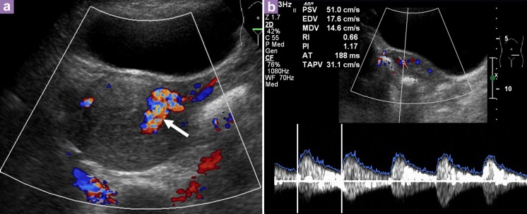
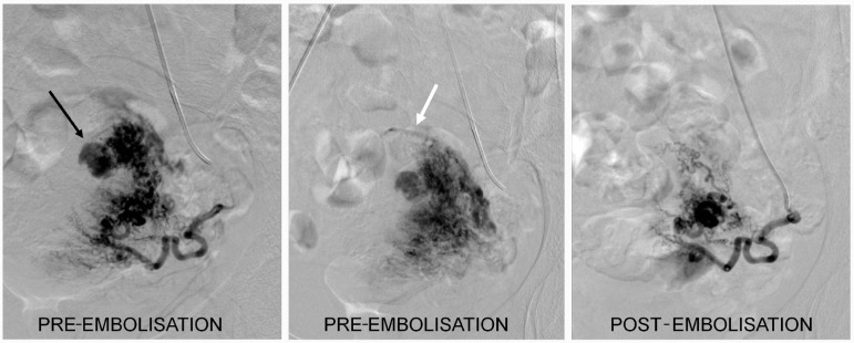
Uterine arteriovenous malformation (AVM) is a rare condition, with fewer than 100 cases reported in the literature. Despite it being rare, it is a potentially life-threatening condition. This case report describes a 33-year-old woman who presented with secondary post-partum hemorrhage. Transabdominal ultrasound (US) of the pelvis showed increased vascularity with multidirectional flow of the uterus and a prominent vessel, located on the left lateral wall. She also had retained product of conception, which complicated the diagnosis. A uterine artery angiogram confirmed an AVM in the fundal region with an early draining vein. Embolisation of the AVM was performed successfully.
Keywords: arteriovenous malformation; embolisation; post-partum hemorrhage; ultrasound; uterine; uterine artery; uterus.
Figures
References
-
- Hickey M, Fraser I. Clinical Implications of Disturbances of Uterine Vascular Morphology and Function. Baillieres Clin ObstetGynaecol. 2000;14(6):937–951. - PubMed
-
- Polat P, Suma S, Kantarcy M, Alper F, Levent A. Colour Doppler Ultrasound in the Evaluation of Uterine Vascular Abnormalities. Radiographics. 2002;22:47–53. - PubMed
-
- Fleming H, Ostor A, Pickel H, Fortune D. Arteriovenous Malformations of the Uterus. Obstet Gynaecol. 1989;73(2):209–213. - PubMed
-
- Grivell R, Reid K, Mellor A. Uterine Arteriovenous Malformations: A review of the Current Literature. Obstet Gynaecol Survey. 2005;60(11):761–767. - PubMed
-
- Huang M, Muradali D, Thurnston W, Burns P, Wilson S. Uterine Arteriovenous Malformations: Gray-Scale and Doppler Ultrasound features with MR Imaging Correlation. Radiology. 1998;206(1):115–123. - PubMed
LinkOut - more resources
Full Text Sources