

doi: 10.12816/0003274.
Epub 2013 Jun 25.
Placental Tumour: What could it be?
Affiliations
- PMID: 23984037
- PMCID: PMC3749036
- DOI: 10.12816/0003274
Item in Clipboard
Placental Tumour: What could it be?
Sultan Qaboos Univ Med J.
2013 Aug.
Abstract
Placental tumours include placental chorioangiomas, teratomas, haemangiomas, and haematomas. Placental chorioangiomas are benign vascular tumours and are the most common placental tumours, with a prevalence of 1%. Large placental chorioangiomas are rare and may lead to pregnancy complications and poor perinatal outcomes. These complications include fetal anaemia, hydrops fetalis, fetal growth restriction, polyhydramnios, and preterm delivery. We report a case of a large placental chorioangioma, the antenatal management and the maternal and fetal outcomes.
Keywords: Anemia, fetal; Case Report; Chorioangioma; Oman; Placenta; Polyhydramnios.
Figures
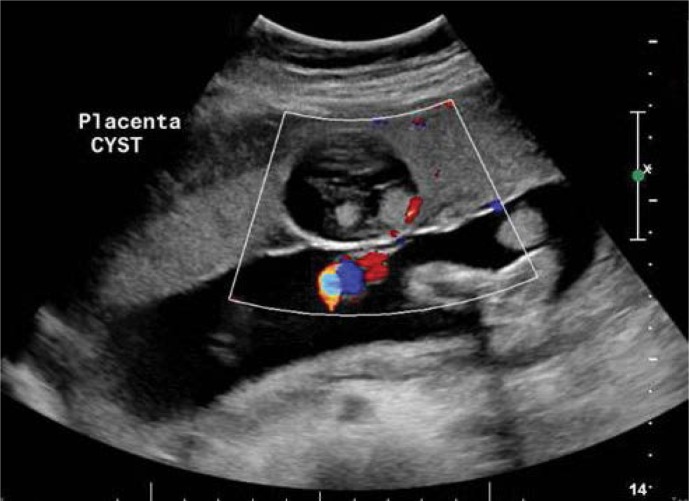
Placental chorioangioma of 6 × 5 cm in size within the centre of the umbilical cord with a feeding vessel.
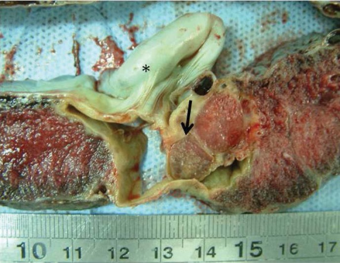
Well-circumscribed lesion (arrow) within the placenta just beneath the insertion of the umbilical cord (asterisk). It had a pale tan cut surface.
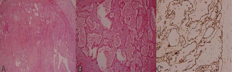
A to C: (A & B) Encapsulated lesion formed of variably-sized dilated and congested vessels. The lesion had undergone infarction. No chorionic villi were seen. (C) The CD-31 immunostain highlights the endothelial cells lining the vascular spaces.
References
-
- Amer HZ, Heller DS. Chorangioma and related vascular lesions of the placenta - a review. Fetal Pediatr Pathol. 2010;29:199–206. - PubMed
-
- Sepulveda W, Aviles G, Carstens E, Corral E, Perez N. Prenatal diagnosis of solid placental masses: The value of color flow imaging. Ultrasound Obstet Gynecol. 2000;16:554–8. - PubMed
-
- Zalwl Y, Weisz B, Gamzu R, Schiff E, Shalmon B, Achiron R. Chorangiomas of the placenta: Sonographic and Doppler flow characteristics. J Ultrasound Med. 2002;21:909–13. - PubMed
-
- Taori K, Patil P, Attarde V, et al. Chorioangioma of placenta: Sonographic features. J Clin Ultrasound. 2008;36:113–15. - PubMed
-
- Wallenburg HCS. Chorioangioma of the placenta: 13 new cases and a review of literature from 1939 to 1970 with special reference to the clinical complications. Obstet Gynecol Surv. 1971;26:411–25. - PubMed
LinkOut - more resources
Full Text Sources