Subfrontal schwannoma mimicking neuroblastoma: case report
- PMID: 23984204
- PMCID: PMC3743587
- DOI: 10.1055/s-0031-1275637
Subfrontal schwannoma mimicking neuroblastoma: case report
Abstract
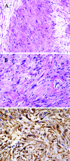
Computed tomography (CT), performed in a healthy 28-year-old man after minor head injury, detected a frontal base tumor. Neurological examination revealed left hyposmia. On magnetic resonance imaging scans, there was a heterogeneously enhanced tumor located in the left paramedian frontal base with extension into the left ethmoid sinus. Angiography showed a hypervascular mass in the left anterior cranial fossa; it was mainly fed by the left ethmoidal artery. Positron emission tomography scanning showed moderate accumulation of 11-methylmethionine and low accumulation of 18-fluorodeoxyglucose (FDG) at the tumor site. Bone image CT disclosed compressive, nondestructive deformation of the left frontal base. The preoperative diagnosis was olfactory neuroblastoma or meningioma. The tumor was totally resected via bifrontal craniotomy. The tumor was histologically diagnosed as typical schwannoma; it was positive for S-100 protein. We report a rare subfrontal schwannoma with extension into the nasal cavity that mimicked neuroblastoma. Low FDG accumulation and compressive deformation of the anterior skull base may help in the differential diagnosis of these tumors.
Keywords: Subfrontal schwannoma; neuroblastoma; olfactory nerve; skull base.
Figures


References
-
- New P FJ. Intracerebral schwannoma. Case report. J Neurosurg. 1972;36:795–797. - PubMed
-
- Russell D S, Rubinstein J L. Baltimore: Williams & Wilkins; 1989. Pathology of Tumors of the Nervous System. 5th ed; pp. 537–560.
-
- Von Strum K W, Bonis G, Kosmaoglou V. Ube rein neurinoma der lamina cribrosa. Zbl Neurochir. 1968;29:217–222. - PubMed
-
- Viale E S, Pau A, Turtas S. Olfactory groove neurinomas: case report. J Neurosurg Sci. 1973;17:193–196.
-
- Auer R N, Budny J, Drake C G, Ball M J. Frontal lobe perivascular schwannoma. Case report. J Neurosurg. 1982;56:154–157. - PubMed
LinkOut - more resources
Full Text Sources
